
Café au lait spots Lisch nodules Axilary freckling Cutes neromatosis Facial angiofibromas~ Hypo / hyper pigmented macules Port wine stain Explain nc markers in detail
neurocutaneous syndromes AND diagnosis
neurocutaneous syndromes diagnostic features cafe au lait Lisch nodules facial angiofibromas port wine stain authoritative
clinical images showing neurofibromatosis type 1 cafe au lait macules axillary freckling Lisch nodules
clinical images showing tuberous sclerosis facial angiofibromas ash leaf hypomelanotic macules
facial port wine stain Sturge Weber syndrome clinical image
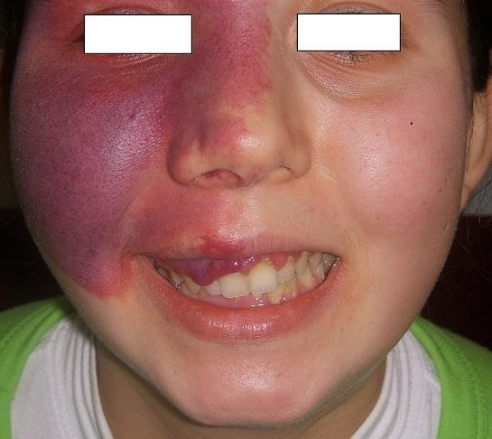
This clinical photograph displays a large, unilateral capillary malformation, commonly known as a port-wine stain (nevus flammeus), on the right side of a patient's face. The lesion is characterized by a deep purple to violaceous hue, with areas of lighter red towards the midline. Its distribution follows the V1 (ophthalmic) and V2 (maxillary) branches of the trigeminal nerve, covering the forehead, periorbital region, right nasal ala, and cheek. The lesion appears congestive and lacks sharply defined borders, blending into the surrounding skin. Additionally, the image shows intraoral manifestations: significant gingival hyperplasia is visible on the right side of the maxillary arch, appearing as bright red, bulbous overgrowths of the gingival tissue. The combination of a facial port-wine stain in the V1/V2 distribution and ipsilateral gingival vascular involvement is a classic clinical presentation of Sturge-Weber syndrome (encephalotrigeminal angiomatosis). This visual is essential for identifying vascular phakomatoses and understanding their dermatological and oral surgical implications.
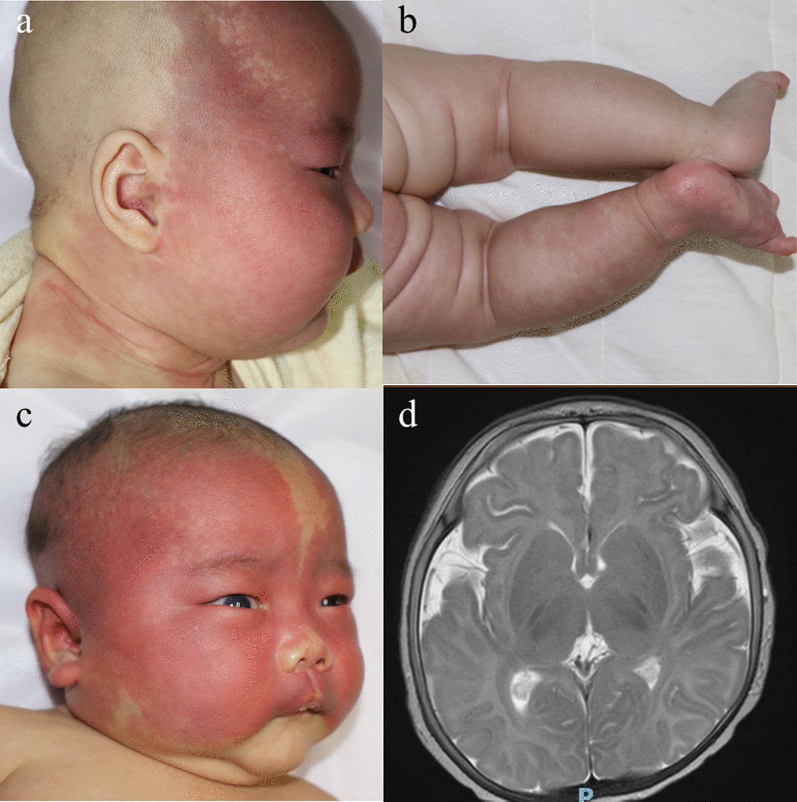
This composite image presents clinical and diagnostic features of Port-Wine Stain (PWS) and Sturge-Weber Syndrome (SWS) in pediatric patients. (a) Clinical photograph of an infant with PWS showing a large, erythematous-to-violaceous capillary malformation on the right side of the face and neck. (b) Clinical photograph showing a mottled capillary malformation on the left lower limb of an infant with symmetric limb length. (c) Clinical photograph of an infant with SWS displaying extensive, bilateral facial capillary malformations, involving the forehead, scalp, and right ear, with associated right-sided proptosis (eyeball protrusion). (d) Axial T2-weighted brain MRI demonstrating neurovascular involvement characteristic of SWS, specifically showing widened subarachnoid spaces (atrophy) and prominent, dilated, and tortuous vascular shadows along the bilateral temporal regions. The collection illustrates the phenotypic spectrum of somatic GNAQ mutations, ranging from isolated cutaneous port-wine stains to syndromic presentations involving ophthalmic and cerebral vascular abnormalities.
https://www.ncbi.nlm.nih.gov/books/NBK537001
1. Café-au-lait macules and axillary freckling: think NF1
Café-au-lait macules (CALMs)
- Flat, uniformly light-brown patches of increased pigment.
- Usually have smooth, well-defined borders, described as “coast of California” borders.
- Often appear in infancy or early childhood.
- A single or a few spots can be normal. The concern is multiple lesions.
Six or more CALMs measuring:
- >5 mm before puberty, or
- >15 mm after puberty.
Axillary or inguinal freckling
- Multiple tiny brown macules in the armpits or groin.
- Called the Crowe sign.
- Typically emerges later than CALMs, often in early childhood.
- It is much more suggestive of NF1 than CALMs alone.
Lisch nodules
- Small, dome-shaped, tan-brown iris hamartomas.
- Seen by slit-lamp ophthalmic examination, not usually obvious to the patient.
- They generally do not impair vision.
- Two or more Lisch nodules support NF1, especially with other NF1 features.
Cutaneous neurofibromas
- Soft, skin-colored to violaceous benign peripheral-nerve-sheath tumors.
- Often develop around adolescence or adulthood.
- A lesion may show the button-hole sign: it invaginates into the skin with pressure.
- Plexiform neurofibroma is a larger, often congenital, diffuse lesion following nerve branches. It feels like a “bag of worms” and is particularly important because it is strongly associated with NF1 and has a risk of malignant peripheral nerve sheath tumor.
NF1 pattern to remember
- Optic pathway glioma
- Learning, attention, or developmental difficulties
- Sphenoid-wing dysplasia, tibial bowing/pseudarthrosis, scoliosis
- Hypertension, including possible renal artery stenosis or pheochromocytoma in selected patients
2. Facial angiofibromas and hypopigmented macules: think tuberous sclerosis complex
Facial angiofibromas
- Multiple small red or pink papules, usually symmetric over the cheeks, nose, and nasolabial folds.
- Commonly appear in childhood, often around ages 2 to 5 years, and increase through adolescence.
- They are benign hamartomas containing fibrous tissue and blood vessels.
- Older term: adenoma sebaceum. This term is inaccurate because they are not sebaceous-gland tumors.
Hypomelanotic macules
- Areas with reduced pigment, not total absence of pigment.
- Often present at birth or in infancy.
- Classical morphology: ash-leaf macule, an oval or lance-shaped pale patch.
- Other patterns include polygonal lesions and tiny “confetti” lesions.
- They can be subtle in fair skin, so examine using a Wood lamp.
Other skin markers of TSC
- Shagreen patch: thickened, leathery, peau-d’orange connective-tissue nevus, commonly over the lumbosacral area.
- Ungual fibromas: fibrous growths around or beneath nails.
- Forehead fibrous plaque: firm plaque on the forehead or scalp.
Why TSC matters
- Brain: cortical tubers, subependymal nodules, subependymal giant-cell astrocytoma, seizures including infantile spasms, developmental or behavioral difficulties
- Kidneys: angiomyolipomas and cysts
- Heart: rhabdomyomas, especially in infants
- Lungs: lymphangioleiomyomatosis, especially in adult women
- Eyes: retinal hamartomas
3. Hyperpigmented versus hypopigmented macules
| Finding | Appearance | Main neurocutaneous association |
|---|---|---|
| Café-au-lait macule | Flat light-brown patch | NF1, Legius syndrome, McCune-Albright syndrome |
| Axillary/inguinal freckles | Tiny brown macules in skin folds | NF1 |
| Ash-leaf macule | Pale lance-shaped hypomelanotic patch | TSC |
| Confetti macules | Numerous tiny hypopigmented spots | TSC |
| Shagreen patch | Rough, thickened lumbosacral plaque | TSC |
- Hypopigmented means less melanin, as in ash-leaf spots.
- Hyperpigmented means more melanin, as in café-au-lait macules.
4. Port-wine stain: consider Sturge-Weber syndrome when high-risk facial distribution is present
Port-wine stain
- Congenital capillary malformation, also called nevus flammeus.
- Flat pink, red, purple, or dark-red patch.
- Unlike many infantile hemangiomas, it does not usually involute; it may darken and thicken over time.
- It is a vascular malformation, not a proliferative tumor.
When to suspect Sturge-Weber syndrome (SWS)
- Forehead
- Upper eyelid/periorbital area
- Upper face, especially extensive or bilateral lesions
- Facial port-wine capillary malformation
- Leptomeningeal vascular malformation on the same side of the brain
- Glaucoma and choroidal hemangioma
- Seizures, focal weakness, visual-field defects, stroke-like episodes, and developmental difficulties
High-yield one-line associations
- Café-au-lait spots + axillary freckles + Lisch nodules + neurofibromas → NF1
- Facial angiofibromas + ash-leaf spots + shagreen patch + seizures → Tuberous sclerosis complex
- Facial port-wine stain, especially forehead/eyelid + seizures or glaucoma → Sturge-Weber syndrome