
Shicks test
The Schick test is a skin test used to determine immunity to diphtheria. A small amount of diphtheria toxin is injected intradermally - if the person lacks sufficient antitoxin antibodies, a red inflammatory reaction appears at the site (positive test), indicating susceptibility. No reaction means immunity.
Searching sources...
Reading File
Finding Sources
Searching the Web
Schick test diphtheria immunity procedure interpretation
Reading File
Finding Sources
Reading File
Here is a complete overview of the Schick Test:
Schick Test
What It Is
The Schick test, developed by Bela Schick in 1913, is an in vivo skin test used to determine whether a person is susceptible to (or immune from) diphtheria. It is historically the in vivo counterpart of the Elek immunodiffusion test (which is in vitro).
- Quick Compendium of Clinical Pathology, 5th ed.: Under diphtheria diagnosis - "Elek immunodiffusion test: in vitro | Schick test: in vivo"
Principle
The test relies on the presence or absence of circulating antitoxin antibodies against diphtheria toxin. If the person has sufficient antitoxin (is immune), the injected toxin is neutralized and there is no skin reaction. If they lack antitoxin (are susceptible), the toxin causes local inflammation.
Procedure
- 0.1 mL of diluted diphtheria toxin (1/50 MLD - minimum lethal dose) is injected intradermally into one forearm.
- Heat-inactivated toxin (control) is injected into the other forearm to detect hypersensitivity reactions unrelated to the toxin itself.
- Results are read at 48-72 hours and again at 5-7 days.
Interpretation
| Result | Toxin Site | Control Site | Meaning |
|---|---|---|---|
| Positive | Red/swollen wheal (5-10 mm), peaks at 4-7 days | No reaction | Susceptible - no antitoxin present |
| Negative | No reaction | No reaction | Immune - adequate antitoxin present |
| Pseudo-reaction | Initial redness (fades within 4 days) | Same initial redness (fades too) | Hypersensitivity to toxin proteins, NOT true susceptibility |
| Combined reaction | Initial redness persists beyond 4 days | Initial redness fades after 4 days | Both susceptible AND hypersensitive |
Clinical Significance
- A positive test = no protective antitoxin = susceptible to diphtheria infection.
- A negative test = protective antibody levels present = immune.
- Historically used for pre-vaccination screening and outbreak contact tracing.
- Has been largely replaced by in vitro antibody assays and universal childhood immunization (DTaP/Tdap vaccines), but retains importance in microbiology teaching and immunology as a classic example of passive immunity assessment.
Organism Context: Corynebacterium diphtheriae
- Humans are the only reservoir.
- The bacterium infects tonsils/oropharynx and produces an exotoxin (only when infected with beta-corynephage).
- The toxin has two fragments: A (active) - inhibits protein synthesis by inactivating EF-2 (elongation factor 2); B (binding) - mediates cell entry.
- Toxin disseminates (not the bacteria) causing damage to the heart, nerves, and kidneys.
- Characteristic pathology: pseudomembrane in the throat.
- Lab diagnosis: Tellurite/Tinsdale media (black colonies), Loeffler serum media (metachromatic granules), Elek test or PCR.
(Quick Compendium of Clinical Pathology, 5th ed., Section 3.5.3.2.1.1)
Western blotting n southern blotting
Western blotting detects specific proteins using antibodies after gel electrophoresis, while Southern blotting detects specific DNA sequences using labeled probes. Both involve separating molecules by size, transferring to a membrane, and probing with a detection molecule.
Searching sources...
Finding Sources
Finding Sources
Reading File
Finding Sources
Reading File
Reading File
Now let me also fetch the Western blotting diagram from the textbook.
Loading Image
Finding Sources
Here is a thorough, side-by-side breakdown of both techniques:
Western Blotting vs Southern Blotting
Western Blotting (Immunoblotting)
What It Detects
Specific proteins in a complex mixture (e.g., cell lysate, serum).
Principle
Proteins are separated by size, transferred to a membrane, and identified using labeled antibodies specific to the target protein.
Step-by-Step Procedure
| Step | Details |
|---|---|
| 1. Sample preparation | Cells are lysed with detergent (e.g., SDS) to solubilize all proteins |
| 2. SDS-PAGE | Proteins are denatured and separated by molecular weight via sodium dodecyl sulfate polyacrylamide gel electrophoresis - smaller proteins migrate faster |
| 3. Transfer (Blotting) | Proteins are transferred (electrophoretically) from the gel to a nitrocellulose or PVDF membrane |
| 4. Blocking | Membrane is blocked (e.g., with milk or BSA) to prevent non-specific antibody binding |
| 5. Primary antibody | Membrane is incubated with a primary antibody specific to the target protein |
| 6. Secondary antibody | A labeled secondary antibody (enzyme-linked or fluorescent) binds to the primary antibody |
| 7. Detection | Enzyme substrate produces a visible/colored/luminescent band at the protein's molecular weight position |
Visual Diagram (HIV Western Blot example):
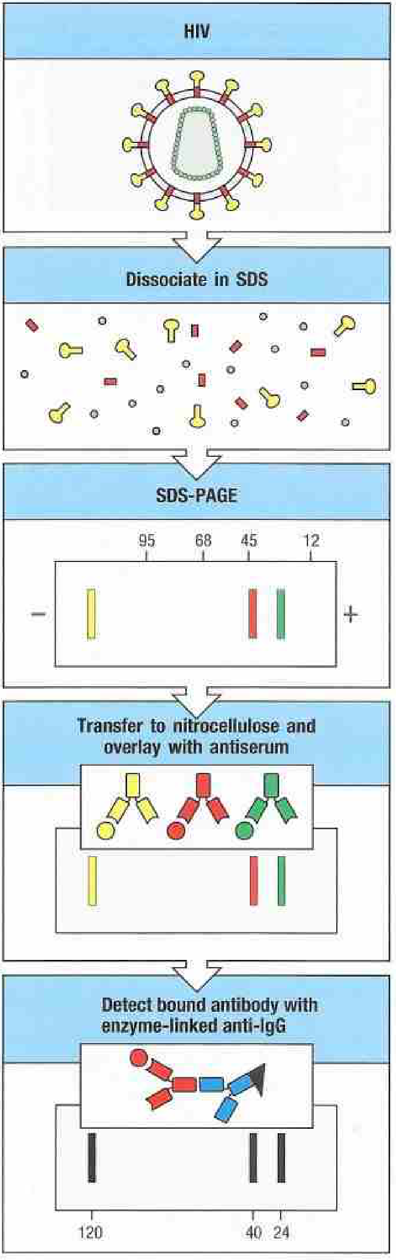
As shown above: HIV proteins are dissociated with SDS, separated by molecular weight, transferred to nitrocellulose, overlaid with test serum, and detected with enzyme-linked anti-IgG - bands appear at 120, 40, and 24 kDa (Janeway's Immunobiology, 10e)
Key Features
- Detects denatured proteins (since SDS unfolds them), so antibodies must recognize linear epitopes
- Result is a band at a specific molecular weight on the membrane
- Both qualitative (is protein present?) and semi-quantitative (how much?)
Clinical Uses
- HIV diagnosis: ELISA screening → confirmed by Western blot (detects antibodies against specific HIV proteins like gp120, p24, gp41)
- Lyme disease confirmation
- Herpes simplex serology
- Research: protein expression analysis
Southern Blotting
What It Detects
Specific DNA sequences (genes, mutations, restriction fragment patterns).
Named after its inventor, Edwin Southern (1975). The name "Western" blot was a pun derived from this - there is no "Mr. Western."
Principle
Genomic DNA is cut with restriction enzymes, fragments are separated by size, transferred to a membrane, and hybridized with a labeled DNA probe complementary to the sequence of interest.
Step-by-Step Procedure
| Step | Details |
|---|---|
| 1. DNA extraction | DNA extracted from patient cells (e.g., WBCs) |
| 2. Restriction digestion | DNA is cut into millions of fragments by a restriction endonuclease (e.g., EcoRI, BamHI) |
| 3. Gel electrophoresis | Fragments separated by size on agarose gel (smaller = faster migration) |
| 4. Denaturation | DNA in gel is denatured (made single-stranded) with NaOH |
| 5. Transfer (Blotting) | Single-stranded DNA fragments are transferred to nitrocellulose membrane |
| 6. Hybridization | A labeled DNA probe (complementary to target sequence) is added; it hybridizes to matching fragments |
| 7. Detection | Probe signal (radioactive, fluorescent, or chemiluminescent) reveals the band(s) |
What It Can Detect
- Large insertions or deletions
- Trinucleotide repeat expansions (e.g., Huntington's, Fragile X)
- Chromosomal rearrangements
- Point mutations that gain or lose restriction sites (RFLP - Restriction Fragment Length Polymorphism)
- Immunoglobulin/T-cell receptor gene rearrangements (lymphoid clonality - gold standard historically)
Clinical Uses
- Gene mutation detection (sickle cell, thalassemia)
- Lymphoma/leukemia clonality assessment
- Forensic DNA analysis (historical - largely replaced by PCR)
- Telomere length measurement
Comparison Table
| Feature | Western Blot | Southern Blot |
|---|---|---|
| Target molecule | Protein | DNA |
| Separation method | SDS-PAGE | Agarose gel electrophoresis |
| Detection probe | Labeled antibody | Labeled complementary DNA probe |
| Transfer membrane | Nitrocellulose or PVDF | Nitrocellulose |
| Denaturation | SDS (chemical) | Alkali (NaOH) |
| Clinical example | HIV confirmation, Lyme disease | Sickle cell disease, lymphoid clonality |
| Named after | Pun on "Southern" | Edwin Southern (inventor) |
The Blotting Family (Memory Aid)
| Blot | Target | Probe |
|---|---|---|
| Southern | DNA | DNA probe |
| Northern | RNA (mRNA) | DNA probe |
| Western | Protein | Antibody |
| Eastern | Post-translational modifications (lipids, carbohydrates) | Various |
"SNoW DRoP" - Southern = DNA, Northern = RNA, Western = Protein (Biochemistry, Lippincott Illustrated Reviews, 8th ed.)
Sources: Janeway's Immunobiology 10e; Biochemistry Lippincott Illustrated Reviews 8e; Jawetz Melnick & Adelberg's Medical Microbiology 28e; Harper's Illustrated Biochemistry 32e
Qbc test
The QBC (Quantitative Buffy Coat) test is a rapid diagnostic method for malaria that uses centrifugation and acridine orange fluorescent staining to concentrate and detect malaria parasites in blood. It is more sensitive than a standard thick blood smear.
Searching sources...
Finding Sources
Finding Sources
Reading File
Reading File
Reading File
Loading Image
Here is a complete overview of the QBC (Quantitative Buffy Coat) Test:
QBC Test (Quantitative Buffy Coat) - Malaria
Full Name
QBC Malaria Test - manufactured by Drucker Diagnostics (Port Matilda, Pennsylvania)
Principle
The QBC test uses acridine orange, a fluorescent dye, combined with microhematocrit centrifugation to concentrate and detect malaria parasites in blood.
- Acridine orange intercalates into parasite nucleic acid (DNA/RNA), causing the parasites to fluoresce bright yellow-green under a fluorescence microscope.
- Centrifugation concentrates parasitized red blood cells at the buffy coat layer (the interface between red cells and plasma), where they are easier to identify.
Procedure
| Step | Details |
|---|---|
| 1. Blood collection | ~55-65 µL of finger-prick capillary blood is collected directly into a special QBC tube pre-coated with acridine orange and heparin (anticoagulant) |
| 2. Centrifugation | Tube is centrifuged in a microhematocrit centrifuge for 5 minutes at high speed |
| 3. Parasite concentration | Blood layers separate; parasitized RBCs concentrate just above the buffy coat layer due to altered density |
| 4. Float insertion | A plastic float is inserted into the tube, which expands the buffy coat region for easier viewing |
| 5. Fluorescence microscopy | The tube is examined under a fluorescence microscope (or a standard microscope with a fluorescence attachment called the ParaLens) |
| 6. Reading | Parasites stained by acridine orange fluoresce brightly against a dark background |
Results
- Positive: Bright fluorescent yellow-green bodies (parasites) visible in the concentrated red cell layer
- Negative: No fluorescent parasites seen
Advantages
- Faster than thick/thin blood smear - results in ~15-20 minutes
- More sensitive than traditional thick and thin smears - detects low-level parasitemia
- Requires smaller blood volume (finger prick only)
- Easy to perform - less skill required than expert smear reading
- Attractive for labs that only occasionally test for malaria
Disadvantages / Limitations
| Limitation | Details |
|---|---|
| Requires fluorescence microscope | Expensive equipment; limits use in resource-poor settings |
| Cannot reliably speciate | Less reliable for identifying the exact Plasmodium species (falciparum vs vivax vs malariae, etc.) |
| Non-specific staining | Fluorescent background can cause false positives; interpretation requires expertise |
| Positive cases still need confirmation | Traditional thick/thin blood films must be examined for species ID and parasite burden calculation |
| Cost | More expensive than conventional smears |
(Henry's Clinical Diagnosis and Management by Laboratory Methods; Tietz Textbook of Laboratory Medicine, 7th ed.)
Comparison with Other Malaria Diagnostic Methods
| Method | Sensitivity | Speciation | Cost | Equipment |
|---|---|---|---|---|
| Thick blood smear (gold standard) | High (expert-dependent) | Yes | Low | Light microscope |
| Thin blood smear | Lower than thick | Best | Low | Light microscope |
| QBC test | Higher than smear | Unreliable | Moderate | Fluorescence microscope |
| RDT (Rapid Antigen) e.g., ParaSight F (HRP-2) | >95% for P. falciparum | Limited | Low | None |
| PCR | Highest | Excellent | High | PCR machine |
Clinical Context
- QBC detects all Plasmodium species that infect humans (P. falciparum, P. vivax, P. malariae, P. ovale, P. knowlesi)
- Also detects other blood parasites such as Babesia
- Rapid antigen tests like ParaSight F (detects HRP-2 protein of P. falciparum) and OptiMAL (detects parasite lactate dehydrogenase; distinguishes P. falciparum from P. vivax) are common alternatives in field settings
(Sherris & Ryan's Medical Microbiology, 8th ed.)
Mauntaux test
The Mantoux test (tuberculin skin test) is the standard method to detect TB infection. 0.1 mL of purified protein derivative (PPD) is injected intradermally into the forearm, and the resulting induration (not redness) is measured at 48-72 hours. The size of induration determines positivity based on the patient's risk category.
Searching sources...
Finding Sources
Finding Sources
Reading File
Reading File
Reading File
Reading File
Here is a complete breakdown of the Mantoux Test (you may have heard it called the "Mauntaux" test):
Mantoux Test (Tuberculin Skin Test / TST)
History
- Tuberculin test originally discovered by Von Pirquet in 1907
- A positive reaction indicates past or present infection with Mycobacterium tuberculosis
- It is the only tool for measuring the prevalence of TB infection in a community
What Is PPD (Tuberculin)?
Two WHO-standard tuberculins exist:
- PPD-S (5 TU = 0.1 µg/0.1 mL)
- PPD-RT 23 (1 TU = equivalent to 5 TU of PPD-S; used in India with Tween 80 as a detergent to prevent adsorption)
In India: 1 TU of PPD-RT 23 in 0.1 mL is used. Internationally: 5 TU of PPD-S in 0.1 mL is used.
Procedure
| Step | Detail |
|---|---|
| Site | Flexor (inner) surface of left forearm, midway between elbow and wrist |
| Injection | 0.1 mL of PPD injected intradermally using a tuberculin syringe, bevel facing upward |
| Wheal | A pale, blanched wheal of 6-10 mm diameter should appear immediately if correctly placed |
| Reading time | 48-96 hours after injection; 72 hours (3rd day) is ideal |
| What is measured | Induration only (palpable, raised hardened area) - erythema (redness) alone is NOT measured |
| How measured | Transverse (horizontal) diameter of induration in millimetres using a transparent ruler or calipers |
Interpretation of Results
General cutoffs (Park's Preventive Medicine):
| Induration | Result |
|---|---|
| 0 mm | Record as '0' |
| < 6 mm | Negative |
| 6-9 mm | Doubtful (may be due to M. tuberculosis or atypical mycobacteria) |
| ≥ 10 mm | Positive |
Risk-Based Positive Cutoffs (CDC Classification)
| Induration Size | Positive In |
|---|---|
| ≥ 5 mm | HIV-positive persons; recent close contacts of active TB case; fibrotic chest X-ray changes suggesting old TB; organ transplant/immunosuppressed patients (>15 mg/day prednisone for >1 month) |
| ≥ 10 mm | Recent immigrants (<5 yrs) from high-prevalence countries; HIV-negative IV drug users; lab personnel; residents/staff of prisons, nursing homes, hospitals, homeless shelters; medical risk conditions (diabetes, silicosis, CKD, leukemia, lymphoma, gastrectomy, >10% below ideal body weight); children <4 years or those exposed to high-risk adults |
| ≥ 15 mm | Persons with no risk factors for TB |
(Park's Textbook of Preventive & Social Medicine; Rosen's Emergency Medicine)
Immunological Basis
The Mantoux test is a Type IV (delayed-type) hypersensitivity reaction (cell-mediated immunity):
- Sensitized T lymphocytes from prior TB exposure recognize tuberculin antigens
- Cytokines are released → recruitment of macrophages → local induration
- Reaction peaks at 48 hours
(Histology: A Text and Atlas, 10e)
Causes of False Positive
- BCG vaccination (produces tuberculin hypersensitivity)
- Infection with atypical (non-tuberculous) mycobacteria
- Boosting effect (second TST boosting a waned response from old infection)
Causes of False Negative (Anergy)
| Cause |
|---|
| HIV infection / AIDS |
| Malignancy (Hodgkin's disease, leukemia) |
| Malnutrition |
| Severe bacterial infection (including active TB itself) |
| Viral infections (measles, chickenpox, glandular fever) |
| Recent live-virus vaccination (measles) |
| Immunosuppressive drugs (steroids) |
| Incorrect injection of PPD |
A negative Mantoux test does NOT rule out TB - particularly in immunocompromised patients.
Two-Step Testing
Used for periodic re-testing (e.g., healthcare workers) to distinguish true new conversion from a boosted old reaction:
- First test read at 48-72 hours
- If positive → consider infected
- If negative → proceed to step 2
- Second test given 1-3 weeks later, read at 48-72 hours
- If positive → consider previously infected (boosted reaction)
- If negative → consider truly uninfected
Prognostic Significance
- ≥ 20 mm induration (strong reactors): highest risk of developing active TB
- < 5 mm: greater risk than 6-9 mm group
- 92% of new TB cases occur in persons who are already tuberculin reactors
Comparison: Mantoux vs IGRA (Interferon-Gamma Release Assay)
| Feature | Mantoux TST | IGRA (QuantiFERON-TB Gold / T-SPOT) |
|---|---|---|
| Type | Skin test | Blood test |
| Visits required | 2 (injection + reading) | 1 |
| BCG false positive | Yes | No |
| Result time | 48-72 hours | Within 24 hours |
| Boosting effect | Yes | No |
| Use | Latent + active TB screening | Latent TB only |
| Cost | Low | Higher |
| Equipment | None | Lab required |
(Park's Textbook of Preventive & Social Medicine; Goldman-Cecil Medicine; Bailey & Love's Surgery)
"The tuberculin test must be approached with respect, administered with care, read with deliberation, and interpreted with sentient discrimination." - Park's Preventive Medicine
Elisa n types n test
ELISA (Enzyme-Linked Immunosorbent Assay) is a plate-based immunoassay that detects and quantifies antigens or antibodies using enzyme-labeled reagents. The four main types are direct, indirect, sandwich, and competitive ELISA. It is widely used for HIV, hepatitis, allergy testing, and many other conditions.
Searching sources...
Finding Sources
Finding Sources
Reading File
Finding Sources
Reading File
Reading File
Loading Image
Loading Image
Here is a complete breakdown of ELISA - its principle, all four types, and clinical applications:
ELISA (Enzyme-Linked Immunosorbent Assay)
Full Name
Enzyme-Linked Immunosorbent Assay
Basic Principle
ELISA detects and quantifies antigens or antibodies by using an enzyme-labeled reagent that converts a colorless substrate into a colored product. The intensity of color is measured by a spectrophotometer (at a specific absorbance wavelength) and is proportional to the amount of antigen/antibody present.
Key elements:
- Solid phase: Plastic microtitre (96-well) plate - antigen or antibody is coated onto wells
- Enzyme label: Usually horseradish peroxidase (HRP) or alkaline phosphatase
- Substrate: Colorless → produces color when cleaved by the enzyme
- Detection: Optical density (OD) measured by a plate reader
The Four Types of ELISA
1. Direct ELISA
What it detects: Antigen directly
How it works:
- Antigen from the sample is coated directly onto the well
- Enzyme-labeled primary antibody is added and binds to the antigen
- Unbound antibody is washed away
- Substrate is added → color change measured
Diagram:
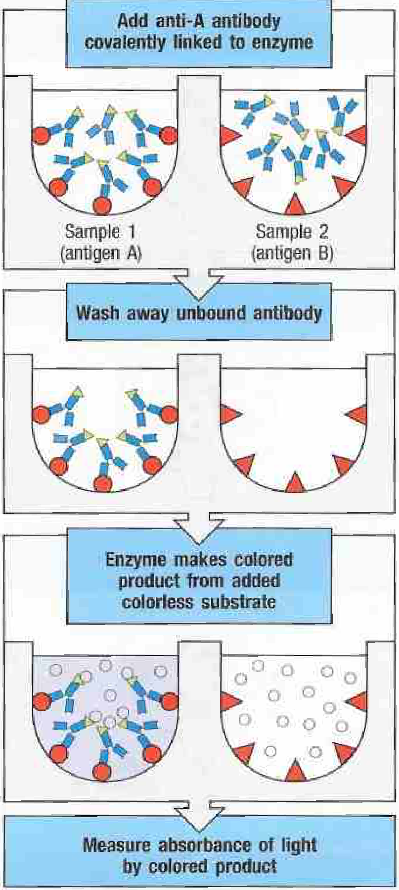
Sample 1 (antigen A) binds the enzyme-linked anti-A antibody → color produced. Sample 2 (antigen B) does not bind → no color. (Janeway's Immunobiology, 10e)
| Feature | Detail |
|---|---|
| Advantage | Simple, fast, no secondary antibody needed |
| Disadvantage | Less sensitive; primary antibody must be individually labeled; high background |
| Use | Drug/toxin detection (e.g., amphetamine assays) |
2. Indirect ELISA
What it detects: Antibody in patient sample (or antigen indirectly)
How it works:
- Known antigen is coated onto the well
- Patient serum (containing unknown antibody) is added → antibody binds antigen
- Enzyme-labeled secondary antibody (anti-human IgG) is added → binds patient antibody
- Substrate → color change
| Feature | Detail |
|---|---|
| Advantage | More sensitive than direct (signal amplified by secondary antibody); one secondary antibody works for many assays |
| Disadvantage | Extra step; possible non-specific secondary antibody binding |
| Use | HIV antibody screening, Lyme disease antibody detection, anti-hCG antibody testing |
3. Sandwich ELISA (Capture ELISA) - Most Commonly Used
What it detects: Antigen (even at very low concentrations)
How it works:
- Capture antibody (specific for antigen) is coated onto the well
- Sample containing antigen is added → antigen is captured
- Enzyme-labeled detection antibody (recognizes a DIFFERENT epitope of the same antigen) is added
- The antigen is sandwiched between two antibodies
- Substrate → color change
The antigen acts as a bridge - the more antigen present, the more detection antibody binds, the stronger the color.
| Feature | Detail |
|---|---|
| Advantage | Highest sensitivity and specificity; can detect very low antigen concentrations; works even in complex mixtures |
| Disadvantage | Requires two antibodies recognizing different (non-overlapping) epitopes on the same antigen |
| Use | Cytokine detection (TNF, IL-6, etc.); HIV p24 antigen detection; isoenzyme measurement (troponin, CK-MB); pregnancy tests (lateral flow); TNF antagonist drug monitoring |
4. Competitive ELISA (Inhibition ELISA)
What it detects: Antigen in samples of unknown composition; especially small antigens/haptens
How it works:
- Antibody is coated onto the well
- Labeled (known) antigen + unlabeled sample antigen are added simultaneously - they COMPETE for antibody binding sites
- The more unlabeled antigen in the sample, the less labeled antigen binds
- After washing, substrate is added
Inverse relationship: More antigen in sample = LESS color (weaker signal = positive/high concentration)
Diagram:
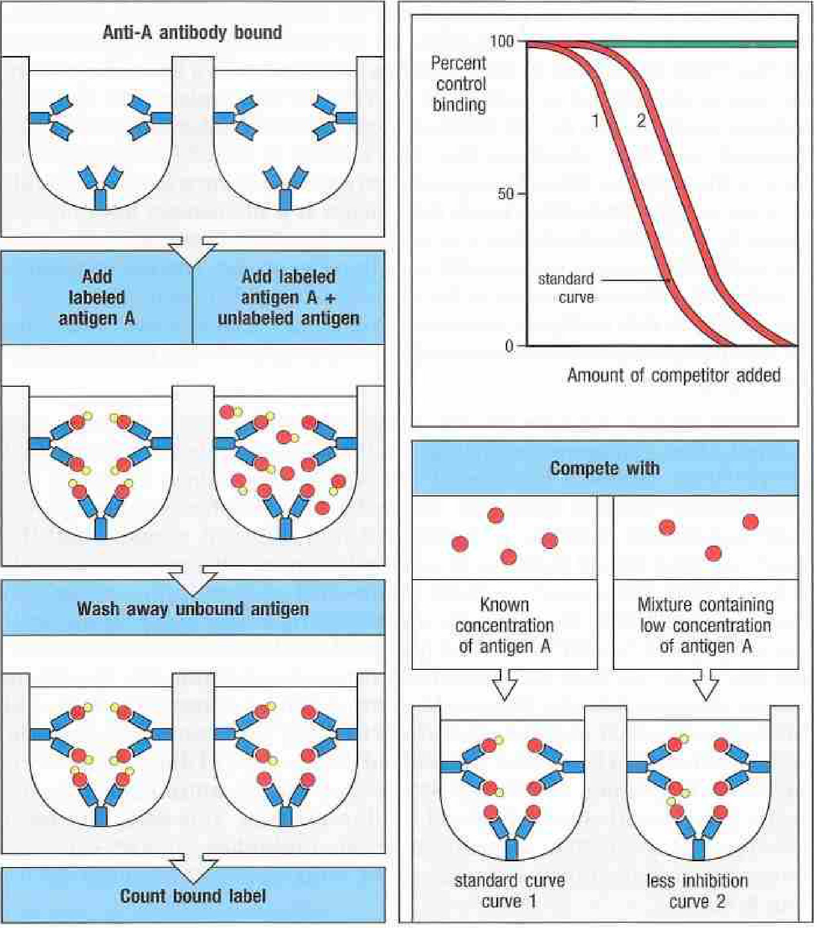
Labeled antigen + unlabeled sample antigen compete for antibody binding. High unlabeled antigen → less labeled antigen bound → less color. Standard curve used to calculate concentration. (Janeway's Immunobiology, 10e)
| Feature | Detail |
|---|---|
| Advantage | Works for small molecules (haptens) that cannot be "sandwiched" with two antibodies; can measure antigen in complex/impure samples |
| Disadvantage | Inverse result (higher antigen = lower signal); technically more complex |
| Use | Drug level monitoring (DPD for bone resorption); hormone assays; small molecule detection |
Summary Comparison Table
| Type | Coated on plate | What is detected | Signal direction | Sensitivity |
|---|---|---|---|---|
| Direct | Antigen | Antigen | Direct (more Ag = more color) | Lowest |
| Indirect | Antigen | Antibody in serum | Direct | Moderate |
| Sandwich | Capture antibody | Antigen | Direct | Highest |
| Competitive | Antibody | Antigen | Inverse (more Ag = LESS color) | Moderate-high |
Common Enzymes Used in ELISA
| Enzyme | Substrate | Color produced |
|---|---|---|
| Horseradish peroxidase (HRP) | TMB (tetramethylbenzidine) | Blue → Yellow (after stop solution) |
| Alkaline phosphatase | pNPP | Yellow |
Clinical Applications of ELISA
| Disease/Condition | Type of ELISA used |
|---|---|
| HIV screening | Indirect (detects anti-HIV antibodies); 4th-gen detects p24 Ag + Ab |
| Hepatitis B/C | Indirect |
| Lyme disease (screening) | Indirect |
| Cytokine measurement | Sandwich |
| Pregnancy test (hCG) | Sandwich (lateral flow format) |
| Troponin / CK-MB | Sandwich |
| Drug monitoring | Competitive |
| Autoantibody detection (ANA, anti-dsDNA) | Indirect |
| Pemphigus / Pemphigoid | Indirect/direct - preferred over immunofluorescence (more sensitive + quantitative) |
| Allergen-specific IgE | Indirect |
ELISA vs Western Blot (for HIV as example)
| Test | Purpose | Method |
|---|---|---|
| ELISA | Screening (high sensitivity) | Detects antibodies to HIV |
| Western Blot | Confirmation (high specificity) | Detects antibodies to specific HIV protein bands |
ELISA first (cheap, fast, sensitive) → if positive → Western blot to confirm (more specific, avoids false positives)
(Janeway's Immunobiology 10e; Cellular and Molecular Immunology; Tietz Textbook of Laboratory Medicine, 7th ed.; Rosen's Emergency Medicine)
Widal test
The Widal test is a serological test for typhoid fever that detects agglutinating antibodies against Salmonella typhi O (somatic) and H (flagellar) antigens in patient serum. A rising titer between two samples is more significant than a single result.
Searching sources...
Finding Sources
Finding Sources
Reading File
Reading File
Reading File
Here is a complete overview of the Widal Test:
Widal Test (Felix-Widal Test)
What It Is
The Widal test (also called the Felix-Widal test) is a tube dilution agglutination test used to detect agglutinating antibodies (both O and H) in the serum of patients suspected of having typhoid fever (Salmonella Typhi) or paratyphoid fever (S. Paratyphi A and B).
- Named after Georges-Fernand Widal (French physician, 1896)
- Still widely used in the Indian subcontinent and other resource-limited settings, though considered obsolete in many developed countries
Antigens Tested
| Antigen | Type | Antibody appears |
|---|---|---|
| O antigen | Somatic (cell wall LPS) | Day 6-8 after onset of fever |
| H antigen | Flagellar protein | Day 10-12 after onset |
| Vi antigen | Capsular (virulence) | Found in some chronic carriers |
Both S. Typhi and S. Paratyphi A, B antigens are tested separately
Procedure
Two formats are used:
1. Slide Agglutination Test (Rapid/Screening)
- Patient serum is mixed with known antigen suspension on a slide
- Clumping (agglutination) visible within a few minutes
- Only a qualitative/screening test
2. Tube Dilution Agglutination Test (Standard/Quantitative)
- Patient serum is serially diluted (1:20, 1:40, 1:80, 1:160, 1:320, 1:640...)
- Each dilution is mixed with a known suspension of S. Typhi O and H antigens in separate tubes
- Tubes are incubated (overnight at 37°C)
- Read for agglutination (clumping) - the highest dilution showing visible agglutination = the titer
Interpretation of Results
| Antibody | Titer considered Positive (single sample) |
|---|---|
| O antigen (Somatic) | ≥ 1:160 (some use ≥ 1:80 in high-endemic areas) |
| H antigen (Flagellar) | ≥ 1:640 |
| Vi antigen | Elevated titer suggests chronic carrier state |
Most significant finding: A 4-fold rise in titer between two sera collected 7-10 days apart (acute and convalescent) is considered strong evidence of active typhoid infection.
(Jawetz Melnick & Adelberg's Medical Microbiology 28e; Sleisenger & Fordtran's GI Disease)
Timing of the Test
- Best time: 2nd and 3rd week of illness (when antibody titers peak)
- Antibodies may be absent or very low in the 1st week → false negative
- Two specimens 7-10 days apart are ideal to demonstrate rising titer
Interpretation of O vs H Antibody Pattern
| Pattern | Interpretation |
|---|---|
| High O, low H | Active/early infection (O antibodies rise first) |
| High H, low O | Past infection or previous vaccination (H antibodies persist longer) |
| High O + High H | Active infection |
| Elevated Vi antibody | Chronic carrier state |
Limitations / False Results
False Positives
- Cross-reactions with other Salmonella serotypes (share O and H antigens)
- Cross-reactions with other Enterobacteriaceae
- Malaria, typhus, bacteremia by other organisms
- Liver cirrhosis
- Previous vaccination against typhoid (H antibodies persist)
- Endemic populations (baseline antibody levels already elevated)
False Negatives (up to 30% of culture-proven cases)
- Early disease (antibodies not yet raised)
- Prior antibiotic therapy (blunts antibody response)
- Immunocompromised patients
- Testing in the first week of illness
Comparison with Other Typhoid Diagnostic Methods
| Method | Sensitivity | Notes |
|---|---|---|
| Blood culture | 60-80% | Primary diagnostic test; lower if prior antibiotics given |
| Bone marrow culture | >90% | Gold standard; even in treated patients |
| Stool culture | Positive in weeks 2-3 | |
| Widal test | Moderate (~70%) | Widely used in endemic areas; many false +/- |
| Typhidot® | Higher | Detects IgM + IgG vs 50 kD S. Typhi antigen; takes 3 hours |
| Typhidot-M® | Better specificity | Detects IgM only (avoids IgG from old infection) |
| IDL Tubex® | Good | Detects IgM anti-O9; results in minutes |
| Dipstick test | Good | Detects S. Typhi-specific IgM against LPS; developed in Netherlands |
| PCR | High | Expensive; not widely available |
Current Status / Recommendations
- CDC does not recommend the Widal test for diagnosing acute typhoid - difficult to interpret in endemic populations and where prior vaccination or Salmonella infection may cause false positives
- Still widely used in India, South Asia, and Africa where blood cultures are not readily available
- Results of serologic tests for Salmonella infection cannot be relied upon to establish a definitive diagnosis - culture isolation remains the standard
(Park's Textbook of Preventive & Social Medicine; Jawetz Melnick & Adelberg; Red Book 2021; Sabiston Textbook of Surgery)
Quick Memory Aid
| Mnemonic | Meaning |
|---|---|
| "O" = Ongoing (active) | High O titer = active current infection |
| "H" = History/past | High H alone = past infection or vaccination |
| "Vi" = carVIer | High Vi = chronic carrier |
Immunity n types of immunity
Immunity is the body's ability to resist or overcome infection and disease. It is broadly divided into innate (non-specific, present from birth) and adaptive (specific, acquired). Adaptive immunity is further divided into humoral (antibody-mediated, B cells) and cell-mediated (T cells). Immunity can also be natural or artificial, and active or passive.
Searching sources...
Finding Sources
Finding Sources
Finding Sources
Finding Sources
Reading File
Reading File
Reading File
Loading Image
Here is a complete, well-structured overview of Immunity and its Types:
Immunity - Definition and Types
Definition
Immunity is the ability of the body to resist or overcome infection, disease, and foreign substances (antigens). It involves recognition of "self" vs "non-self" and elimination of what is foreign or harmful.
Master Classification of Immunity
IMMUNITY
│
├── 1. INNATE (Natural / Non-specific)
│
└── 2. ADAPTIVE (Acquired / Specific)
        ├── Humoral (B cell / Antibody-mediated)
        └── Cell-mediated (T cell)
            │
            Both can be:
            ├── ACTIVE (host generates own response)
            │     ├── Natural active (after infection)
            │     └── Artificial active (vaccination)
            └── PASSIVE (preformed antibodies given)
                  ├── Natural passive (maternal antibodies)
                  └── Artificial passive (antiserum/Ig given)
1. INNATE IMMUNITY (Natural / Non-specific)
Also called natural immunity or native immunity.
Key Features
| Feature | Detail |
|---|---|
| Present | From birth; always "on" |
| Specificity | Low - recognizes broad patterns shared by groups of microbes (PAMPs) |
| Memory | None (or very limited) |
| Speed | Very fast - responds within hours |
| Diversity | Low - germline-encoded receptors |
Components
| Component | Examples |
|---|---|
| Physical/Mechanical barriers | Skin, mucous membranes, cilia, respiratory epithelium |
| Chemical barriers | Stomach acid (HCl), lysozyme in tears/saliva, defensins |
| Cellular components | Phagocytes (neutrophils, macrophages), dendritic cells, NK cells, mast cells, innate lymphoid cells (ILCs) |
| Soluble proteins | Complement system, lectins, acute-phase proteins (CRP, fibrinogen) |
| Inflammatory response | Cytokines (IL-1, TNF, IL-6, interferons) |
Pattern Recognition
Innate immunity uses PRRs (Pattern Recognition Receptors) like Toll-like receptors (TLRs) to recognize PAMPs (Pathogen-Associated Molecular Patterns) - conserved structures on microbes.
2. ADAPTIVE IMMUNITY (Acquired / Specific)
Also called specific immunity or acquired immunity.
Key Features
| Feature | Detail |
|---|---|
| Develops | After exposure to antigen |
| Specificity | Very high - specific to a single antigen/epitope |
| Memory | Yes - faster and stronger on re-exposure |
| Speed | Slower - takes days to weeks to develop |
| Diversity | Extremely high - somatic gene recombination generates millions of antigen receptors |
The Classic Diagram:
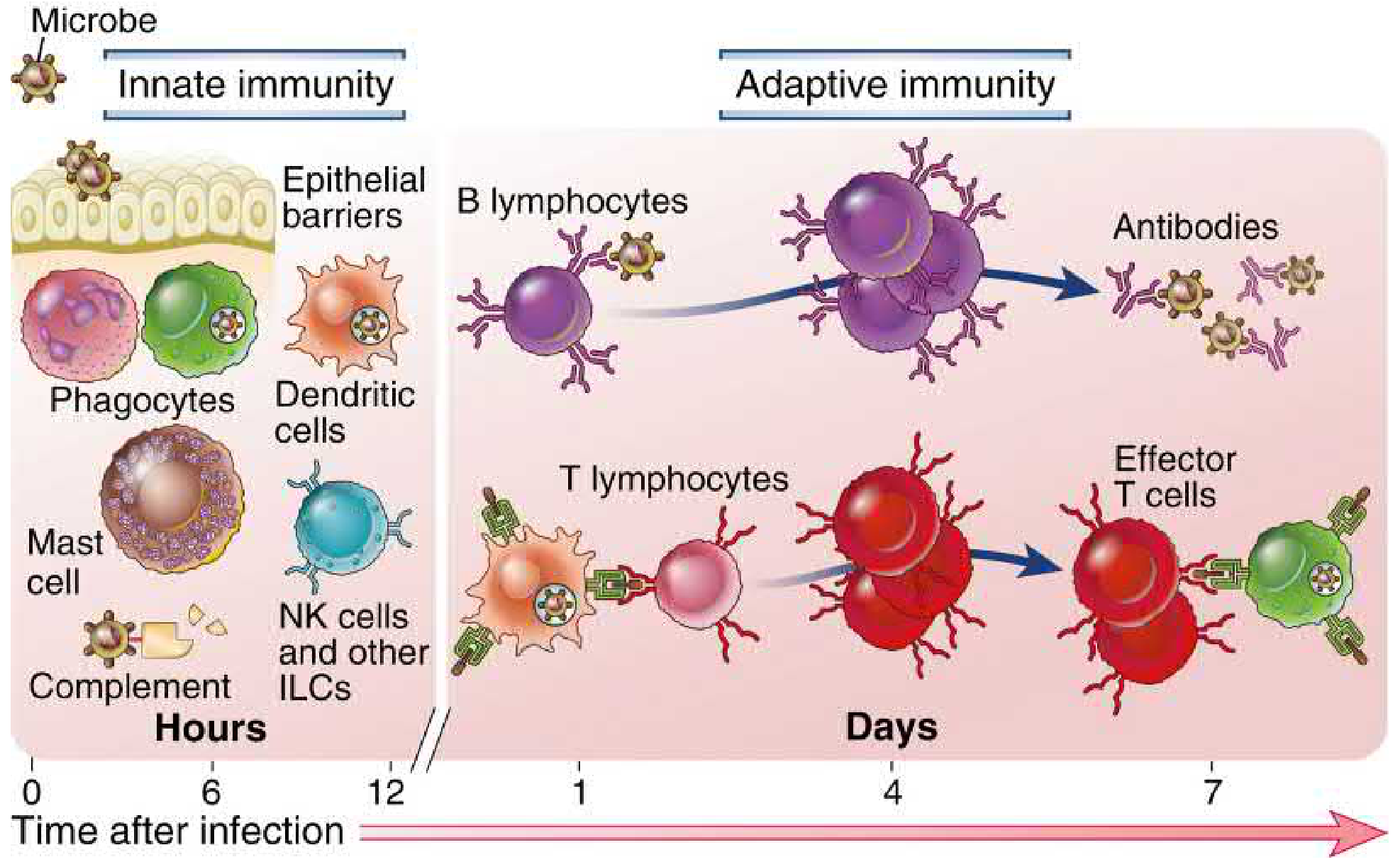
Cellular and Molecular Immunology - Innate immunity acts in hours; Adaptive immunity develops over days after lymphocyte activation
Adaptive Immunity is divided into two arms:
2A. Humoral Immunity (Antibody-Mediated Immunity)
- Mediated by B lymphocytes (B cells)
- B cells are activated by antigen → differentiate into plasma cells → secrete antibodies (immunoglobulins)
- Antibodies circulate in blood and body fluids (humors = fluids, hence the name)
Functions of antibodies:
- Neutralization (block toxins/viruses)
- Opsonization (coat bacteria for phagocytosis)
- Complement activation
- Antibody-dependent cellular cytotoxicity (ADCC)
Effective against: Extracellular bacteria, toxins, viruses (before cell entry)
2B. Cell-Mediated Immunity (CMI)
- Mediated by T lymphocytes (T cells)
- T cells do NOT produce antibodies but recognize antigens presented by MHC molecules
- On contact with antigen, T cells initiate: activation of macrophages, release of cytotoxic factors, delayed hypersensitivity reactions, secretion of lymphokines/cytokines
T cell subtypes:
| T Cell | Function |
|---|---|
| CD4+ Helper T cells (Th) | Help B cells make antibodies; activate macrophages; coordinate immune response |
| CD8+ Cytotoxic T cells (CTL) | Kill virus-infected cells, tumor cells, transplanted cells |
| Regulatory T cells (Treg) | Suppress immune response; prevent autoimmunity |
Effective against: Intracellular pathogens (M. tuberculosis, M. leprae, S. typhi, Candida, viruses), tumor cells, transplanted organs
A child born with a defect in humoral immunity may survive 6 years without therapy; a severe defect in cell-mediated immunity causes death within the first 6 months of life - illustrating the fundamental importance of CMI. (Park's Preventive & Social Medicine)
Innate vs Adaptive - Comparison Table
| Feature | Innate | Adaptive |
|---|---|---|
| Also called | Natural, non-specific | Acquired, specific |
| Speed | Hours | Days-weeks |
| Specificity | Broad (PAMPs) | Highly specific (single epitope) |
| Memory | No | Yes |
| Key cells | Neutrophils, macrophages, NK, mast cells | B cells, T cells |
| Key molecules | Complement, cytokines, defensins | Antibodies, TCR |
| Diversity | Low (germline-encoded) | Very high (somatic recombination) |
(Cellular and Molecular Immunology; Janeway's Immunobiology, 10e)
3. ACTIVE vs PASSIVE IMMUNITY
Active Immunity
- The host's own immune system generates the response
- Takes time to develop (days to weeks)
- Long-lasting (memory cells formed)
| Type | How acquired | Example |
|---|---|---|
| Natural active | After actual infection | Recovery from measles, chickenpox |
| Artificial active | After vaccination | DPT, MMR, polio vaccines |
Advantages of active immunity:
- Long duration (often lifelong)
- Severe reactions rare
- Protective efficacy approaches 100% in some vaccines
- Less expensive than passive
Passive Immunity
- Preformed antibodies are transferred to the host (no immune response generated)
- Established rapidly (immediate protection)
- Short-lived (days to months - until antibodies are cleared)
- No immunological memory; no education of the reticuloendothelial system
| Type | How acquired | Example |
|---|---|---|
| Natural passive | Maternal antibodies via placenta (IgG) or breast milk (IgA) | Neonatal protection in first 6 months |
| Artificial passive | Administration of antiserum/immune globulin | Anti-tetanus serum (ATS), anti-rabies Ig, IVIG |
(Park's Textbook of Preventive & Social Medicine; Guyton & Hall Medical Physiology; Jawetz Melnick & Adelberg's Medical Microbiology 28e)
4. HERD IMMUNITY (Community Immunity)
- When a sufficiently large proportion of a population is immune, susceptible individuals are indirectly protected because chains of infection cannot be maintained
- Elements that build herd immunity:
- Clinical and subclinical infections in the population
- Immunization programmes
- Herd structure (birth rate, death rate, population mobility)
- Basis for elimination and eradication of diseases (polio, diphtheria, smallpox)
(Park's Textbook of Preventive & Social Medicine)
Quick Memory Summary
| Type | Key feature | Duration | Example |
|---|---|---|---|
| Innate | Non-specific, fast, no memory | Immediate | Fever, phagocytosis |
| Adaptive - Humoral | B cells, antibodies, specific | Long (with memory) | Anti-HIV antibodies |
| Adaptive - CMI | T cells, specific, kills infected cells | Long (with memory) | TB immunity |
| Active natural | Own response after infection | Long (lifelong) | Post-measles immunity |
| Active artificial | Own response after vaccination | Long | Post-DPT immunity |
| Passive natural | Maternal antibodies | Short (6 months) | Neonatal IgG/IgA |
| Passive artificial | Antiserum given | Short (weeks-months) | Anti-tetanus serum |
This is a shared conversation. Sign in to Orris to start your own chat.