
Heart sounds
Reading File
Finding Sources
Finding Sources
Searching PubMed
"heart sounds"
Searching the Web
heart sounds S1 S2 S3 S4 murmurs clinical examination authoritative
Reading File
Reading File
Reading File
Reading Web Page
https://www.ncbi.nlm.nih.gov/books/NBK541010
Searching Images
diagram timing of S1 S2 S3 S4 heart sounds cardiac cycle phonocardiogram
Heart sounds
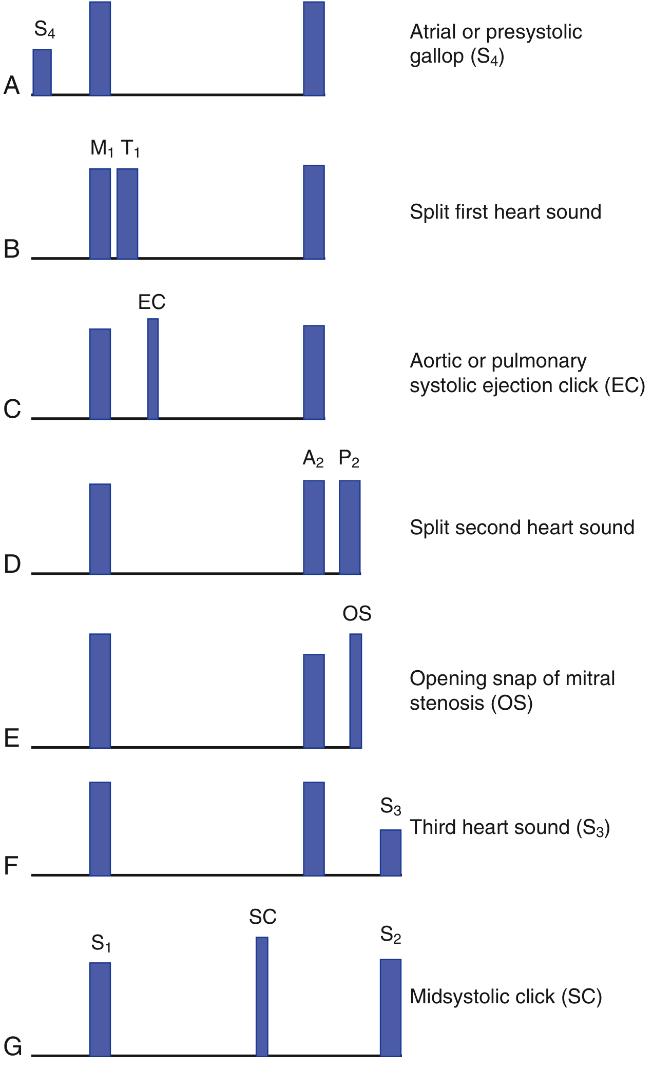
Heart sounds are vibrations produced mainly by valve closure and blood-flow related vibrations during the cardiac cycle. Normal auscultation gives “lub-dub”:
| Sound | Timing | Main cause | Usual significance |
|---|---|---|---|
| S1 | Start of systole | Closure of mitral and tricuspid valves | Normal “lub”; loudest at apex |
| S2 | End of systole, start of diastole | Closure of aortic and pulmonary valves | Normal “dub”; loudest at base |
| S3 | Early diastole, just after S2 | Rapid ventricular filling | Can be normal in children, young adults, and pregnancy; in older adults may suggest volume overload or heart failure |
| S4 | Late diastole, just before S1 | Atrial contraction into a stiff ventricle | Usually abnormal, seen with reduced ventricular compliance such as hypertension, aortic stenosis, or ischemic heart disease; absent in atrial fibrillation |
S1
- Produced when ventricular pressure exceeds atrial pressure and the AV valves close.
- M1 is mitral closure; T1 is tricuspid closure.
- Best heard at the apex.
- A loud S1 may occur with mitral stenosis or tachycardia. A soft S1 can occur with mitral regurgitation, long PR interval, or poor LV contractility.
S2
- A2 is aortic valve closure and normally occurs before P2, pulmonary valve closure.
- Best heard at the upper sternal borders.
- Physiological splitting: during inspiration, P2 is delayed because increased venous return prolongs right-ventricular ejection. This is normal. The MSD auscultation guide describes this normal inspiratory S2 split and important abnormal patterns.
Abnormal S2 splitting
- Wide split: delayed P2, for example right bundle branch block or pulmonary stenosis.
- Fixed wide split: classically atrial septal defect.
- Paradoxical split: delayed A2, for example left bundle branch block or severe aortic stenosis; splitting is heard on expiration and narrows with inspiration.
Gallop rhythm
A three-sound rhythm resembling a gallop:
- S3 gallop:
S1-S2-S3, early diastolic. - S4 gallop:
S4-S1-S2, late diastolic. - Summation gallop: S3 and S4 merge in tachycardia.
Where to listen
Valve areas indicate where sounds are best transmitted, not the anatomical positions of the valves:
- Aortic: right 2nd intercostal space, parasternal
- Pulmonic: left 2nd intercostal space, parasternal
- Tricuspid: left lower sternal border, around 4th-5th intercostal space
- Mitral: apex, left 5th intercostal space at the midclavicular line
Practical examination points
- Use the diaphragm for high-pitched sounds: S1, S2, murmurs of aortic regurgitation and mitral regurgitation.
- Use the bell lightly for low-pitched sounds: S3, S4, and mitral stenosis.
- Listen at the apex in the left lateral position for S3/S4 and mitral stenosis.
- Listen at the left upper sternal border during inspiration to assess S2 splitting.
A new S3 or S4, a diastolic murmur, syncope, breathlessness, chest pain, or leg swelling should be assessed clinically, often with echocardiography.
This is a shared conversation. Sign in to Orris to start your own chat.