
Complete notes on atherosclerosis
- Definition, epidemiology and risk factors (modifiable and non-modifiable)
- Pathogenesis (response-to-injury, lipid hypothesis, inflammation)
- Morphology: fatty streak, fibrofatty plaque, complicated plaque
- Vulnerable vs stable plaque and plaque rupture
- Clinical consequences by vascular bed (coronary, cerebral, aortic, renal, peripheral, mesenteric)
- Diagnosis: lipids, biomarkers, imaging, risk scores
- Prevention and management: lifestyle, statins and other lipid-lowering drugs, antiplatelets, revascularization
- Recent evidence and guidelines
- Endothelial dysfunction, from hemodynamic stress, hyperlipidemia or toxins, increases permeability and leukocyte adhesion.
- LDL accumulates in the intima and becomes oxidized.
- Monocytes migrate in and become macrophages, which take up oxidized LDL and turn into foam cells.
- T cells and macrophages release cytokines and growth factors.
- Smooth muscle cells migrate into the intima, proliferate, and lay down extracellular matrix and a fibrous cap.
Which topic should I cover in full detail first?
- Definition, epidemiology and risk factors (modifiable and non-modifiable)
- Pathogenesis (response-to-injury, lipid hypothesis, inflammation)
- Morphology: fatty streak, fibrofatty plaque, complicated plaque
- Vulnerable vs stable plaque and plaque rupture
- Clinical consequences by vascular bed (coronary, cerebral, aortic, renal, peripheral, mesenteric)
- Diagnosis: lipids, biomarkers, imaging, risk scores
- Prevention and management: lifestyle, statins and other lipid-lowering drugs, antiplatelets, revascularization
- Recent evidence and guidelines
- Endothelial dysfunction, from hemodynamic stress, hyperlipidemia or toxins, increases permeability and leukocyte adhesion.
- LDL accumulates in the intima and becomes oxidized.
- Monocytes migrate in and become macrophages, which take up oxidized LDL and turn into foam cells.
- T cells and macrophages release cytokines and growth factors.
- Smooth muscle cells migrate into the intima, proliferate, and lay down extracellular matrix and a fibrous cap.
"atherosclerosis"[MeSH Terms]
atherosclerotic plaque morphology fibrous cap lipid core foam cells
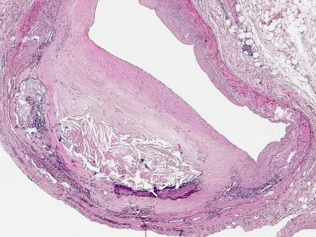
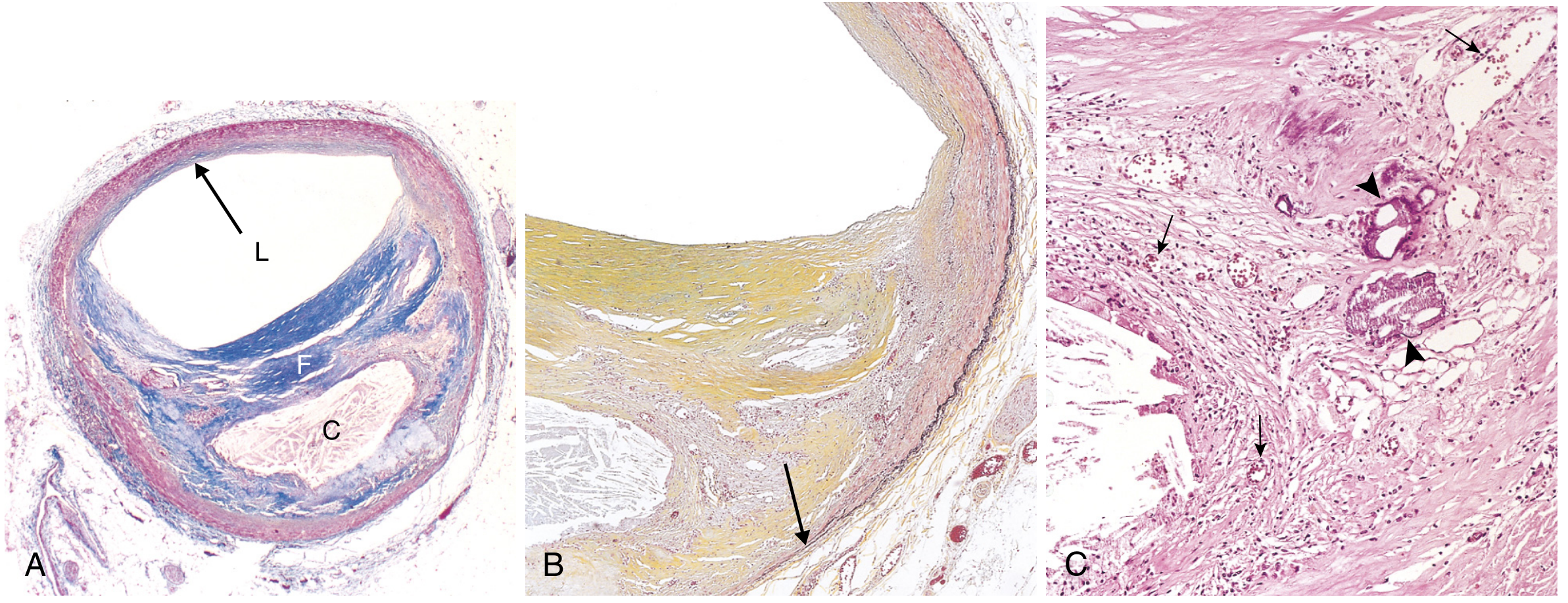
Light microscopy of a human coronary artery cross-section (Left Anterior Descending, LAD). The specimen shows an atherosclerotic plaque within the intima and media with a thick fibrous cap overlying a lipid-rich core. Foam cells, lipid droplets, and extracellular cholesterol crystals are present, indicating intra- and extracellular lipid deposits. The fibrous cap consists of dense connective tissue with smooth muscle cells and collagen, separating the luminal blood from the necrotic/lipid-rich core. The lumen is constricted by the plaque but not completely occluded in this section, consistent with a stable phenotype. Calcification may be present as small, basophilic granules in the plaque. Overall morphology reflects chronic atherogenesis due to long-standing hypertension with intimal thickening and macrophage infiltration. Notable features include macrophage-derived foam cells, cholesterol clefts, and a relatively thick fibrous cap, which reduces acute rupture risk compared with unstable plaques that display thinning, fissuring, cap rupture, hemorrhage, and luminal thrombosis. Clinically, such plaques can cause effort-induced chest pain (stable angina) and may precipitate myocardial ischemia if flow is compromised. This histology image aids in education, illustrating plaque composition, spatial relationships between lipid core and fibrous cap, and the pathophysiology of coronary artery disease.
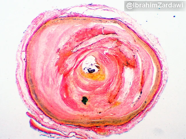
Comprehensive description: This histology slide uses light microscopy with Verhoeff-Van Gieson elastic stain to visualize the coronary artery wall in cross-section. The specimen displays an advanced atherosclerotic lesion within a epicardial coronary artery, with marked intimal thickening and concentric to eccentric plaque encroaching on the lumen, resulting in near-total luminal occlusion. The elastin-rich internal elastic lamina is variably preserved but disrupted in regions where the plaque expands; elastic fibers appear red with the stain, delineating media and intima boundaries. Within the intima there are lipid-laden foam cells and smooth muscle cells clustered in a fibrous matrix, consistent with a lipid-rich atherosclerotic core. The plaque shows a dense collagenous cap in places, creating a fibrous component, while possible microcalcifications are not conspicuous in this field. The vascular media remains evident around the lesion, but the lumen is severely narrowed, explaining ischemic risk. Clinically, such lesions underlie coronary artery disease and can precipitate angina or myocardial infarction. This image is valuable for teaching coronary atherosclerosis morphology, plaque composition, and the role of elastic staining in outlining elastic lamina continuity and plaque architecture. Potential applications include histopathology education, surgical planning discussions, and research on plaque stability and coronary occlusion. Enhancing diagnostic value globally.
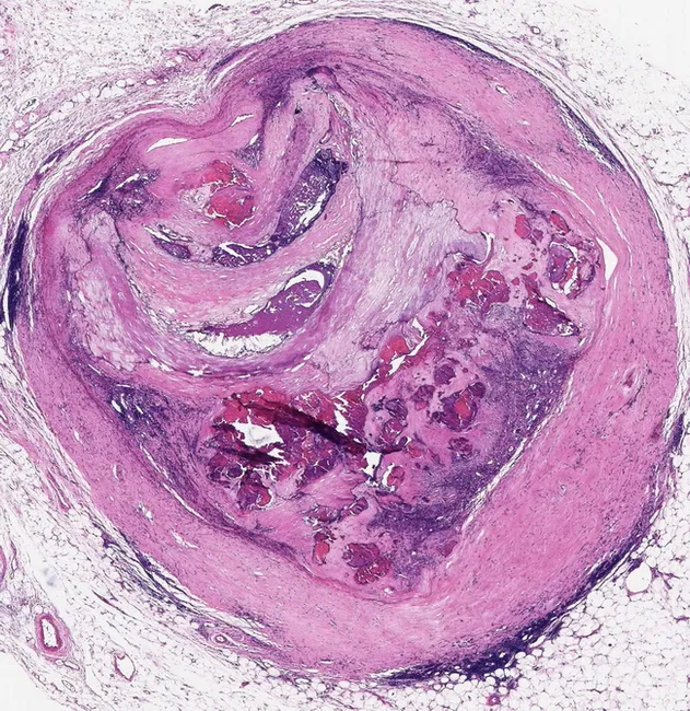
This histology image depicts a complex atheromatous plaque within the right coronary artery from a 70-year-old male, analyzed by light microscopy on paraffin-embedded tissue stained with hematoxylin and eosin. The cross-sectional view reveals an advanced atherosclerotic lesion characterized by a lipid-rich necrotic core (foam cell-laden debris) encircled by a thick fibrous cap. The plaque geometry shows concentric remodeling with calcific deposits and cholesterol clefts embedded within the intima. Notable features include ongoing cycles of injury and repair: medial smooth muscle cells appear to migrate into the intima and participate in fibrous cap formation and extracellular matrix deposition, contributing to cap thickening and structural integrity but potentially promoting vascular stiffness. Inflammatory infiltrates and microvessels within the plaque indicate ongoing chronic inflammation. The lesion resembles a healed, yet vulnerable, atherosclerotic plaque with potential fissuring, hemorrhage, or rupture risk in vivo, consistent with the patient’s history of recurrent myocardial infarctions. Surrounding media shows degenerative changes; the adventitia contains adipose tissue and loose connective tissue. Clinically, such plaques are associated with ischemic events, thrombosis, and restenosis; histology underscores the role of smooth muscle cell proliferation in repair and plaque stabilization paradoxically linked to luminal narrowing. This image serves as a teaching example of complex plaque morphology and remodeling in advanced coronary atherosclerosis.
2026 ACC/AHA dyslipidemia guideline LDL targets atherosclerotic cardiovascular disease
"Fatty streaks" in /textbooks/9780443264528
"## Atherosclerotic Plaque" in /textbooks/9780443264528
Atherosclerosis: Complete Notes
1. Definition and epidemiology
- Atherosclerosis is an intimal-based lesion made of a fibrous cap and an atheromatous (lipid necrotic) core. The plaque contains smooth muscle cells (SMCs), extracellular matrix (ECM), inflammatory cells, calcification, lipid and necrotic debris. It affects large elastic arteries (aorta, carotid, iliac) and large and medium muscular arteries (coronary, popliteal).
- It is the most frequent and clinically important vasculopathy. It underlies coronary, cerebral and peripheral vascular disease, and it causes roughly half of all deaths in the Western world. Myocardial infarction (MI) alone causes almost a quarter of US deaths. - Robbins, Cotran & Kumar Pathologic Basis of Disease, p. 465
- Mortality has fallen sharply in high-income countries because of risk reduction and better therapy. Coronary death rates in Africa, India and Southeast Asia now exceed those in the US. - Robbins, Cotran & Kumar Pathologic Basis of Disease, p. 465
2. Risk factors
| Group | Factors |
|---|---|
| Non-modifiable | Family history (the most important independent risk factor; usually polygenic, rarely Mendelian such as familial hypercholesterolemia), age, male sex (women are relatively protected until menopause) |
| Major modifiable | Hyperlipidemia (raised LDL, low HDL), hypertension, cigarette smoking, diabetes mellitus |
| Additional or emerging | Lipoprotein(a), C-reactive protein (CRP) and inflammation, hyperhomocysteinemia, hemostatic factors (for example raised PAI-1), obesity and metabolic syndrome, physical inactivity, stress |
- Risk factors act multiplicatively, not additively. - Robbins, Cotran & Kumar Pathologic Basis of Disease, p. 465
- Hypercholesterolemia alone is enough to start lesions. LDL is the atherogenic fraction. HDL moves cholesterol from the periphery (including atheromas) to the liver, and higher HDL is associated with lower risk. However, therapies that only raise HDL have not been effective. - Robbins, Cotran & Kumar Pathologic Basis of Disease, p. 466
- Omega-3 fatty acids are beneficial. Industrial trans fats worsen the lipid profile.
- Statins inhibit HMG-CoA reductase and also have anti-inflammatory "off-target" effects.
- PCSK9 binds the LDL receptor and speeds its degradation. PCSK9 inhibitors lower LDL markedly.
- Lp(a) is LDL with apo(a) attached to apoB-100. It is an independent risk factor that also affects fibrinolysis and inflammation.
- CRP adds prognostic information at every level of Framingham risk.
3. Pathogenesis: "response to injury" hypothesis
- Endothelial injury or dysfunction. This causes increased permeability, leukocyte adhesion and thrombosis.
- Lipoprotein accumulation in the intima, mainly LDL and its oxidized forms.
- Monocyte adhesion and migration into the intima, where they become macrophages and foam cells.
- Platelet adhesion.
- Factor release from platelets, macrophages and wall cells, which recruits SMCs from the media or from circulating precursors.
- SMC proliferation, ECM production and T-cell recruitment.
- Lipid accumulation inside and outside cells.
- Robbins, Cotran & Kumar Pathologic Basis of Disease, p. 467
-
Hemodynamic disturbance. Plaques form at vessel ostia, branch points and the posterior abdominal aorta, where flow is non-laminar. Laminar flow induces KLF2, which turns on atheroprotective genes and turns off inflammatory ones. Turbulent flow makes a site atheroprone. Statins partly act through KLF2.
-
Hypercholesterolemia. It raises reactive oxygen species, which impair endothelial function and speed nitric oxide (NO) decay. LDL becomes aggregated or oxidized in the intima. Macrophages take it up through scavenger receptors (not the LDL receptor) and become foam cells. SMCs can also become foam cells.
-
Inflammation. Smoking toxins, hypertriglyceridemia and cytokines contribute. Activated endothelium expresses adhesion molecules (VCAM-1) that recruit monocytes and T cells.
-
Robbins, Cotran & Kumar Pathologic Basis of Disease, p. 468-469
- The dominant plaque lipids are cholesterol and cholesterol esters.
- Familial hypercholesterolemia (LDL receptor loss of function) can cause MI before age 20 in homozygotes.
- Plasma cholesterol and LDL levels correlate with disease severity.
- Lowering cholesterol slows progression, causes regression of some plaques and reduces events.
- Activated macrophages and T cells release cytokines and growth factors (PDGF, FGF, TGF-α).
- SMCs lay down collagen, which stabilizes the plaque. Inflammatory cells release proteases that break down the matrix, which destabilizes it and makes rupture more likely. - Robbins, Cotran & Kumar Pathologic Basis of Disease, p. 470
4. Morphology
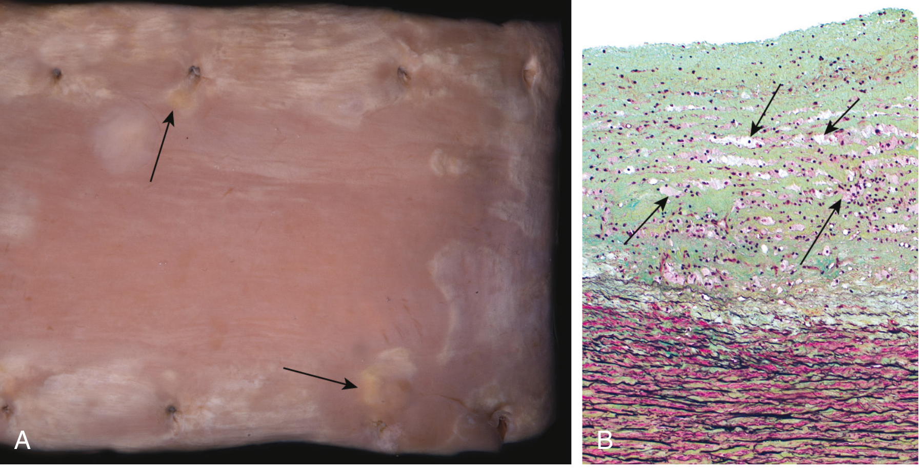
- This is the earliest lesion. It consists of lipid-filled foamy macrophages in the intima. Grossly it appears as small flat yellow macules that may coalesce into streaks 1 cm or longer. It is slightly raised and causes no flow disturbance.
- It is present in the aortas of infants and in virtually all adolescents. Coronary fatty streaks form at the same sites that later develop plaques, which suggests a precursor relationship. Not all fatty streaks progress. - Robbins, Cotran & Kumar Pathologic Basis of Disease, p. 471
-
Grossly it is yellow-tan and raised. An overlying thrombus on an ulcerated plaque looks red-brown.
-
Lesions are patchy and eccentric on cross-section. They are rarely circumferential, because of local hemodynamics.
-
Lesions at different stages often coexist in the same vessel.
-
Structure, from lumen to media:
- Fibrous cap: SMCs, collagen, elastin, proteoglycans, and some inflammatory cells.
- Shoulder region: the junction of cap and wall, rich in macrophages and T cells. This is the usual site of rupture.
- Necrotic core: lipid, cholesterol clefts, foam cells and debris.
- Periphery: neovascularization, calcification, and thinning of the media with attenuated elastic laminae.
-
Common sites, in decreasing order: lower abdominal aorta, coronary arteries, popliteal arteries, descending thoracic aorta, internal carotid arteries, and the circle of Willis.
-
Robbins, Cotran & Kumar Pathologic Basis of Disease, p. 471-473
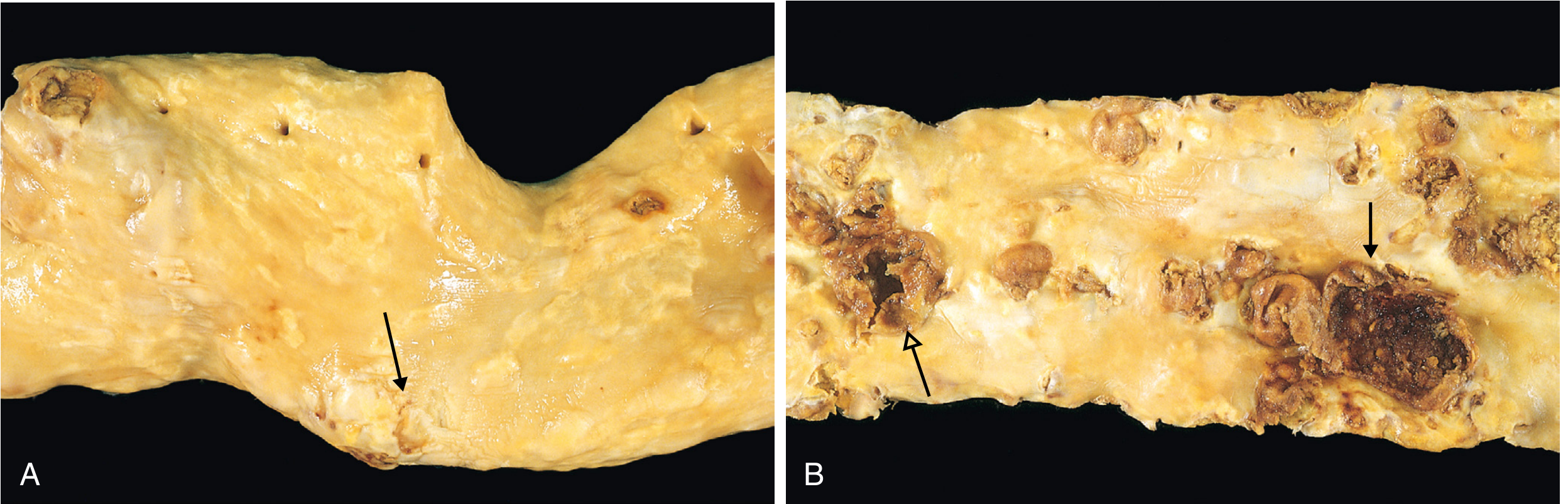
- Rupture or fissuring
- Erosion or ulceration
- Hemorrhage into the plaque
- Atheroembolism (cholesterol emboli)
- Calcification
- Aneurysm formation (the plaque causes ischemic atrophy of the media)
5. Stable vs vulnerable plaque
| Feature | Stable | Vulnerable (unstable) |
|---|---|---|
| Fibrous cap | Thick, dense collagen | Thin |
| Lipid core | Small | Large |
| Inflammation | Minimal | Dense macrophage and T-cell infiltrate |
| SMCs | Many | Few (apoptosis) |
| Typical consequence | Chronic ischemia (stable angina, claudication) | Acute thrombosis (MI, stroke, sudden death) |
- Robbins, Cotran & Kumar Pathologic Basis of Disease, p. 474-475
- Rupture or fissuring. This exposes thrombogenic contents and often causes a fully occlusive thrombus.
- Erosion or ulceration. This exposes the basement membrane and less often causes full occlusion.
- Hemorrhage into the atheroma. This expands the plaque volume.
- Intrinsic: plaque structure, collagen loss and protease activity.
- Extrinsic: blood pressure surges, platelet reactivity and vasospasm.
6. Clinical consequences
- Chronic stenosis. Symptoms depend on the degree of narrowing (roughly 70% or more is flow-limiting), the collateral supply and tissue demand. Slow stenosis allows collateral growth. - Robbins, Cotran & Kumar Pathologic Basis of Disease, p. 474
- Coronary: stable angina, acute coronary syndromes, ischemic cardiomyopathy and sudden cardiac death.
- Cerebral: carotid and intracranial disease causing TIA and stroke, and multi-infarct dementia.
- Aorta: abdominal aortic aneurysm (AAA), which can rupture, and aortic atheroemboli.
- Renal: renal artery stenosis with renovascular hypertension and ischemic nephropathy.
- Mesenteric: chronic mesenteric ischemia ("abdominal angina") and bowel infarction.
- Lower limb: intermittent claudication, rest pain, ulcers and gangrene.
7. Diagnosis and risk assessment
- Lipid profile: LDL-C, non-HDL-C, HDL, triglycerides, and apoB. Lp(a) should be measured once in adults.
- Other labs: HbA1c or glucose, renal function, and hs-CRP as a risk enhancer.
- Risk calculators: Framingham, ASCVD pooled cohort equations, and the AHA PREVENT equations.
- Noninvasive tests:
- Ankle-brachial index for peripheral arterial disease.
- Carotid duplex and carotid intima-media thickness.
- Coronary artery calcium (CAC) score.
- CT coronary angiography.
- Stress testing.
- Invasive tests: invasive coronary angiography, with IVUS or OCT for plaque characterization.
8. Management
- Stop smoking.
- Eat a Mediterranean or DASH-type diet.
- Do regular exercise.
- Lose weight.
- Limit alcohol.
- Control blood pressure.
- Control diabetes. Consider SGLT2 inhibitors or GLP-1 receptor agonists, which have proven cardiovascular benefit.
- High-intensity statin as first line.
- Add ezetimibe if the LDL target is not reached.
- Add a PCSK9 inhibitor (evolocumab, alirocumab) or inclisiran, or bempedoic acid.
- Icosapent ethyl for selected patients with high triglycerides.
- Aspirin for secondary prevention.
- P2Y12 inhibitors (dual antiplatelet therapy) after acute coronary syndrome or stenting.
- Low-dose rivaroxaban plus aspirin in selected stable coronary artery disease or peripheral arterial disease.
- It uses the PREVENT-ASCVD risk equations.
- It restores explicit LDL-C and non-HDL-C goals.
- It makes more use of apoB and Lp(a) as markers.
- It recognizes triglyceride-rich remnants and Lp(a) as independent risk contributors.
9. Recent evidence check
- Subclinical atherosclerosis risk is increased in primary Sjogren's syndrome (PMID 37977997).
- Different exercise modes affect arteriosclerosis risk factors in postmenopausal women (PMID 40858980).
- Small-vessel "microatherosclerosis" is an often overlooked cause of ischemic stroke (PMID 42898271).
10. High-yield exam points
- Earliest lesion: the fatty streak (foam cells). It is reversible and can occur in childhood.
- Main cell types: endothelial cells, monocytes and macrophages, T cells and SMCs.
- Most important modifiable factors: LDL, hypertension, smoking and diabetes.
- Most dangerous lesion: the thin-cap, lipid-rich, inflamed plaque. The usual rupture site is the shoulder.
- Most acute events come from plaques with less than 70% stenosis.
- Most commonly involved vessel: the abdominal aorta, then the coronary arteries.
- Atherosclerosis weakens the media and so predisposes to aneurysm, especially in the abdominal aorta.