
Diagnosis of anasarca
anasarca diagnosis etiology
anasarca generalized edema clinical photo
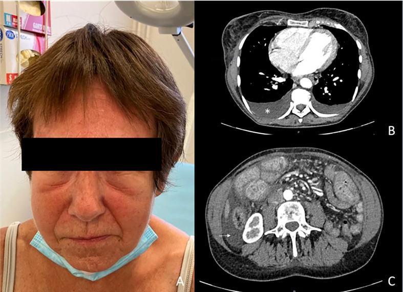
This composite medical image displays clinical signs of generalized edema (anasarca). Panel A is a clinical photograph of a patient's face showing significant facial edema, characterized by periorbital swelling, puffiness of the cheeks, and fullness of the soft tissues, leading to a narrowed appearance of the eyes. Panel B is an axial CT scan of the thorax demonstrating mild bilateral pleural effusion, visible as dependent, low-density crescentic fluid collections in the posterior pleural spaces (indicated by a white star). Panel C is an axial abdominal CT scan showing mild ascites, identified as low-density fluid surrounding abdominal viscera, with a white arrow specifically pointing to fluid in the perihepatic/paracolic gutter area. These findings collectively illustrate the multisystemic fluid accumulation associated with conditions like systemic capillary leak syndrome or profound hypoalbuminemia. The images are highly relevant for medical education concerning the physical and radiological manifestations of fluid overload and serous cavity effusions.
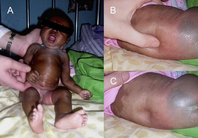
This set of clinical photographs illustrates a pediatric patient presenting with severe generalized edema (anasarca). Panel A displays a frontal view of an infant with significant swelling affecting the face, periorbital region, neck, trunk, and extremities. The skin appears tense, glossy, and exhibits areas of pallor and hyperpigmentation. Panels B and C demonstrate the physical examination for pitting edema on the patient's back. Panel B shows a clinician's thumb applying pressure to the edematous tissue, while Panel C reveals a deep, persistent indentation (pitting) remaining after the pressure is released. This clinical presentation is associated with systemic fluid overload or severe hypoalbuminemia, documented here in the context of an atypical parasitic infection. The images serve as a teaching tool for identifying the visual signs of gross edema and the technique for eliciting and documenting pitting in soft tissues.
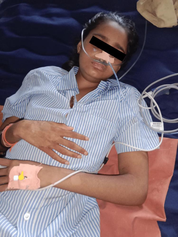
This clinical photograph shows a young female patient in a supine position, illustrating the resolution phase of generalized edema (anasarca). The patient exhibits a visible reduction in swelling around the facial region, specifically the periorbital and lip areas, compared to a state of acute exacerbation. Clinical support is evident via a nasal cannula for supplemental oxygen and a peripheral intravenous (IV) catheter secured on the dorsal aspect of the left hand. Notable dermatological findings include patches of hypopigmentation or depigmentation on the right forearm and wrist. The image serves as an educational representation of the clinical response to corticosteroid therapy in a pediatric patient with systemic swelling. The patient's upper extremities show improved anatomical definition in the fingers and wrists, indicative of successful diuresis or resolution of interstitial fluid accumulation. The image captures the patient's status post-treatment for progressive body swelling and dysphagia, demonstrating partial recovery of normal tissue contours.

This clinical photograph displays an infant in a supine position, demonstrating generalized edema (anasarca) and features suggestive of Cushingoid facies. The patient exhibits significant facial puffiness with full, rounded cheeks and obscured facial contours, characteristic of 'moon facies.' There is evidence of generalized swelling throughout the upper and lower extremities, with visible skin distention and increased circumference of the limbs. The skin appears taut and smooth over the edematous areas. Clinical markers of a medical setting are present, including pink identification bands on the right wrist and ankle, and a white bandage on the left hand. The image serves as an educational illustration of systemic fluid retention and physical manifestations of hypercortisolism in a pediatric patient, likely in the context of iatrogenic Cushing syndrome. The infant is shown on a hospital bed, and the photograph is focused on the morphology and distribution of the edema across the entire body.
anasarca diagnosis causes investigation approach clinical
Diagnosis of Anasarca
Definition
Pathophysiologic Mechanisms
| Mechanism | Example Conditions |
|---|---|
| Decreased plasma oncotic pressure (hypoalbuminemia) | Nephrotic syndrome, liver cirrhosis, malnutrition |
| Increased capillary hydrostatic pressure | Congestive heart failure, venous obstruction |
| Increased endothelial permeability | Sepsis, capillary leak syndrome |
| Impaired lymphatic drainage | Malignancy, filariasis |
| Sodium and water retention | Heart failure, CKD, SIADH |
Clinical Diagnosis
- Tachycardia, tachypnea, reduced O2 saturation (signs of fluid overload)
- Hypotension or hypertension depending on the etiology
- Pitting edema - dependent areas (legs, sacrum if bedridden); bilateral symmetric pitting edema is typical in cardiac and hepatic causes
- Facial and periorbital edema - prominent in nephrotic syndrome
- Ascites - shifting dullness, fluid thrill
- Pleural effusions - dullness to percussion, reduced breath sounds at bases
- Scrotal/labial edema
- Skin changes - taut, shiny, stretched skin; later brawny non-pitting edema in chronic cases
- Press firmly with thumb for 5 seconds over bony prominence (tibia, dorsum of foot, sacrum)
- A persistent pit indicates pitting edema (excess interstitial water)
- Non-pitting edema suggests lymphedema or myxedema
Current Surgical Therapy 14e notes anasarca-related morbidities include extremity ischemia (requiring escharotomy), retrobulbar edema (lateral canthotomy), intracompartmental edema (fasciotomy), and abdominal compartment syndrome.
Clinical image
Causes to Identify (Differential Diagnosis)
The "Big Three" (Most Common)
-
Congestive Heart Failure (CHF)
- Bilateral pitting edema, raised JVP, S3 gallop, bilateral crackles
- Right HF features: hepatomegaly, hepatojugular reflux, tricuspid regurgitation murmur
- Harrison's 22e: severe tricuspid stenosis associated with marked hepatic congestion, cirrhosis, jaundice, malnutrition, anasarca, and ascites
-
Nephrotic Syndrome
- Massive proteinuria (>3.5 g/day), hypoalbuminemia, hypercholesterolemia
- Facial/periorbital edema prominent, frothy urine
- Comprehensive Clinical Nephrology 7e: fluid retention can be of variable severity, sometimes resulting in anasarca; amyloidosis increases thromboembolic risk
-
Hepatic Cirrhosis
- Portal hypertension, ascites, low albumin
- Signs of chronic liver disease: jaundice, spider angiomata, palmar erythema, caput medusae, gynecomastia
Other Causes
- Malnutrition / Kwashiorkor - severe protein deficiency
- Malignancy - hypoalbuminemia, lymphatic obstruction
- Capillary leak syndrome - protein leaks from vessels into tissues
- Sepsis / severe infection - cytokine-mediated permeability increase
- Hypothyroidism (myxedema) - non-pitting, periorbital
- Excessive IV fluid administration - iatrogenic
- Drugs - calcium channel blockers (dihydropyridine class), corticosteroids, NSAIDs, chemotherapy (gemcitabine)
- Parasitic infections - heavy Fasciolopsis buski infection (malabsorption, anasarca, ascites per Tietz Textbook of Laboratory Medicine 7e)
- iMCD-TAFRO syndrome - rare; anasarca + thrombocytopenia + fever + reticulin fibrosis (Fishman's Pulmonary Diseases and Disorders)
- Burns - massive fluid shifts from resuscitation
Diagnostic Workup
Laboratory Studies
| Test | What It Detects |
|---|---|
| Serum albumin | Hypoalbuminemia (nephrotic, hepatic, malnutrition) |
| Urine protein (spot protein:creatinine ratio or 24-hr) | Nephrotic syndrome (>3.5 g/day) |
| Urine microscopy | Casts - nephritic vs. nephrotic pattern |
| LFTs, PT/INR, bilirubin | Hepatic cause (cirrhosis) |
| Serum creatinine, BUN, eGFR | Renal failure |
| BNP / NT-proBNP | Heart failure (elevated) |
| CBC | Anemia, infection, malignancy |
| TSH | Hypothyroidism |
| Serum lipid panel | Hyperlipidemia of nephrotic syndrome |
| Blood culture | Sepsis |
| ANA, anti-dsDNA, complement (C3/C4) | Lupus nephritis |
| SPEP / UPEP | Paraproteinemia (AL amyloidosis, multiple myeloma) |
Imaging Studies
| Modality | Findings |
|---|---|
| Chest X-ray | Cardiomegaly, pulmonary congestion, pleural effusions |
| Echocardiogram | LV/RV function, valvular disease, constrictive pericarditis |
| Abdominal ultrasound | Ascites, liver architecture (cirrhosis), portal hypertension |
| CT chest/abdomen/pelvis | Extent of effusions, malignancy, lymphadenopathy |
| Renal ultrasound | Kidney size, echogenicity (CKD), obstructive uropathy |
Renal Biopsy
- Indicated when nephrotic syndrome is identified to clarify the specific glomerular lesion (minimal change disease, FSGS, membranous nephropathy, amyloidosis, diabetic nephropathy)
Additional Tests
- Liver biopsy (cirrhosis confirmation, NASH, amyloidosis)
- Bone marrow biopsy (iMCD-TAFRO, multiple myeloma)
- Bone marrow reticulin staining (iMCD-TAFRO: reticulin fibrosis is a major criterion)
Diagnostic Approach Algorithm
Anasarca
    ↓
History + Physical Examination
    ↓
↙         ↓         ↘
Raised JVP    Facial edema    Signs of liver disease
+ S3 gallop   + proteinuria   + ascites
    ↓              ↓                ↓
   CHF        Nephrotic         Cirrhosis
              Syndrome
    ↓              ↓                ↓
Echo           Renal biopsy     LFTs, USS
BNP            24-hr protein    liver
               Albumin

    → If all three negative: consider malnutrition,
      malignancy, capillary leak, hypothyroidism,
      drugs, paraprotein, iMCD-TAFRO
Key Distinguishing Features at a Glance
| Feature | CHF | Nephrotic | Cirrhosis |
|---|---|---|---|
| JVP | Elevated | Normal/low | Normal/low |
| Ascites | Less prominent | Less prominent | Prominent |
| Proteinuria | Mild/absent | Massive (>3.5 g/d) | Mild |
| Serum albumin | Normal/low | Very low | Low |
| BNP | Markedly elevated | Normal | Normal/elevated |
| Edema distribution | Bilateral legs, sacrum | Periorbital, legs | Legs, ascites |
| Liver signs | Hepatomegaly (R HF) | Absent | Spider naevi, jaundice |