
Cytotoxic cerebral edema
Reading File
Finding Sources
Finding Sources
Searching PubMed
"cytotoxic edema" AND "brain edema"
Reading File
Reading File
Searching Images
cytotoxic versus vasogenic edema schematic
Cytotoxic (cellular) cerebral edema is intracellular swelling of neurons, astrocytes and endothelial cells. The blood-brain barrier (BBB) is initially intact, so the swelling comes from failure of cellular ion pumps. It is not caused by leakage of plasma into the extracellular space.
Definition
Robbins describes it as "an increase in intracellular fluid secondary to neuronal and glial cell injury, as might follow a generalized hypoxic or ischemic insult or exposure to certain toxins." - Robbins & Kumar Basic Pathology (Robbins Pathology), Cerebral Edema
Mechanism
- Energy failure. Ischemia or hypoxia depletes ATP, so the Na+/K+-ATPase stops working.
- Ion shift. Neurons depolarize and cannot repolarize. Intracellular Na+ (and Ca2+) rises and K+ leaks out.
- Water follows sodium. Water moves from the extracellular to the intracellular compartment, mainly through aquaporin-4 on astrocyte end-feet.
- Net effect. Total brain water and volume increase, but the extracellular space shrinks. This adds mass effect, and Plum and Posner note it further compresses adjacent tissue.
Plum and Posner explain: "When neurons are injured, they can no longer maintain ion gradients. The increased intracellular sodium causes a shift of fluid from the extracellular to the intracellular compartment, resulting in cytotoxic edema. Cytotoxic edema is not affected by corticosteroids." - Plum and Posner's Diagnosis and Treatment of Stupor and Coma, Fig. 3.1
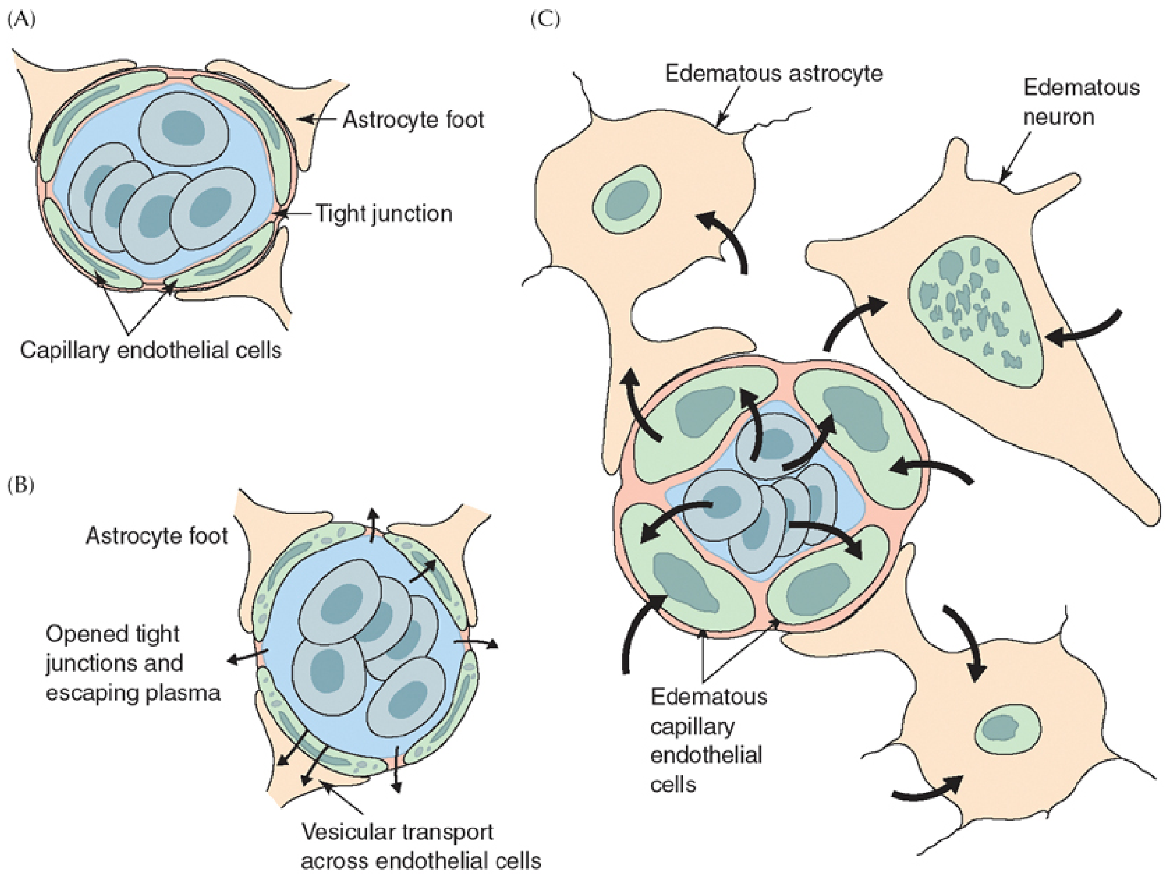
Common causes
- Ischemic stroke (the classic cause, especially in the first minutes to hours)
- Global hypoxia-ischemia, such as cardiac arrest
- Traumatic brain injury
- Osmotic or metabolic shifts: hyponatremia and water intoxication, and the dialysis disequilibrium syndrome
- Acute liver failure (ammonia causes astrocyte swelling via glutamine)
- Toxins and metabolic disorders: methanol, carbon monoxide, mitochondrial disease, diabetic ketoacidosis in children, and Reye syndrome
- Status epilepticus and severe infections such as meningitis (mixed with vasogenic edema)
Cytotoxic versus vasogenic edema
| Feature | Cytotoxic | Vasogenic |
|---|---|---|
| Fluid compartment | Intracellular | Extracellular |
| BBB | Intact initially | Disrupted |
| Main tissue involved | Gray and white matter | Predominantly white matter |
| Typical causes | Ischemia, hypoxia, toxins | Tumor, abscess, trauma, inflammation |
| Corticosteroids | No benefit | Often effective |
| Reversibility | Usually not, if cells die | Can resolve once the vessel damage heals |
| DWI / ADC on MRI | Restricted diffusion (bright DWI, low ADC) | Increased diffusion (high ADC) |
In practice the two are often mixed. Robbins notes that conditions with generalized edema often have both components, and Harrison's says clinically significant brain edema "usually represents a combination of vasogenic and cytotoxic components." Pure cytotoxic edema progresses to vasogenic edema as ischemia damages the BBB over hours to days, as in malignant MCA infarction.
Imaging and pathology
- MRI. Restricted diffusion (bright on DWI, low on ADC) appears within minutes of onset. It is the earliest imaging sign of infarction and the key way to separate cytotoxic from vasogenic edema.
- CT. There is loss of gray-white differentiation, sulcal effacement and hypodensity, usually later than MRI changes.
- Gross pathology. The brain is softer than normal and fills the cranial vault. In generalized edema the gyri are flattened, the sulci narrowed and the ventricles compressed (Robbins).
Clinical consequences
- Raised intracranial pressure and mass effect
- Midline shift and herniation (subfalcine, uncal, tonsillar)
- Reduced cerebral perfusion pressure, which worsens ischemia
Management
Treatment targets the cause and the raised ICP, because steroids do not help.
- Basic measures. Elevate the head of the bed to 30 degrees, keep the patient normothermic, avoid hypotonic fluids, correct hyponatremia carefully, and treat hypoxia and hypercapnia.
- Hyperosmolar therapy. Use mannitol or hypertonic saline.
- Other ICP measures. Brief hyperventilation as a bridge only, sedation, and CSF drainage if there is hydrocephalus.
- Decompressive craniectomy. Consider it for malignant infarction or refractory ICP.
- Cause-specific treatment. Reperfusion in stroke, and treatment of hyperammonemia in liver failure.
- Steroids. Avoid them for ischemic or traumatic cytotoxic edema. Dexamethasone is reserved for vasogenic edema from tumor.
Recent evidence
A PubMed search of the last 2 years found no meta-analyses or systematic reviews that contradict the textbook account. One 2025 systematic review and meta-analysis of animal studies (NMDA antagonists in TBI edema, PMID 39138715) covers experimental anti-edema therapies. Those are preclinical, not yet standard care.
If you want more detail, I can cover the molecular mechanisms (AQP4, SUR1-TRPM4 channels), the MRI findings, or stroke-specific management.
This is a shared conversation. Sign in to Orris to start your own chat.