
Disorders of olfaction
olfactory disorders anosmia
olfactory pathway anatomy diagram olfactory bulb cribriform plate
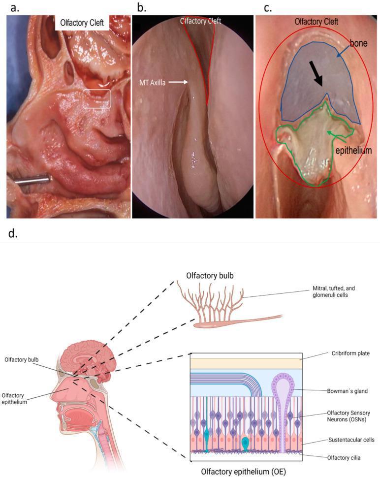
This composite educational graphic illustrates the anatomy and histology of the human olfactory system. Figure (a) is a sagittal anatomical specimen showing the olfactory cleft in the cranial nasal cavity. Figure (b) provides an endonasal endoscopic view of the olfactory cleft and the axilla of the middle turbinate (MT Axilla). Figure (c) displays a surgical or dissection view highlighting the relationship between the olfactory epithelium and the underlying bone, with a black arrow indicating the olfactory fibers. Figure (d) features a multi-level schematic: a sagittal head diagram showing the olfactory bulb's position, a zoomed histological view of the olfactory epithelium (OE), and a neural pathway diagram. The OE schematic identifies key cellular components: Olfactory Sensory Neurons (OSNs) with distal cilia, sustentacular cells, and basal cells, as well as Bowman’s glands and the cribriform plate. The neural diagram depicts OSN axons synapsing with mitral, tufted, and glomeruli cells in the olfactory bulb. This resource is designed for teaching the physiological structure and microanatomy of the human olfactory network.
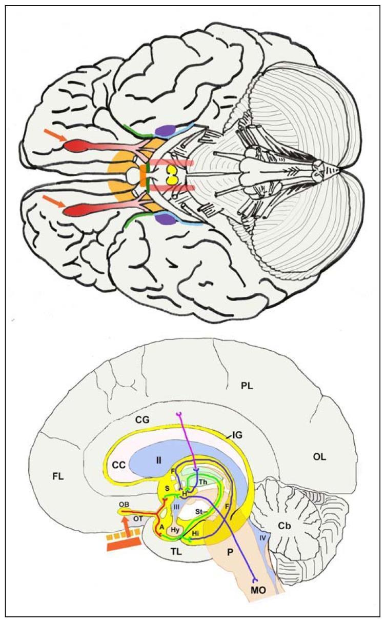
This medical illustration depicts the human olfactory pathway through dual neuroanatomical perspectives: a ventral view (top) and a composite sagittal view (bottom). The ventral image highlights key structures using a color-coded schema: the olfactory bulbs and tracts (red/pink), anterior perforated substance and septal area (orange-yellow), anterior commissure (orange bar), pre-piriform cortex (green), entorhinal cortex (pale blue), and amygdala (purple). Yellow mammillary bodies and pale red medial forebrain bundles are also shown centrally. The sagittal view illustrates the pathway starting from the olfactory epithelium (red bar) through the cribriform plate to the olfactory bulb (OB). It maps secondary and tertiary projections (coded in green, dark blue, and purple) to various regions including the thalamus (Th), hypothalamus (Hy), hippocampus (Hi), and cingulate gyrus (CG). Major landmarks such as the corpus callosum (CC), ventricles (II, III, IV), and brainstem (Pons, Medulla Oblongata) provide anatomical context. This diagram is designed for neuroanatomical education, specifically illustrating the limbic connections of the olfactory system and its relative spatial distribution within the brain.
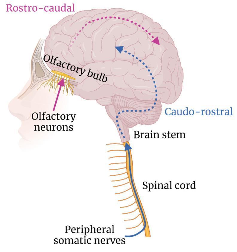
This medical illustration depicts hypothesized transneural neuroinvasion pathways within the human central nervous system (CNS). The diagram highlights two primary anatomical routes: the olfactory route and the spinal cord route. The olfactory route begins at the olfactory neurons in the nasal cavity, passing through the cribriform plate to the olfactory bulb. A dashed pink line indicates subsequent 'rostro-caudal' spread from the anterior to the posterior brain regions. The spinal route illustrates 'caudo-rostral' neuroinvasion, originating from peripheral somatic nerves that enter the spinal cord. A dashed blue line traces the ascending pathway from the spinal cord through the brain stem and into the deep cortical structures. The illustration serves as an educational tool for understanding how neurotropic pathogens, such as arboviruses or rabies, utilize existing neural architecture to bypass the blood-brain barrier and disseminate throughout the CNS.
Disorders of Olfaction
Anatomy of the Olfactory System (Overview)
- Bipolar receptor cells - project 3-30 receptor-bearing cilia into the olfactory mucus; act as both receptor and first-order neuron; can regenerate from basal cells
- Sustentacular cells - support cells
- Microvillar cells
- Basal cells - precursors for regeneration
- Bowman's glands - major source of olfactory mucus (in lamina propria)
- Anterior olfactory nucleus
- Piriform cortex (encodes odor quality and identity)
- Anterior cortical nucleus of the amygdala
- Periamygdaloid complex
- Rostral entorhinal cortex
Terminology and Classification of Olfactory Disorders
| Term | Definition |
|---|---|
| Anosmia | Total loss of smell |
| Hyposmia / Microsmia | Partial (less than total) smell loss |
| Hyperosmia | Increased sensitivity to odorants (many such patients are actually experiencing dysosmia with decrements on testing) |
| Dysosmia / Parosmia | Distorted smell when an odorant or warm air is present; often described as "chemical-like or garbage-like" |
| Phantosmia | Smell perception in the absence of any stimulus |
| Cacosmia | Parosmia with fecal/putrid character |
| Olfactory agnosia | Inability to recognize odors despite an intact olfactory system; due to some brain lesions |
- Bradley and Daroff's Neurology in Clinical Practice, p. 329-333
Epidemiology
-
Overall prevalence of olfactory dysfunction: ~13.5% (NHANES 2013-2014)
-
>50% of people aged 65-80 have significant smell decrements
-
75% of those aged ≥80 are affected (presbyosmia)
-
Women outperform men on olfactory tests and retain function to a later age
-
Presbyosmia causes elderly patients to report food as flavorless, risking nutritional deficiency, and is responsible for a disproportionate number of accidental gas poisonings in the elderly
-
Harrison's Principles of Internal Medicine 22E, p. 284
Causes of Olfactory Dysfunction
Three Most Common Causes of Long-Lasting or Permanent Smell Loss (in order of frequency)
- Severe upper respiratory infections (URIs)
- Head trauma
- Chronic rhinosinusitis
1. Upper Respiratory Infections
- Directly damage the olfactory epithelium
- Decrease receptor cell number
- Damage cilia on remaining receptor cells
- Induce replacement of sensory epithelium with respiratory epithelium
2. Head Trauma
- Mechanism: shearing and subsequent scarring of the olfactory fila as they pass through cribriform plate foramina (the cribriform plate does not have to be fractured)
- Severity correlates with poor Glasgow Coma Scale score and length of posttraumatic amnesia
- <10% of posttraumatic anosmic patients recover age-related normal function over time (rises to ~25% for those with partial rather than total loss)
3. Chronic Rhinosinusitis
- Smell loss correlates with disease severity
- Most loss occurs when rhinosinusitis + polyposis coexist (5-10% experience total loss)
- Systemic glucocorticoids can induce short-term improvement but do not return scores to normal on average, implying chronic permanent neural loss
Systemic and Metabolic Causes
- Diabetes, hypertension, hypothyroidism, kidney disease, liver disease
- Most immune-mediated diseases: allergic rhinitis, asthma, autoimmune pancreatitis, Behcet disease, Churg-Strauss syndrome, Crohn disease, fibromyalgia, giant cell arteritis, lupus, MS, myasthenia gravis, rheumatoid arthritis, Sjogren syndrome, scleroderma, psoriasis
Olfactory Dysfunction in Neurological Disease
Relative Severity of Smell Loss by Disease (Bradley's scale):
| Severity (relative) | Disease |
|---|---|
| ++++ (Most severe) | Idiopathic Parkinson disease (PD), Alzheimer disease (AD), Dementia with Lewy bodies (DLB), Guam PD-dementia complex, Idiopathic REM sleep behavior disorder (iRBD) |
| +++ | Huntington disease, Down syndrome, PARK8 PD |
| ++ | Multiple system atrophy (type-P), PARK1 PD, schizophrenia, narcolepsy |
| + (Mild) | Motor neuron disease, SCA2 PD, Friedreich ataxia, PARK3, corticobasal degeneration, frontotemporal dementia |
| 0 (Normal) | Essential tremor, vascular parkinsonism, MPTP-induced parkinsonism, progressive supranuclear palsy, PARK2 |
Key clinical points:
- Parkinson disease: Olfactory impairment often predates clinical diagnosis by years. Olfactory bulbs may be - along with the dorsomotor nucleus of the vagus - the first site of neural damage (based on alpha-synuclein/Lewy body staging)
- Alzheimer disease: Poor smell correlates with higher AD-related pathology even in presymptomatic stages
- DLB: Smell loss is more marked in early DLB than in mild AD
- Progressive supranuclear palsy & MPTP-parkinsonism: Minimal or no smell loss - useful differentiating feature
- iRBD: Same magnitude of smell loss as PD; iRBD patients frequently go on to develop PD and hyposmia
- Narcolepsy with cataplexy: Olfactory impairment linked to loss of orexin (hypocretin) neurons; intranasal orexin-A improves olfactory function
- Multiple sclerosis: Smell loss related to demyelinating lesions within olfaction-related structures
- Idiopathic intracranial hypertension (IIH): Associated with impaired olfactory sensitivity
- Migraine: Osmophobia during attacks; episodic migraineurs may show microsmia/hyposmia during acute attacks
Broad List of Associated Conditions (Table 35-1, Harrison's)
Clinical Assessment
Objective Olfactory Testing
- University of Pennsylvania Smell Identification Test (UPSIT) - most widely used; a scratch-and-sniff test with forced-choice odor identification
- Scores interpreted relative to age- and sex-matched normative data
- Results are classified as: normosmia, mild microsmia, moderate microsmia, severe microsmia, anosmia
- The UPSIT figure in Harrison's shows that women consistently identify odorants better than men at all ages, and performance declines progressively with age
- History: onset, duration, quality (lost vs. distorted), triggering events (trauma, URI, surgery)
- Nasal endoscopy and imaging (CT/MRI for sinonasal disease, intracranial lesions)
- Neurological examination: especially in the context of neurodegenerative disease suspicion
- Laboratory tests: thyroid function, renal/hepatic function, zinc levels
Management
| Cause | Approach |
|---|---|
| Rhinosinusitis / polyposis | Systemic glucocorticoids (short-term improvement); topical corticosteroids; surgery (functional endoscopic sinus surgery) |
| Post-URI smell loss | Olfactory training (smell retraining); alpha-lipoic acid (some evidence); spontaneous recovery possible |
| Head trauma | Mainly expectant; spontaneous partial recovery in ~25% with partial loss |
| COVID-19 anosmia | Olfactory training; intranasal corticosteroids (limited evidence); most recover within months; 30% may have persistent loss |
| Medications | Discontinue offending drug where possible |
| Neurodegenerative disease | No effective treatment for smell loss itself; treat underlying disease |
Recent Evidence
- Harrison's Principles of Internal Medicine 22E (2025), Chapter 35 - Disorders of Smell, Taste, and Hearing
- Bradley and Daroff's Neurology in Clinical Practice, Chapter 19 - Olfaction and Gustation
- Cummings Otolaryngology Head and Neck Surgery - Olfaction Rehabilitation