
Meyer Bakshi pedicle graft
Meyer Bakshi pedicle graft periodontal surgery technique
Meyer Bakshi double papilla pedicle graft mucogingival technique steps indications
pedicle graft root coverage gingival recession lateral
https://www.mitalpatel.co.uk/wp-content/uploads/2020/12/publ…
lateral pedicle graft root coverage mucogingival surgery
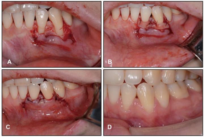
This composite of four clinical intraoral photographs documents a mucogingival surgery for root coverage of the mandibular left lateral incisor and canine. Image A illustrates the initial surgical phase with a full-thickness flap detachment extending beyond the mucogingival junction, exposing the underlying alveolar bone and root surfaces. Image B displays the placement of a subepithelial connective tissue graft, which is secured to the recipient site using fine resorbable sutures to ensure graft stability and close adaptation to the root surfaces. Image C shows the coronally advanced flap positioned over the graft and fixed with sling sutures at the interdental papillae, which provides tension-free closure and coronal positioning. Image D captures the post-operative outcome two months later, demonstrating successful healing, significantly increased zones of keratinized tissue, and successful root coverage with healthy gingival contours and integration of the grafted tissue.

This sequence of four intraoral clinical photographs documents a periodontal mucogingival surgery for root coverage using a subepithelial connective tissue graft (SCTG) and a double lateral sliding bridge flap (DLSBF) technique on the mandibular anterior teeth. (a) Baseline: Shows significant gingival recession on the mandibular incisors with exposed root surfaces and thin biotype. (b) Immediate Postoperative: Displays the surgical site with a coronally repositioned flap covering the SCTG, secured with multiple black silk interrupted sutures; the tissue is erythematous and edematous, characteristic of immediate surgical trauma. (c) Four-Month Follow-Up: Shows successful root coverage with healthy, pink, keratinized gingiva and a stable mucogingival junction. (d) Two-Year Follow-Up: Demonstrates long-term stability of the gingival margin with a visible apical scar in the vestibule corresponding to the previous surgical incision. The patient is also undergoing active orthodontic treatment, indicated by fixed metal brackets and an archwire. This progression illustrates the surgical management of Cairo class recessions and the aesthetic integration of grafted tissue over time.
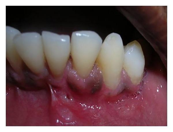
This intraoral clinical photograph shows a postoperative view of the mandibular left quadrant, focusing on the successful surgical treatment of gingival recession on the lower left lateral incisor (tooth 32). The image demonstrates complete root coverage following a periodontal plastic surgery procedure, likely involving a subepithelial connective tissue graft and a double pedicle flap. The gingival margin at tooth 32 is positioned at the cemento-enamel junction (CEJ), matching the level of adjacent teeth. The interdental papillae are well-formed and fill the proximal spaces. The newly formed keratinized tissue appears pink, healthy, and firm, with a smooth texture and a clearly defined mucogingival junction. No clinical signs of inflammation, such as edema or erythema, are visible. The neighboring mandibular teeth appear clean with smooth enamel surfaces and an absence of plaque or calculus deposits. This visual serves as a clinical example of regenerative periodontal therapy achieving both functional attachment and esthetic harmony in the treatment of Miller's Class II recession.
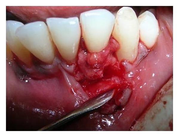
This intraoral clinical photograph illustrates an intermediate stage of a periodontal plastic surgery procedure for root coverage. The image shows the mandibular anterior region, specifically tooth number 32 (lower left lateral incisor), which presents with Miller's class II gingival recession. A subepithelial connective tissue graft (SCTG) has been harvested and positioned over the denuded root surface. The surgical site displays a reflected partial-thickness flap with mesial and distal vertical releasing incisions. The adjacent papillary grafts from the interdental spaces are being manipulated and sutured midbuccally over the graft to provide a double pedicle covering. A metallic surgical periosteal elevator is visible at the inferior aspect of the site, reflecting the mucosal flap. The surrounding gingiva appears erythematous and edematous, consistent with acute surgical trauma and inflammation. Visible 5-0 sutures are used to stabilize the graft and approximate the tissue margins. This image demonstrates the clinical application of a combined SCTG and double pedicle flap technique for correcting localized gingival recession and increasing the zone of keratinized tissue.
Bakshi double lateral pedicle graft palatal recession
Meyer-Bakshi Pedicle Graft
Concept
Indications
- Single tooth gingival recession defect (Miller's Class I or II)
- Adequate keratinized gingiva adjacent to the recession site (donor zone)
- Shallow to moderate recession depth (< 5 mm)
- No or minimal bone loss at the recession site
- When aesthetics and root sensitivity are the primary concerns
- When avoiding a second surgical (palatal harvest) site is preferred
Contraindications / Limitations
- Narrow zones of keratinized attached gingiva adjacent to the recipient tooth
- Wide recession defects where the pedicle cannot cover without tension
- Deep pockets or bone loss at the adjacent donor site
- Frenum attachments or muscle pull near the donor area
- Shallow vestibular depth limiting pedicle mobilization
Surgical Steps
- Scale and root plane the exposed root surface
- Root conditioning with citric acid (pH 1.0) for 2-3 minutes or EDTA to remove the smear layer and expose collagen fibers
- Make a V-shaped or crescent incision at the gingival margin to remove surface epithelium and expose the underlying connective tissue ("raw" bed for the graft to adhere to)
- In deep pockets or frenum cases, extend the V incision apically and laterally
- Measure the width of the recession defect; the pedicle should be at least twice the width of the recession
- Make an oblique incision away from the recession, leaving 1-2 mm of keratinized gingiva around the adjacent tooth (to prevent donor site recession)
- A second oblique distal relieving incision is made apically
- In the double papilla variant: two interdental papillae (mesial and distal to the recession tooth) are raised separately as bilateral pedicles
- Raise the flap using a split-thickness (partial thickness) dissection at ~0.75 mm depth to include only connective tissue without stripping periosteum from the donor alveolar bone
- For the double papilla approach: full or split thickness flap can be used depending on tissue thickness
- Ensure the pedicle base is wider than the apex to maintain adequate blood supply
- Reflect the flap free of muscle attachments and tension
- If residual tension exists, place a periosteal releasing incision (Corn's cutback incision) at the base of the flap
- The flap must move passively to the recipient site with no tension
- Rotate (or laterally slide) the pedicle over the denuded root surface
- Position the flap 1-2 mm coronal to the CEJ (cementoenamel junction)
- For the double papilla technique: suture the two papillae together over the root with interrupted sutures, then advance the combined tissue coronally
- Use 4-0, 5-0, or 6-0 sutures
- Place interrupted sutures at the margins of the flap
- A sling suture is used to pull the papilla interproximally and hold tissue tightly against the tooth
- The donor site is left to heal by secondary intention
- Periodontal dressing (e.g., Coe-Pak) placed over the surgical site
- 0.12-0.2% chlorhexidine rinse twice daily for 2-3 weeks
- Avoid brushing the operated site for 3 weeks
- Antibiotics and analgesics as indicated
- Review at 1 week and 4 weeks; reassess at 3 and 6 months
Outcomes
| Outcome measure | Expected result |
|---|---|
| Mean root coverage | 34-81% |
| Complete root coverage | 40-50% of treated sites |
| Donor site recession risk | ~1 mm average |
| Keratinized tissue gain | Moderate |
Advantages
- No second surgical site (no palatal harvest) - less patient morbidity
- Preserved blood supply from the pedicle base - lower graft failure risk
- Good color match with adjacent tissue
- Can augment keratinized tissue width simultaneously
Disadvantages
- Limited to single (or at most two) adjacent teeth
- Risk of donor site recession
- Requires adequate adjacent keratinized tissue
- Not suitable for wide or multiple recession defects
- "Tire patch" appearance if full-thickness free graft is used instead
Clinical images showing pedicle root coverage procedures:
Note: The specific eponym "Meyer-Bakshi" is used in Indian periodontology teaching. "Meyer" refers to modifications of the original Grupe-Warren (1956) lateral sliding flap, and "Bakshi" to contributions from the Indian periodontal literature. The core technique is the lateral/rotational pedicle flap with double papilla variations as described above.