
Explain the mechanism of acute respiratory distress syndrome
Reading File
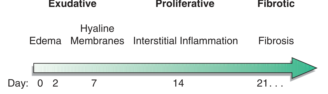
ARDS is a syndrome of acute hypoxemic respiratory failure caused by diffuse alveolar damage. The core mechanism involves an uncontrolled inflammatory response that disrupts the alveolar-capillary membrane, leading to protein-rich edema flooding the air spaces, surfactant dysfunction, and severe ventilation-perfusion mismatch. Key phases are exudative (0-7 days), proliferative (7-21 days), and fibrotic (>21 days).
Searching sources...
Finding Sources
Searching PubMed
"acute respiratory distress syndrome"[MeSH Terms] AND pathogenesis
Good - the best sources are Murray & Nadel's (dedicated ARDS chapter in block29/30/32) and Goldman-Cecil (block12). Let me read these in parallel.
Reading File
Reading File
Searching PubMed
"acute respiratory distress syndrome"[MeSH Terms] AND mechanism
The pancreatitis block was from a different chapter. Let me get the dedicated ARDS mechanism chapter from Murray & Nadel block32, and also the Sabiston block which should have mechanism detail.
Reading File
Reading File
Searching PubMed
"acute respiratory distress syndrome"[MeSH Terms] AND "pathophysiology"[MeSH Terms]
That was the HFNC/NIV section. Let me search for the core ARDS pathophysiology/mechanism content using paragraphs_fts, and also find the dedicated ARDS chapter in Murray & Nadel.
Finding Sources
Finding Sources
Excellent - I found rich sources. Now let me read the most relevant sections: Robbins Pathology (dedicated ARDS section) and Harrison's (clinical course/pathophysiology), plus Goldman-Cecil ARDS pathobiology.
Reading File
Reading File
Reading File
Harrison's has the best phase-by-phase mechanism description. Let me read more of that section and also search for the neutrophil/cytokine/coagulation cascade detail.
Reading File
Finding Sources
Now I have enough rich content. Let me also get the full exudative phase mechanism detail from Harrison's which was cut off, and fetch the key ARDS phase diagram image.
Reading File
Loading Image
I now have comprehensive information. Let me also read the exudative phase cytokine detail from Harrison's that was referenced earlier.
Reading File
I have extensive information from Harrison's (primary source), Goldman-Cecil, Robbins, and Sabiston. I now have everything needed for a comprehensive answer.
Acute Respiratory Distress Syndrome (ARDS): Mechanism
ARDS is a clinical syndrome of severe, rapid-onset hypoxemia and diffuse pulmonary infiltrates that leads to respiratory failure. The underlying lesion is diffuse alveolar damage (DAD) - injury to both the vascular endothelium and the alveolar epithelium, resulting in a non-cardiogenic, protein-rich pulmonary edema. The pathophysiology unfolds in three sequential phases.
Triggers and Initial Insult
Injury can arrive via two routes:
| Direct Lung Injury | Indirect Lung Injury |
|---|---|
| Pneumonia | Sepsis (most common overall) |
| Aspiration of gastric contents | Severe trauma / multiple fractures |
| Pulmonary contusion | Multiple transfusions (TRALI) |
| Near-drowning | Pancreatitis |
| Toxic inhalation | Drug overdose |
80% of ARDS cases are caused by pneumonia, sepsis, aspiration, or major trauma. The risks multiply when multiple predisposing conditions coexist. - Harrison's Principles of Internal Medicine, 22E
Phase 1: Exudative Phase (Days 0-7)
This is the core mechanistic phase and contains the most important pathophysiology.
1. Alveolar-Capillary Barrier Disruption
The initial insult - whether direct (e.g., inhaled toxin) or indirect (e.g., circulating endotoxin from sepsis) - activates the innate immune system. Proinflammatory cytokines, particularly IL-8, TNF-α, and IL-1, are released from resident macrophages and injured epithelial cells. These cytokines:
- Upregulate adhesion molecules (ICAM-1, E-selectin) on pulmonary capillary endothelium
- Drive massive sequestration and activation of neutrophils within alveolar and interstitial spaces
Neutrophils are the central effector cells of ARDS. Once activated, they release:
- Proteases (elastase, matrix metalloproteinases) that degrade the extracellular matrix and tight junctions
- Reactive oxygen species (ROS) that damage lipid membranes
- Platelet-activating factor and additional cytokines that amplify the inflammatory cascade
The result is destruction of type I pneumocytes (alveolar epithelial cells) and capillary endothelial cells, breaching the normally tight alveolar barrier. - Goldman-Cecil Medicine; Harrison's 22E
2. Protein-Rich Edema Floods the Alveoli
Once the barrier is disrupted, fluid and proteins pour freely from the vasculature into the interstitium and alveolar spaces. This edema is non-cardiogenic (pulmonary capillary wedge pressure is normal) and protein-rich, distinguishing it from hydrostatic edema. The heavy, fluid-filled lung behaves like a sponge - edema is distributed preferentially in dependent lung zones, causing collapse and atelectasis of large dependent sections. - Harrison's 22E
3. Surfactant Dysfunction
Type I pneumocyte death is accompanied by injury to type II pneumocytes, which normally synthesize and secrete surfactant. Phospholipase A2 (released especially in pancreatitis-associated ARDS) enzymatically degrades surfactant. Loss of surfactant dramatically increases alveolar surface tension, promoting alveolar collapse and further atelectasis. - Murray & Nadel's Textbook of Respiratory Medicine
4. Hyaline Membrane Formation
Protein-rich exudate that pools in the alveoli undergoes partial organization and deposited fibrin combines with necrotic cellular debris to form hyaline membranes - the histologic hallmark of DAD. These membranes line the alveolar walls and further impair gas diffusion. - Robbins & Kumar Basic Pathology
5. Microvascular Occlusion and Dead Space
Simultaneously, activation of the coagulation cascade in injured capillaries leads to in situ microthrombi in small pulmonary arteries. This diverts blood away from ventilated alveoli (increasing dead space and pulmonary vascular resistance), resulting in pulmonary hypertension. - Goldman-Cecil Medicine
Net Physiologic Consequences
Two major gas exchange abnormalities result:
- Intrapulmonary shunt - collapsed, edema-filled alveoli are perfused but not ventilated → severe refractory hypoxemia (PaO₂/FiO₂ <300)
- Increased dead space - ventilated alveoli are not perfused → hypercapnia
Decreased lung compliance (the stiff, waterlogged lung requires higher pressures to ventilate), dramatically increased work of breathing, and rapid deterioration to respiratory failure follow. - Harrison's 22E; Goldman-Cecil Medicine
Phase 2: Proliferative Phase (Days 7-21)
Most patients enter a period of recovery beginning around day 7. However, the lung is remodeling under injury conditions:
- Interstitial inflammation becomes prominent
- Type II pneumocyte hyperplasia develops as a repair response - these cells proliferate to repopulate the denuded alveolar basement membrane and eventually differentiate into type I cells
- Fibroblast infiltration begins along the interstitium and alveolar spaces, laying down new connective tissue
- The organizing fibrous tissue and proliferating cells gradually clear the hyaline membranes
Many patients improve during this phase, but oxygenation and pulmonary compliance may still be impaired. - Goldman-Cecil Medicine; Harrison's 22E
Phase 3: Fibrotic Phase (>21 Days)
In patients who do not recover fully, the proliferative changes progress to established fibrosis:
- Alveolar septal thickening from organizing fibrosis
- Bullae formation from destruction and coalescence of damaged alveoli
- Loss of pulmonary architecture
- Persistent hypoxemia, reduced diffusing capacity, and exercise limitation
The fibrotic phase is not universal - most patients recover without permanent fibrosis if the initial insult is controlled. Temporal uniformity of the pathologic changes (all areas at the same stage) is characteristic and reflects the single acute event. - Goldman-Cecil Medicine; Harrison's 22E
Summary Cascade
Trigger (sepsis / trauma / aspiration...)
        ↓
Macrophage activation → IL-8, TNF-α, IL-1
        ↓
Neutrophil sequestration in lung capillaries
        ↓
Neutrophil activation → proteases, ROS, PAF
        ↓
Type I pneumocyte + endothelial cell death
        ↓
Alveolar-capillary barrier breakdown
        ↓
Protein-rich flood of alveoli + interstitium
        ↓
Surfactant loss (Type II pneumocyte injury)
        ↓
Hyaline membrane formation + microthrombi
        ↓
↑ Intrapulmonary shunt + ↑ Dead space
        ↓
Severe refractory hypoxemia (PaO₂/FiO₂ <300)
        ↓
Respiratory failure → mechanical ventilation
Berlin Definition (2012, Updated 2023)
| Severity | PaO₂/FiO₂ |
|---|---|
| Mild | 200-300 mmHg |
| Moderate | 100-200 mmHg |
| Severe | <100 mmHg |
All require: bilateral opacities on imaging, onset within 1 week of a known insult, and exclusion of purely cardiogenic edema. The 2023 global update allows SpO₂/FiO₂ ratios and high-flow nasal oxygen as alternative criteria to improve applicability in resource-limited settings. - Harrison's Principles of Internal Medicine, 22E
Sources: Harrison's Principles of Internal Medicine 22E | Goldman-Cecil Medicine (International Ed.) | Murray & Nadel's Textbook of Respiratory Medicine | Robbins & Kumar Basic Pathology | Sabiston Textbook of Surgery 11E
This is a shared conversation. Sign in to Orris to start your own chat.