
About hamstring muscle
Reading File
The hamstring is a group of three muscles at the back of the thigh - biceps femoris, semitendinosus, and semimembranosus - that flex the knee and extend the hip. Let me pull comprehensive textbook coverage for you.
Searching sources...
Finding Sources
Finding Sources
Good hits from Gray's Anatomy and Campbell's. Let me read the key anatomy sections now.
Reading File
Finding Sources
Excellent data. Let me read more of the Gray's anatomy section (semimembranosus continuation + hamstring injury + blood supply/nerve supply) and also get the injury section:
Reading File
Reading File
I have rich content from Gray's Anatomy for Students. Let me now fetch the two key diagrams and also look up the hamstring strain section from a clinical/orthopaedic reference:
Loading Image
Loading Image
Reading File
I now have everything needed for a comprehensive, well-illustrated answer. Let me compile it.
Hamstring Muscles - Complete Overview
(Source: Gray's Anatomy for Students; Miller's Review of Orthopaedics 9th Ed.)
What Are the Hamstrings?
The hamstrings are a group of three muscles in the posterior compartment of the thigh. They are collectively responsible for knee flexion and hip extension. The term "hamstring" is sometimes applied to four muscles when the hamstring part of adductor magnus is included, because it shares the same origin and nerve supply.
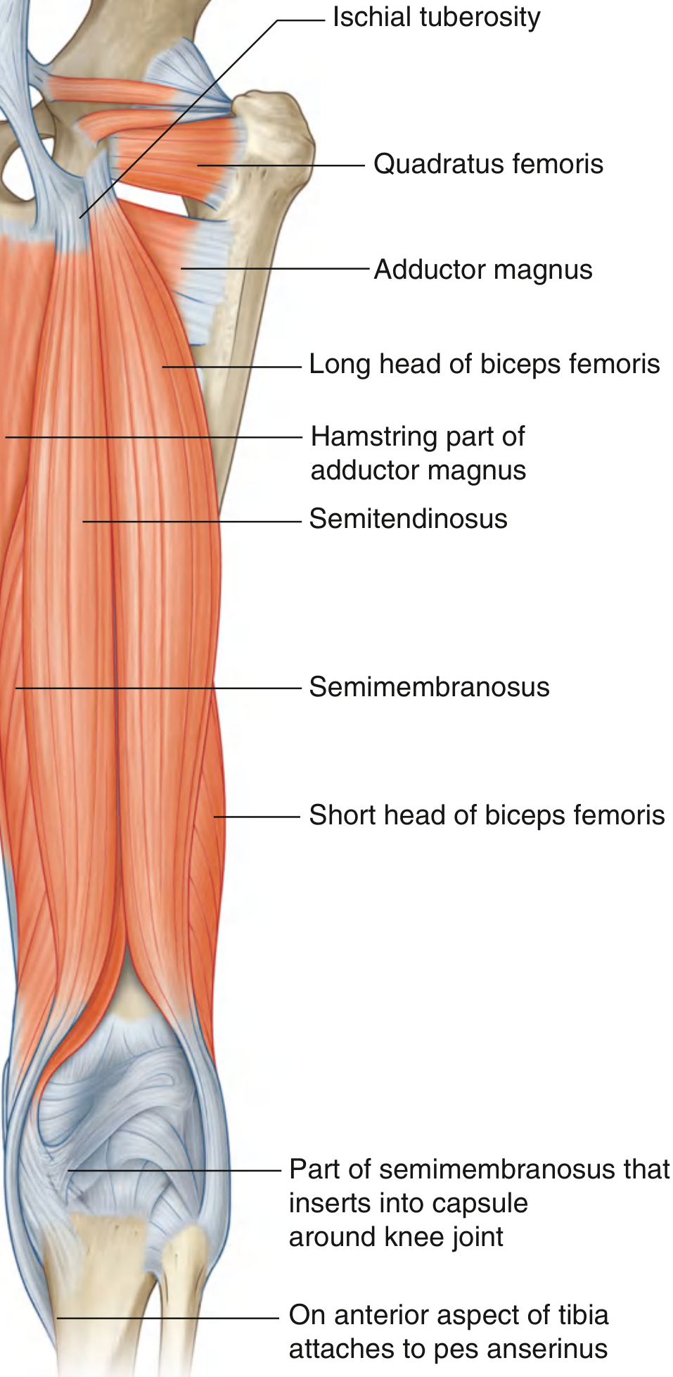
The Three Hamstring Muscles
1. Biceps Femoris
The biceps femoris lies on the lateral side of the posterior compartment and has two heads:
| Head | Origin |
|---|---|
| Long head | Inferomedial part of the upper ischial tuberosity (shared with semitendinosus) |
| Short head | Lateral lip of the linea aspera on the femoral shaft |
- Insertion: Head of the fibula (main tendon). Extensions blend with the fibular collateral ligament and lateral knee ligaments.
- Actions:
- Flexes the leg at the knee
- Long head: extends and laterally rotates the hip
- With knee partly flexed: laterally rotates the leg
- Innervation:
- Long head: tibial division of sciatic nerve (L5, S1, S2)
- Short head: common fibular (peroneal) division of sciatic nerve
Note: The short head is the only hamstring muscle NOT innervated by the tibial division.
2. Semitendinosus
Located medial to biceps femoris, the semitendinosus has a distinctive long cord-like tendon that forms in the lower half of the thigh.
- Origin: Inferomedial part of the upper ischial tuberosity (shared with long head of biceps femoris)
- Insertion: Medial surface of the tibia, posterior to sartorius and gracilis tendons - forming part of the pes anserinus ("goose foot")
- Actions:
- Flexes the leg at the knee
- Extends the thigh at the hip
- Medially rotates the thigh at the hip and the leg at the knee
- Innervation: Tibial division of the sciatic nerve
3. Semimembranosus
The deepest of the hamstrings, lying deep to semitendinosus.
- Origin: Superolateral impression on the ischial tuberosity
- Insertion: Groove on the medial and posterior surfaces of the medial tibial condyle. Tendon expansions contribute to ligaments and capsule around the knee joint.
- Actions:
- Flexes the leg at the knee
- Extends the thigh at the hip
- Medially rotates the thigh and leg (working with semitendinosus)
- Innervation: Tibial division of the sciatic nerve
Summary Table
| Muscle | Origin | Insertion | Innervation | Actions |
|---|---|---|---|---|
| Biceps femoris (long head) | Ischial tuberosity (inferomedial) | Head of fibula | Tibial (L5, S1, S2) | Knee flexion, hip extension, lateral rotation |
| Biceps femoris (short head) | Lateral lip of linea aspera | Head of fibula | Common fibular (L5, S1, S2) | Knee flexion, lateral rotation of leg |
| Semitendinosus | Ischial tuberosity (inferomedial) | Medial tibia (pes anserinus) | Tibial (L5, S1, S2) | Knee flexion, hip extension, medial rotation |
| Semimembranosus | Ischial tuberosity (superolateral) | Medial tibial condyle | Tibial (L5, S1, S2) | Knee flexion, hip extension, medial rotation |
Innervation and Blood Supply
- All hamstrings are innervated by branches of the sciatic nerve (L5, S1, S2).
- The tibial division supplies the long head of biceps femoris, semitendinosus, and semimembranosus.
- The common fibular division supplies only the short head of biceps femoris.
- Blood supply comes primarily from the perforating branches of the profunda femoris artery (deep artery of the thigh), which is the main arterial supply to the posterior thigh.
Pes Anserinus
The semitendinosus tendon, along with gracilis and sartorius, inserts into the medial surface of the tibia as the pes anserinus (Latin: "goose foot"). This is a clinically important landmark - pes anserinus bursitis causes medial knee pain, especially in obese or osteoarthritic patients.
Clinical Relevance: Hamstring Injuries
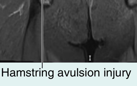
Hamstring Strains
Hamstring injuries are among the most common musculoskeletal injuries in athletes, particularly in sprinting, football, and track-and-field sports. They occur most often during:
- Late swing phase of running, when the hamstrings contract eccentrically to decelerate the leg
- Sudden accelerations/decelerations or rapid changes of direction
Most common site of injury: The musculotendinous junction - a wide transition zone between muscle and tendon.
Grading
- Grade 1 (mild strain): Minor fiber disruption, focal edema on MRI
- Grade 2 (partial tear): More fibers disrupted, significant weakness
- Grade 3 (complete tear/rupture): Full disruption of muscle or tendon
Special Populations
- Adults: Musculotendinous junction is the most vulnerable zone.
- Adolescents: Avulsion of the ischial tuberosity apophysis is more common - the growth plate (apophysis) is the weakest link in the proximal hamstring unit in skeletally immature patients.
Imaging
- Ultrasound: Can identify fluid within muscle, tears
- MRI: Gold standard - shows extent of injury and predicts prognosis (risk of re-tear, functional loss)
Management
| Injury | Treatment |
|---|---|
| Mild-moderate strain | Rest, ice, compression, elevation; then physiotherapy (stretching + strengthening) |
| Return to sport | Delayed until strength reaches ~90% of the contralateral side |
| Single tendon rupture | Nonoperative |
| Multiple tendon ruptures with <2 cm retraction | Nonoperative |
| Three tendon ruptures | Surgical direct repair to bone |
Hamstring Tendons as Surgical Grafts
The semitendinosus and gracilis tendons (medial hamstring tendons) are commonly harvested as autografts for ACL reconstruction due to their length, strength, and acceptable donor-site morbidity. This is a standard technique in orthopaedic sports medicine.
Sources: Gray's Anatomy for Students (p.700-702); Miller's Review of Orthopaedics 9th Ed. (p.10931-10944); Campbell's Operative Orthopaedics 15th Ed. 2026
This is a shared conversation. Sign in to Orris to start your own chat.