
Gi examination in CLD patient detailed with possible viva questions
spider naevi palmar erythema chronic liver disease CLD signs
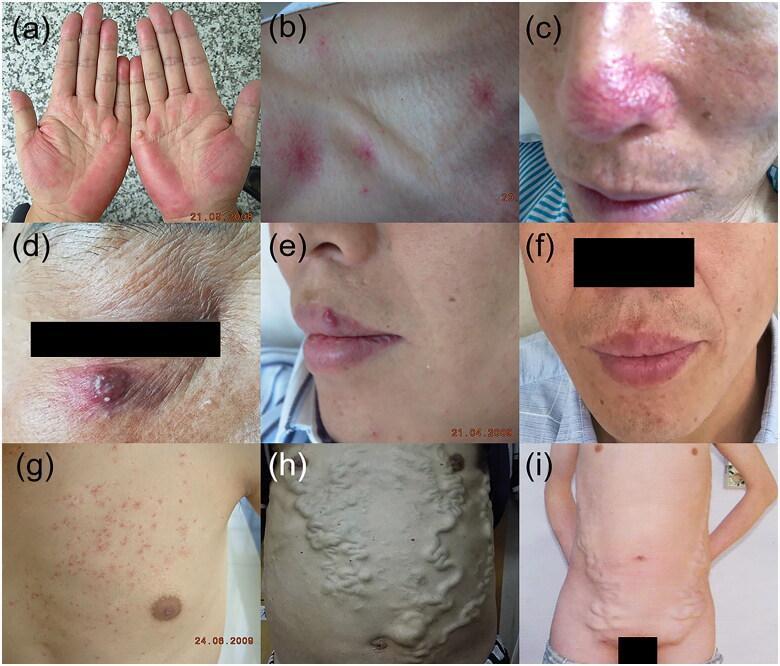
This composite of clinical photographs illustrates various cutaneous vascular manifestations of chronic liver disease and portal hypertension. (a) Palmar erythema, showing bilateral symmetric redness over the thenar and hypothenar eminences. (b, c) Spider angiomata on the chest and nose, appearing as central red arterioles with radiating capillary 'legs'. (d, e, f) Arteriovenous haemangioma presenting as a raised, reddish-purple nodule on the face, with (e) and (f) demonstrating lesion regression over time. (g) Paper money skin on the upper torso, characterized by numerous fine, thread-like telangiectasias reminiscent of security threads in currency. (h) Caput medusa, exhibiting severe, tortuous dilatation of the paraumbilical veins radiating from the umbilicus due to portal-systemic shunting. (i) Abdominal varicose veins associated with inferior vena cava obstruction syndrome, showing prominent, vertically-oriented collateral veins on the lateral and lower abdominal wall. These images serve as diagnostic markers for hepatobiliary pathology and related hemodynamic alterations.
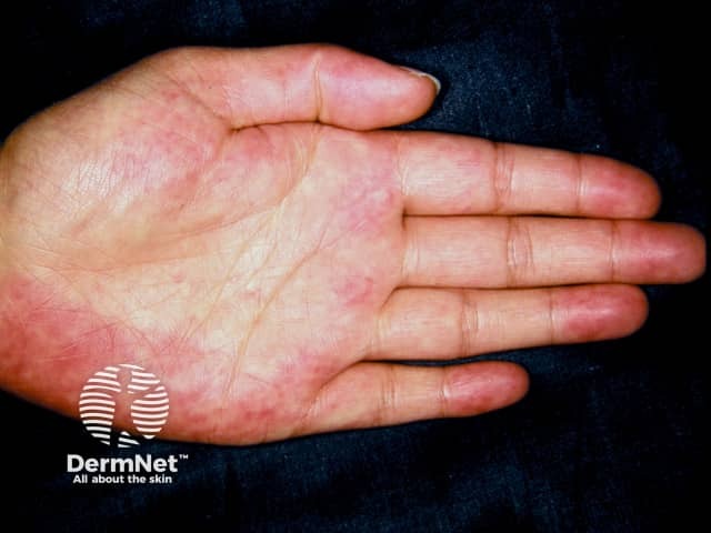
Clinical photography of the palmar surface (volar aspect) of the hand demonstrating palmar erythema, a cutaneous vascular sign often associated with liver disease. Modality: Clinical photography; color image captured under bright, diffuse illumination to show true erythema without shadowing. Anatomical location: palm (thenar and hypothenar regions) of the hand; symmetric or asymmetric involvement may be observed, though the provided image shows patchy diffuse redness along the palmar creases. Visual features include diffuse, blanchable erythema most prominent on the central palmar regions; warm appearance; minimal or absent edema; no vesicles or scaling evident. Pathophysiology: estrogen-mediated cutaneous vascular dilation; may reflect hepatic dysfunction with portal circulation alterations or hyperestrogenism. Diagnostic significance: Palmar erythema can indicate liver disease (cirrhosis, chronic hepatitis, alcoholic liver disease) or conditions with elevated estrogen; in pregnancy or with estrogen therapy it may appear similarly. Differential considerations include pregnancy-related erythema, drug-induced flushing, autoimmune or rheumatologic conditions, and vasculitis; clinical correlation with liver function tests (ALT/AST, ALP, bilirubin, albumin), coagulation profile, and portal hypertension signs is essential. Clinical relevance: serves as a non-invasive bedside sign prompting hepatology evaluation, risk stratification, and longitudinal monitoring; useful in medical education to illustrate cutaneous markers of systemic disease and to teach differential diagnosis in dermatology and internal medicine.
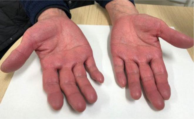
This clinical photograph displays the bilateral palmar surfaces of a patient's hands, illustrating a pronounced, diffuse erythema. The redness is uniform across the palms, thenar and hypothenar eminences, and extends into the distal phalanges of all fingers. The skin appears intact without evidence of scaling, ulceration, or hyperkeratosis. While palmar erythema is frequently associated with chronic liver disease, this specific presentation serves as a clinical sign of hyperviscosity or erythrocytosis, specifically in the context of polycythemia vera. The image demonstrates how the correction of underlying iron deficiency anemia can unmask the plethoric appearance of polycythemia vera as hemoglobin levels rise. This visual is a key educational resource for identifying secondary skin manifestations of myeloproliferative neoplasms and differentiating them from other causes of palmar flushing.
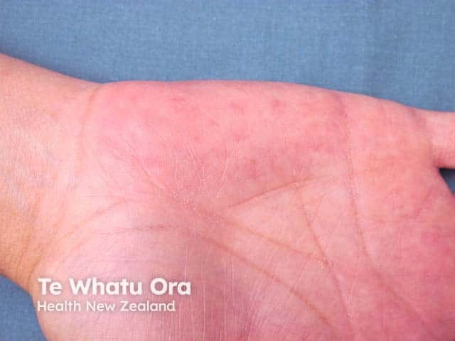
This is a clinical photograph of the palmar (ventral) surface of the hand, captured in high resolution with even illumination to document skin color. The primary subject is diffuse palmar erythema: uniform reddening of the palm, most prominent on the thenar and mid-portion of the hand, with preserved skin texture and fine palmar creases visible. No discrete lesions, vesicles, or edema are evident in the image. The redness appears transiently blanchable under pressure and may be warm to the touch, though this can only be inferred from appearance. The finding is non-specific but classically associated with systemic conditions such as chronic liver disease (cirrhosis, hepatitis), pregnancy, estrogen therapy, hyperthyroidism, autoimmune disorders, and drug reactions. Clinically relevant diagnostic significance is that palmar erythema warrants evaluation for hepatic function, hormonal status, and medication history, especially in patients with risk factors for liver disease or pregnancy. Differential diagnoses include physiologic flushing, contact dermatitis, and other causes of cutaneous hyperemia. This image is valuable for educational and research contexts to illustrate a cutaneous sign that may precede or accompany systemic illness. Potential clinical use cases include medical teaching, triage cue for liver disease assessment, and dermatology training in pattern recognition.
caput medusae ascites flapping tremor asterixis liver cirrhosis
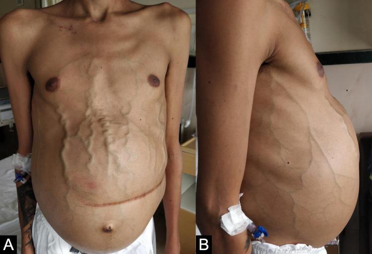
Clinical photograph of a male patient's torso in anterior (A) and lateral (B) views, demonstrating advanced clinical signs of portal hypertension and chronic liver disease. The abdomen is severely distended, consistent with tense ascites. Prominent, tortuous, and dilated superficial veins (caput medusae) are visible across the entire abdominal wall and flanks, radiating from the peri-umbilical region. A well-healed, horizontal surgical scar from a previous splenectomy is present in the mid-abdomen. Additional findings include a small umbilical hernia, bilateral gynecomastia, and significant sarcopenia characterized by muscle wasting in the pectoral region and visible bony landmarks of the clavicles and shoulders. An intravenous cannula is present in the left arm. These visual markers collectively illustrate the systemic manifestations of severe portal venous congestion, commonly associated with conditions such as Gaucher disease or advanced cirrhosis.
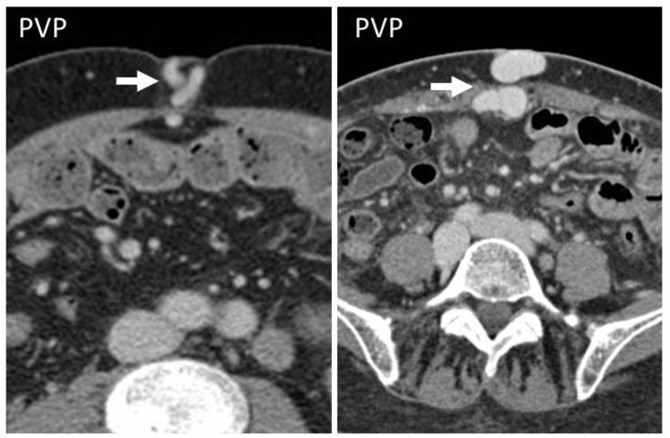
Diagnostic axial CT scans of the abdomen in the portal venous phase (PVP) from two different patients with liver cirrhosis. The images demonstrate the classic CT appearance of 'caput medusae,' characterized by prominent, tortuous, contrast-enhanced collateral vessels (indicated by white arrows) within the subcutaneous fat of the anterior abdominal wall at the level of the umbilicus. These findings result from the recanalization of the umbilical vein as a portosystemic shunt in the setting of portal hypertension. The left image shows a more slender, tortuous morphology of the umbilical collateral, while the right image displays larger, more rounded, hyperattenuating vascular cross-sections in the periumbilical region. Both images show typical abdominal anatomy, including the subcutaneous and mesenteric fat, bowel loops, abdominal wall musculature, and the lumbar vertebrae posteriorly. This visual finding is a crucial diagnostic indicator of advanced liver disease and significant portal venous pressure elevation.
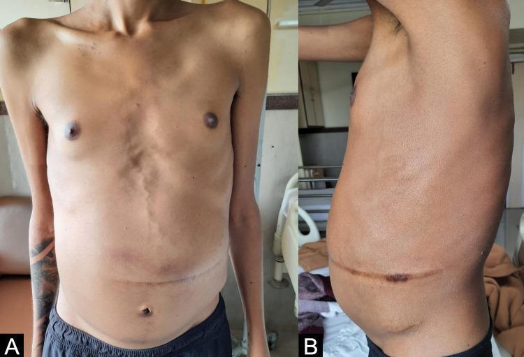
Clinical photograph of a male patient presenting with signs of portal hypertension and chronic systemic illness, captured in anterior (A) and lateral (B) views. The images demonstrate significant abdominal distension consistent with ascites, although according to clinical context, this represents a post-Transjugular Intrahepatic Portosystemic Shunt (TIPS) state with partial resolution. Notable features include prominent vertical venous dilation (caput medusae) along the abdominal midline and a transverse supra-umbilical surgical scar. The patient exhibits a sarcopenic body habitus, characterized by visible clavicles, reduced muscle mass in the upper torso, and a thin appearance relative to the distended abdomen. This presentation is characteristic of complications from Gaucher disease and secondary cirrhosis, demonstrating the physical manifestations of chronic liver disease, portosystemic collateral circulation, and nutritional depletion.
leukonychia terry nails clubbing liver disease hands
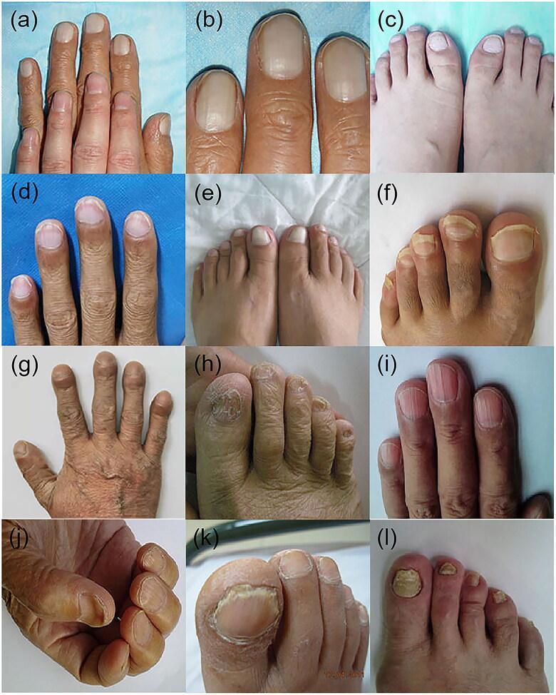
This composite clinical photograph displays multiple nail manifestations associated with systemic conditions, specifically chronic liver disease. The panel includes: (a-c) Terry's nails, showing a ground-glass whitening of the proximal nail bed with a narrow distal pink-brown band and absence of lunulae. (d) Lindsay's nails (half-and-half nails), featuring a sharp demarcation between a white proximal half and a reddish-brown distal half. (e) Leukonychia, characterized by total or partial whitening of the nail plate. (f) Onycholysis, demonstrating separation of the nail plate from the distal nail bed. (g) Clubbing, showing bulbous enlargement of the distal phalanges and increased nail curvature. (h) Brittle nails, exhibiting surface granulation and fragile, worn edges. (i) Longitudinal striations, presenting as prominent vertical ridges (onychorrhexis). (j) Koilonychia, showing characteristic concave or spoon-shaped nail plates. (k, l) Onychomycosis, illustrating fungal infection with subungual hyperkeratosis, yellow-white discoloration, and nail plate thickening. This collection serves as a diagnostic reference for dermatological signs of internal pathology, particularly hepatic cirrhosis and renal dysfunction.
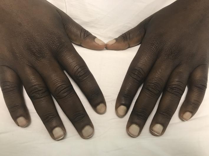
This clinical photograph displays the dorsal surface of both hands in a patient with dark skin pigmentation, demonstrating classic findings of Terry's nails. The fingernails exhibit a characteristic 'ground-glass' appearance, characterized by a diffuse, near-total whitish opacification of the nail bed that obscures the lunula. Distally, a narrow, transverse band of normal pink or brownish-red dermis remains visible (roughly 0.5–3.0 mm wide) just proximal to the free edge of the nail. These changes are indicative of an apparent leukonychia caused by decreased vascularity and increased connective tissue in the nail bed. Clinically, this sign is most strongly associated with hepatic cirrhosis, but may also be seen in chronic heart failure, type 2 diabetes mellitus, and chronic renal failure. The skin over the interphalangeal joints appears slightly thickened with prominent creasing, though no acute dermatological lesions are present. This image is a primary educational resource for internal medicine and dermatology, illustrating nail manifestations of systemic disease.
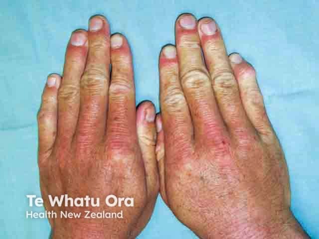
Clinical photograph of bilateral hands (dorsal view) highlighting diffuse leukonychia of the fingernails associated with hypoalbuminaemia. This nail abnormality presents as white, chalky patches and diffuse whitening of the proximal nail plates with variable translucency, contrasting with normally pink nail beds. In hypoalbuminaemic states, decreased circulating albumin can lead to nail bed edema and altered keratinization, producing characteristic leukonychia (white discolouration) that may be punctate or diffuse. The image demonstrates multiple digits with whitish discoloration extending from the lunula toward the distal nail edge, without obvious onycholysis or subungual debris. Secondary vascular and cutaneous changes may accompany malnutrition, including periorbital edema or paler skin. Clinically, this finding is non-specific but serves as a surrogate marker of nutritional status and hepatic or renal protein loss, and it may improve with correction of hypoalbuminaemia. Relevance to practice includes screening for protein-energy malnutrition in hospitalized or chronically ill patients, guiding nutritional intervention, and monitoring response to therapy. Differential considerations include Mees' lines (arsenic exposure), Muehrcke's lines (transverse white bands due to hypoalbuminaemia of edema), and Terry's nails. Documentation aids educational assessment, dermatology and internal medicine training, and research into nail biomarkers of systemic disease.
GI Examination in Chronic Liver Disease (CLD) - Complete Guide with Viva Questions
GENERAL APPROACH TO EXAMINATION
1. GENERAL SURVEY (Before Touching the Patient)
| Finding | Significance |
|---|---|
| Cachexia / muscle wasting | Advanced liver failure, HCC |
| Jaundice (visible at distance) | Bilirubin >2.5 mg/dL |
| Pallor | Anemia (hypersplenism, GI bleed) |
| Pedal edema (visible) | Hypoalbuminemia, portal hypertension |
| Abdominal distension | Ascites |
| Scratch marks on skin | Pruritus from cholestasis |
| Mental status | Hepatic encephalopathy (confusion, drowsiness) |
2. EXAMINATION OF THE HANDS
Nails
- Leukonychia (white nails) - hypoalbuminemia; the entire nail plate appears white due to decreased vascularity and hypoalbuminemia
- Terry's nails - ground-glass whitening of proximal nail with narrow (0.5-3 mm) distal pink band; strongly associated with hepatic cirrhosis
- Clubbing - seen in hepatopulmonary syndrome, biliary cirrhosis, cryptogenic cirrhosis
- Koilonychia (spoon nails) - iron deficiency from chronic GI blood loss
Palm
- Palmar erythema - redness of the thenar and hypothenar eminences; due to estrogen excess and hyperdynamic circulation; seen in cirrhosis, pregnancy, and thyrotoxicosis
Other Hand Signs
- Dupuytren's contracture - fibrosis of palmar fascia; strongly associated with alcohol-related liver disease
- Flapping tremor (Asterixis) - ask patient to dorsiflect wrists with arms outstretched; coarse, irregular flap; HALLMARK of overt hepatic encephalopathy; also seen in renal/respiratory failure
- Muscle wasting of thenar and hypothenar eminences - marker of liver insufficiency
3. EXAMINATION OF THE ARMS
- Spider angiomata (spider naevi) - present on arms, forearms, and upper torso; superficial tortuous arterioles that fill from the center outward (unlike telangiectasias); >5 are significant; due to estrogen excess / hyperdynamic circulation
- Bruising / ecchymoses - coagulopathy (reduced prothrombin synthesis, thrombocytopenia)
- Track marks - IV drug use as etiology of viral hepatitis
4. EXAMINATION OF THE FACE
Eyes
- Scleral icterus - best seen under natural light; requires bilirubin >2.5 mg/dL; check mucous membranes below tongue in dark-skinned individuals
- Kayser-Fleischer rings - golden-brown copper ring at periphery of cornea in Descemet's membrane; pathognomonic of Wilson's disease; requires slit-lamp examination (may be visible to naked eye when advanced)
- Xanthelasma - lipid deposits around eyelids; primary biliary cholangitis, sclerosing cholangitis
- Pallor of conjunctivae - anemia
Mouth/Face
- Fetor hepaticus - sweet, musty ("mousy") odor of breath; due to mercaptans and dimethylsulfide absorbed from gut and exhaled; marker of hepatic encephalopathy
- Parotid enlargement - alcohol-related liver disease
- Oral ulcers, dry mouth - seen in primary biliary cholangitis
Skin of Face
- Spider naevi - on face (nose, cheeks) - same significance as on arms
- Hyperpigmentation - advanced chronic cholestatic diseases (PBC, PSC)
- Slate-gray pigmentation - hemochromatosis
5. EXAMINATION OF THE NECK / SUPRACLAVICULAR
- Left supraclavicular lymphadenopathy (Virchow's node) - suggests intra-abdominal malignancy (e.g., HCC, GI tumor)
- Jugular venous pressure - may be elevated in right heart failure (Budd-Chiari)
6. EXAMINATION OF THE CHEST
- Spider angiomata on chest/upper torso - most commonly found here
- Paper money skin - numerous fine thread-like telangiectasias on upper torso (visible in image above, panel g)
- Gynecomastia - estrogen excess; male patients; especially in alcohol-related cirrhosis
- Loss of male-pattern chest hair - hyperestrogenemia
- Bilateral crackles - hepatic hydrothorax (right-sided pleural effusion from ascitic fluid via diaphragmatic defects)
- Signs of portopulmonary hypertension - loud P2, right ventricular heave
7. ABDOMINAL EXAMINATION (The Core)
Inspection
- Distension - ascites (fullness of flanks)
- Caput medusae - dilated superficial veins radiating from the umbilicus; due to recanalization of the umbilical vein via the paraumbilical vein; portal hypertension
- Direction of flow in dilated veins - critical for diagnosis:
- Caput medusae (portal hypertension): blood flows AWAY from umbilicus in all directions
- IVC obstruction (Budd-Chiari/IVC block): blood flows UPWARD on both sides below umbilicus (and downward above)
- Umbilical hernia - from raised intra-abdominal pressure due to ascites
- Visible pulsation - aortic aneurysm or transmitted hepatic pulsation
Palpation
- Start from right iliac fossa, move upward
- Normal liver may not be palpable
- In early CLD: hepatomegaly (firm, smooth, may be tender)
- In advanced cirrhosis: liver may become small and shrunken (not palpable); a firm, irregular nodular surface suggests cirrhosis or HCC
- Hepatic tenderness - most reliable physical sign (Harrison's)
- Hepatic bruit - auscultate over liver; suggests HCC or alcoholic hepatitis
- Marked hepatomegaly: cirrhosis, sinusoidal obstruction syndrome, amyloidosis, metastatic disease, alcoholic hepatitis
- Start from right iliac fossa diagonally
- Splenomegaly - subtle but significant finding suggesting underlying portal hypertension; can be confirmed with ultrasound
- Shifting dullness - most sensitive; percuss from umbilicus to flank; dullness shifts when patient rolls to opposite side; detects >500 mL of fluid
- Fluid thrill - tap one flank, feel thrill on other; hand of assistant placed vertically in midline to prevent transmission through fat; detects large ascites (>1000 mL)
- Puddle sign - detects as little as 120 mL; patient kneels on all fours
Percussion
- Liver span - upper border by percussion (dullness starts at 5th rib in MCL); lower border by percussion + palpation; normal 6-12 cm
- Traube's space - percussion of left lower costal region; normally resonant (stomach); dullness suggests splenomegaly (or full stomach)
- Flanks - dull in ascites
Auscultation
- Bowel sounds - may be decreased in ileus/peritonitis (SBP), hyperactive in bleeding
- Hepatic bruit - HCC, alcoholic hepatitis
- Venous hum over umbilicus - portal hypertension; Cruveilhier-Baumgarten syndrome (recanalized umbilical vein with audible venous hum)
- Friction rub over liver/spleen - hepatic infarction, perihepatitis, splenic infarct
8. EXAMINATION OF THE GENITALIA
- Testicular atrophy - hyperestrogenemia in male cirrhosis, particularly alcohol-related
- Loss of pubic/axillary hair - feminization in male CLD
9. LOWER LIMBS
- Pitting edema - hypoalbuminemia + portal hypertension + secondary hyperaldosteronism
- Muscle wasting - particularly bitemporal and thigh muscles; protein catabolism
- Loss of leg hair - hyperestrogenemia
- Petechiae - thrombocytopenia (hypersplenism)
10. COMPLETE NEUROLOGICAL ASSESSMENT (Hepatic Encephalopathy)
| Grade | Features |
|---|---|
| 0 (Minimal/Covert) | Subclinical; abnormal neuropsychometric tests only; no asterixis |
| I | Mild confusion, sleep inversion, forgetfulness, distal tremor |
| II | Drowsiness, disorientation, bizarre behavior, asterixis present |
| III | Lethargy, profound disorientation, asterixis (may be absent) |
| IV | Coma, no response to verbal stimuli |
- Asterixis - hallmark of overt HE (Grade II+); also test with tongue, feet
- Trail-making test - connect 25 numbered circles; normal <30s; prolonged in early HE
- Fetor hepaticus - sweet/musty breath
- Constructional apraxia - inability to draw a 5-pointed star or connect dots
CHILD-TURCOTTE-PUGH (CTP) SCORE - Must Know for Viva
| Parameter | 1 point | 2 points | 3 points |
|---|---|---|---|
| Bilirubin (μmol/L) | <34 | 34-50 | >50 |
| Albumin (g/L) | >35 | 25-35 | <25 |
| Ascites | None | Easily controlled | Poorly controlled |
| Encephalopathy | None | Grade I/II | Grade III/IV |
| INR | <1.7 | 1.7-2.2 | >2.2 |
- CTP-A: 5-6 points (compensated); CTP-B: 7-9 points; CTP-C: 10-15 points (decompensated)
- Surgical mortality: CTP-A +10%, CTP-B +30%, CTP-C +80%
VIVA QUESTIONS AND ANSWERS
SIGNS AND MECHANISMS
| Sign | Mechanism |
|---|---|
| Spider naevi | Estrogen excess → arteriolar vasodilation; fill from center out |
| Palmar erythema | Estrogen excess, hyperdynamic circulation, arteriolar dilation |
| Jaundice | Reduced bilirubin conjugation/excretion |
| Paper money skin | Multiple fine telangiectasias - estrogen-related |
| Caput medusae | Portal hypertension → recanalization of umbilical/paraumbilical vein |
| Leukonychia/Terry's nails | Hypoalbuminemia → nail bed edema and altered vascularity |
| Hyperpigmentation | Melanin deposition; PBC/PSC, hemochromatosis (slate-grey) |
- Caput medusae: Blood flows AWAY from umbilicus in all directions (centrifugal); umbilicus may be everted; venous hum may be audible
- IVC obstruction: Blood flows UPWARD on BOTH flanks (i.e., above and below umbilicus, flow is upward); prominent veins on flanks and back; no venous hum; associated with leg edema
ASCITES
- Puddle sign: ~120 mL (most sensitive, rarely used clinically)
- Shifting dullness: ~500 mL
- Fluid thrill: >1000 mL (tense ascites)
- Spontaneous Bacterial Peritonitis (SBP) - most common; presents with fever, abdominal pain, encephalopathy; PMN >250 cells/mm³ in ascitic fluid
- Hepatorenal syndrome
- Umbilical hernia (with risk of rupture)
- Hepatic hydrothorax (right > left pleural effusion)
- Abdominal compartment syndrome (tense ascites)
LIVER PALPATION
- Early/Compensated: Hepatomegaly - firm, smooth, slightly tender edge
- Late/Decompensated: Liver becomes small and hard, may not be palpable; nodular surface suggests established cirrhosis or HCC
- HCC: Firm nodular hepatomegaly; hepatic bruit may be audible
- Hepatocellular carcinoma (HCC) - most important
- Alcoholic hepatitis (hepatic arterial vasodilation)
- Arteriovenous malformation
- Hepatic artery aneurysm
SPLEEN
- Cannot get above it (notch palpable)
- Moves with respiration
- Dull to percussion (Traube's space obliterated)
- No ballottement
- Notch on medial border
HEPATIC ENCEPHALOPATHY
SPECIFIC ETIOLOGY CLUES
| Sign | Etiology |
|---|---|
| Kayser-Fleischer rings | Wilson's disease |
| Dupuytren's, parotid enlargement | Alcohol-related liver disease |
| Slate-gray skin | Hemochromatosis |
| Xanthelasma, hyperpigmentation | Primary biliary cholangitis |
| Tattoos, track marks | Viral hepatitis (HBV, HCV) |
| Cushingoid features | NAFLD/metabolic syndrome |
| Bronze diabetes | Hemochromatosis (iron + diabetes + cirrhosis) |
SUMMARY: RAPID RECALL - Signs by Region
HANDS: Leukonychia, Terry's nails, clubbing, palmar erythema,
       Dupuytren's, asterixis, muscle wasting

ARMS:  Spider naevi, bruising

FACE:  Scleral icterus, KF rings, xanthelasma, parotid enlargement,
       fetor hepaticus, paper money skin

CHEST: Spider naevi, gynecomastia, loss of chest hair,
       pleural effusion (hydrothorax)

ABDOMEN: Distension (ascites), caput medusae, umbilical hernia,
         hepatomegaly (or small liver), splenomegaly, shifting
         dullness, fluid thrill, hepatic bruit, venous hum

GENITALIA: Testicular atrophy, loss of pubic hair

LEGS:  Pitting edema, muscle wasting, petechiae