
Anesthetic consideration in siabetic mellitus
Anesthetic Considerations in Diabetes Mellitus
1. Preoperative Assessment
Cardiovascular
- Diabetics have a significantly elevated risk of coronary artery disease (CAD), hypertension, congestive heart failure, and perioperative myocardial infarction.
- Silent ischemia is more common because autonomic neuropathy blunts anginal symptoms - this is a critical point for anesthetic risk stratification.
- The 2014 ACC/AHA guideline classifies insulin-dependent diabetics at elevated cardiac risk. ECG is mandatory preoperatively.
- β-blockers should be considered if the patient has ≥2 risk factors for adverse cardiac events; they do not worsen glucose intolerance or mask hypoglycemic symptoms.
Renal
- DM is the leading cause of renal failure requiring dialysis.
- Check serum creatinine and electrolytes; renal impairment alters drug dosing and fluid management.
Neurologic
- Autonomic neuropathy may cause hemodynamic instability under anesthesia (exaggerated hypotension with induction) and gastroparesis, which significantly increases aspiration risk.
- Peripheral neuropathy and vascular disease increase susceptibility to positioning injuries - document pre-existing deficits before anesthesia begins.
- Cerebrovascular and peripheral vascular disease are more prevalent.
Airway
- Stiff joint syndrome - due to glycosylation of proteins and abnormal collagen cross-linking - can affect the temporomandibular, atlanto-occipital, and cervical spine joints in longstanding Type 1 diabetics, leading to difficult tracheal intubation.
- A thorough airway examination is mandatory. The "prayer sign" (inability to fully oppose palms with fingers extended) suggests stiff joint involvement at other joints, including those relevant to airway manipulation. Maintain a high index of suspicion for difficult airway.
Laboratory Work-up
- Blood glucose, HbA1c, serum electrolytes, creatinine, urinalysis (ketonuria), and ECG.
- Target HbA1c: <8% for Type 1 and <7% for Type 2 (American Diabetes Association). If above target, elective surgery should be delayed for optimization.
2. Preoperative Optimization
- Schedule the diabetic patient as the first case of the day to minimize fasting duration and glucose variability.
- Have the patient arrive early and check blood glucose on arrival, then hourly if there is a delay.
- If blood glucose is <70 mg/dL on arrival, administer a glucose tablet or clear juice (or IV dextrose if IV is in place).
3. Medication Management on the Day of Surgery
Oral Antidiabetics
- Hold all oral antidiabetic agents on the day of surgery to avoid reactive hypoglycemia.
- Metformin must be stopped due to the risk of perioperative lactic acidosis (especially with renal hypoperfusion or contrast use).
- Non-insulin injectables (GLP-1 agonists, etc.) are also held.
Insulin
| Insulin Type | Day-of-Surgery Management |
|---|---|
| Rapid/short-acting | Hold on morning of surgery (unless hyperglycemia requires correction) |
| Intermediate/long-acting | Give a reduced dose (typically 50-80% of usual basal dose) |
| Insulin pumps | Continue at sleep (basal) rates for short procedures; convert to IV insulin infusion for major/prolonged surgery |
- Type 1 diabetics must continue basal insulin even while NPO to prevent diabetic ketoacidosis (DKA).
- Once insulin is given to an NPO patient, glucose must be provided in IV fluids or blood glucose closely monitored.
4. Perioperative Glucose Targets
| Society | Target (mg/dL) |
|---|---|
| Society for Ambulatory Anesthesia (SAMBA) | <180 |
| American Diabetes Association | 140-180 |
| Society of Critical Care Medicine | <150 |
| American College of Physicians | 140-200 |
5. Intraoperative Management
- Check blood glucose every 1-2 hours intraoperatively (or more frequently if on IV insulin infusion).
- For short/minor procedures in well-controlled diabetics: subcutaneous sliding-scale insulin + 1-2 hourly glucose checks may suffice.
- For major/prolonged surgery: intravenous insulin infusion (regular insulin) is preferred over subcutaneous or pump insulin.
- Administer insulin infusion via a side port into the same IV line as the glucose infusion (for safety).
- Use a separate non-glucose isotonic solution for replacing fluid deficits and intraoperative losses.
- Concurrent separate infusions of insulin and glucose allow easier adjustment and better control than combined glucose/insulin/potassium (GIK) infusions.
- For cardiac surgery patients and the critically ill, tight glycemic control has the most evidence for reducing morbidity and mortality.
Choice of Anesthetic Agents
- No single anesthetic technique has been proven superior in diabetics.
- Autonomic neuropathy increases sensitivity to vasodilatory and myocardial depressant effects of volatile agents and IV induction agents - anticipate and prepare for hemodynamic instability.
- Regional anesthesia may be preferred where appropriate, as it blunts the stress response and avoids issues of gastroparesis/aspiration with general anesthesia. However, peripheral neuropathy must be documented beforehand.
- Gastroparesis mandates aspiration precautions for general anesthesia: RSI (rapid sequence induction), H2 blocker/proton pump inhibitor, and prokinetic agents preoperatively.
6. Postoperative Care
- Prevent and treat postoperative nausea and vomiting (PONV) aggressively - particularly important because vomiting disrupts oral intake and insulin resumption.
- Encourage early resumption of diet to allow return to the patient's home insulin regimen.
- Check blood glucose before discharge.
- Perioperative insulin requirements vary by: body weight, liver disease, steroid use, infection, and whether cardiopulmonary bypass is used.
7. Special Considerations
Diabetic Ketoacidosis (DKA) Risk
- Type 1 diabetics: must never be left without basal insulin, even while NPO. Omitting basal insulin in a stressed surgical patient is a common trigger for DKA.
Hyperosmolar Hyperglycemic State (HHS) Risk
- Type 2 diabetics under severe physiologic stress (sepsis, major surgery) are more prone to non-ketotic HHS - severe dehydration, hyperglycemia, and hyperosmolarity.
Pediatric Patients
- The Society for Pediatric Anesthesia recommends similar perioperative principles, though no consensus HbA1c threshold exists for pediatric patients.
Positioning
- Peripheral neuropathy and vascular disease increase the risk of pressure injuries and nerve damage from positioning - use extra padding and document baseline neurological status.
- Barash, Cullen, and Stoelting's Clinical Anesthesia, 9e - Chapters 23 and 15 (Diabetes Mellitus, Perioperative Glucose Management)
- Sabiston Textbook of Surgery, 11e - Chapter 20 (Endocrinology, Metabolism, and Nutrition: Diabetes Mellitus)
Find research on autonomic neuropathy complications in diabetic patients under anesthesia
diabetic autonomic neuropathy anesthesia perioperative complications
diabetic autonomic neuropathy cardiovascular perioperative
cardiac autonomic neuropathy diabetes surgery hemodynamic
PMID: 30166651
PMID: 30788010
diabetic autonomic neuropathy gastroparesis aspiration anesthesia
cardiac autonomic neuropathy intraoperative hypotension sudden death
PMID: 29559406
PMID: 39834981
Diabetic Autonomic Neuropathy (DAN): Complications Under Anesthesia
Overview
1. Cardiovascular Autonomic Neuropathy (CAN)
Epidemiology
Intraoperative Hypotension at Induction
- Autonomic reflex dysfunction in surgical patients is directly associated with hypotension after anesthesia induction. (Latson et al., Anesthesiology. 1994;80(2):326-337 - cited in Barash Clinical Anesthesia, 9e)
- Patients with CAN have impaired baroreceptor reflex and sympathetic vasomotor tone, leaving them unable to compensate for the vasodilation from induction agents.
- Vohra et al. (Br J Anaesth. 1993;71(2):258-261) demonstrated that diabetes mellitus specifically affects cardiovascular responses to induction of anaesthesia and tracheal intubation - blunted tachycardia and impaired pressor response.
- Miller's Anesthesia (10e) notes: autonomic neuropathy causes attenuated hyperadrenergic signs during hypoglycemia under anesthesia, making it difficult to detect low blood glucose on clinical grounds alone.
Silent Myocardial Ischemia
- Silent ischemia is present in 30-50% of Type 2 diabetics - a direct consequence of CAN blunting pain sensation. (Cheisson et al., Anaesth Crit Care Pain Med, 2018 - PMID 29559406, Practice Guideline)
- Patients may sustain perioperative myocardial infarction without classic chest pain, making ECG and troponin monitoring more important in this population.
Sudden Cardiac Death
- Page & Watkins (Lancet. 1978;1:14-16) - a landmark early paper - documented cardiorespiratory arrest as a complication of diabetic autonomic neuropathy under anesthesia. This risk arises from:
- Impaired cardiorespiratory reflexes
- Prolonged QT interval (a recognized feature of CAN)
- Increased susceptibility to arrhythmias
Clinical Implications
- Cheisson et al. (Practice Guideline, 2018) recommend that CAN must be investigated preoperatively because it causes an increased risk of cardiovascular events and sudden death, even when clinically asymptomatic.
- Signs suggestive of CAN mandate close perioperative cardiovascular surveillance.
- Regional/neuraxial anesthesia can worsen hypotension in patients with autonomic insufficiency due to additional sympathetic blockade - Miller's Anesthesia 10e states: "Regional and neuraxial techniques have the advantage of reducing sympathetic stimulation but can become problematic in patients with autonomic insufficiency where hypotension may be exacerbated."
2. Gastroparesis and Aspiration Risk
Mechanism
- DAN directly impairs gastric motility through vagal dysfunction, producing diabetic gastroparesis.
- Barash Clinical Anesthesia (9e) is explicit: "The extent of delayed gastric emptying with diabetes mellitus correlates well with the presence of autonomic neuropathy but not with age, duration of disease, HbA1c, or peripheral neuropathy."
- The difference in gastric emptying times between healthy patients and those with Type 1 diabetes ranges from 30 minutes to 2 hours.
Evidence of Elevated Gastric Volume
- Grade 2 antrum (solids/fluid in both supine and lateral positions): 18% of diabetic patients vs. 3% in non-diabetics
- Mean gastric volume was significantly higher in diabetics (34.52 mL vs controls)
- 37.2% of diabetic patients had a high-risk antrum meeting criteria for aspiration precautions
- Risk increased with female gender and older age
- [Velayudhan et al., Cureus, 2024 (PMID 39834981)]
Anesthetic Management
- Cheisson et al. (2018 Practice Guideline, PMID 29559406) recommend rapid sequence induction (RSI) for diabetic patients with gastroparesis undergoing general anesthesia.
- Barash places DM (with peripheral neuropathy) on its "At-Risk for Aspiration" list alongside full stomach, bowel obstruction, and peritonitis.
- Pre-induction pharmacologic measures: H2 blockers (famotidine), PPIs, sodium citrate ± metoclopramide.
- GLP-1 receptor agonists (now widely used in Type 2 diabetes) cause further slowing of gastric emptying - Miller's Anesthesia notes patients held off long-acting GLP-1 agonists for >7 days and fasted for >8 hours may still have residual gastric contents. The ASA recommends stopping injectable GLP-1 agonists for at least 1 week before elective surgery.
3. Blunted Hypoglycemia Detection
- Under general anesthesia, all the adrenergic symptoms of hypoglycemia (tachycardia, diaphoresis, tremor) are absent or attributed to other causes.
- Patients with CAN or on beta-blockers have additionally attenuated hyperadrenergic signs, meaning:
- Hypoglycemia can reach dangerously low levels before being detected
- Clinical diagnosis under anesthesia is essentially impossible
- Miller's Anesthesia mandates: "close monitoring of glucose concentrations intraoperatively (every 1-2 hours in patients receiving insulin perioperatively) either by frequent fingersticks or a continuous glucose monitor."
4. Thermoregulatory Dysfunction
- Kitamura et al. (Anesthesiology. 2000;92(5):1311-1318) demonstrated that patients with diabetic neuropathy are at risk of a greater intraoperative reduction in core temperature due to impaired thermoregulatory responses.
- DAN disrupts sympathetic control of peripheral vasomotion and sweating, reducing the ability to maintain normothermia under anesthesia.
- Perioperative hypothermia independently increases cardiac complications, bleeding risk, and wound infection.
5. Urologic and Genitourinary Autonomic Dysfunction
- DAN causes neurogenic bladder (detrusor hypotonicity/atonicity), which can result in urinary retention postoperatively, especially with neuraxial anesthesia - worsening a pre-existing dysfunctional bladder.
Summary of Key Anesthetic Risks and Management
| Complication | Mechanism | Management |
|---|---|---|
| Induction hypotension | Impaired baroreceptor/sympathetic tone | Reduce induction dose; pre-load; vasopressors on hand |
| Silent MI / cardiac arrest | CAN + blunted angina | Continuous ECG; troponin monitoring; avoid tachycardia |
| Pulmonary aspiration | Gastroparesis from vagal DAN | RSI; H2 blocker/PPI; sodium citrate; NG if needed |
| Undetected hypoglycemia | CAN blunts adrenergic response | 1-2 hourly glucose monitoring; continuous glucose monitor |
| Intraoperative hypothermia | Impaired vasomotor/sweating | Active warming; forced-air blankets |
| Exaggerated neuraxial hypotension | Additive sympathetic blockade | Use cautiously; titrate slowly; vasopressors ready |
| Positioning injury | Peripheral neuropathy + vascular disease | Document pre-op deficits; extra padding |
Key References
-
Moningi S, Nikhar S, Ramachandran G. Autonomic disturbances in diabetes: Assessment and anaesthetic implications. Indian J Anaesth. 2018 Aug. PMID 30166651 - Dedicated review of DAN and all its anaesthetic implications.
-
Cheisson G et al. Perioperative management of adult diabetic patients. Preoperative period. Anaesth Crit Care Pain Med. 2018. PMID 29559406 - Practice Guideline; covers CAN screening and RSI for gastroparesis.
-
Agashe S, Petak S. Cardiac Autonomic Neuropathy in Diabetes Mellitus. Methodist DeBakey Cardiovasc J. 2018. PMID 30788010 - Prevalence 2.5-90%, clinical manifestations from orthostasis to sudden death.
-
Velayudhan S et al. Preoperative Ultrasonographic Assessment of the Gastric Antrum in Diabetic Patients. Cureus. 2024. PMID 39834981 - 37.2% of diabetic patients had high-risk antrum on ultrasound.
- Barash, Cullen, and Stoelting's Clinical Anesthesia, 9e - Chapter 23 (Diabetes Mellitus, perioperative glucose management, aspiration risk)
- Miller's Anesthesia, 10e - Intraoperative diabetes management, autonomic neuropathy and hypoglycemia masking, GLP-1 agonist considerations
- Sabiston Textbook of Surgery, 11e - Chapter 20 (Endocrinology, Metabolism)
Eclampsia in details
eclampsia management magnesium sulfate
PMID: 37815037
PMID: 34187284
Eclampsia - Detailed Overview
1. Definition
Eclampsia can occasionally present with seizures in the absence of hypertension or proteinuria - making vigilance necessary in any pregnant/postpartum woman with unexplained seizures.
2. Epidemiology
- Hypertensive disorders of pregnancy (including eclampsia) affect approximately 85 per 1,000 deliveries in the United States.
- Seizures complicate approximately 2% of preeclampsia cases in the US.
- Up to one-third of eclampsia occurs postpartum, sometimes days to weeks after delivery - making it a common emergency medicine presentation.
- Internationally, eclampsia remains a leading cause of maternal mortality, especially in low- and middle-income countries.
- 30-50% of eclamptic women have no evidence of severe hypertension preceding their seizure.
3. Risk Factors
| Risk Factor | Relative Risk (vs. reference) |
|---|---|
| Antiphospholipid antibodies | 9.72 (95% CI 4.34-21.75) |
| Previous preeclampsia | 7.19 (5.85-8.83) |
| Preexisting diabetes mellitus | 3.56 (2.54-4.99) |
| Twin pregnancy | 2.93 (2.04-4.21) |
| Nulliparity | 2.91 (1.28-6.61) |
| Maternal age ≥40 (multiparous) | 1.96 (1.34-2.87) |
| Obesity | ~3x increased risk |
| Chronic hypertension | 25% develop superimposed preeclampsia |
| Hydatidiform mole | 70% develop preeclampsia |
| Systemic lupus erythematosus | Elevated, especially with nephropathy |
4. Pathophysiology
4a. Placental Origin - The Core Mechanism
- In normal pregnancy, trophoblasts invade the muscular walls of spiral arteries, converting them into wide, low-resistance sinusoids that can accommodate increased blood flow.
- In preeclampsia/eclampsia, this remodeling is impaired - the musculoelastic walls are retained, vessels remain narrow (~40% the normal diameter), and uteroplacental perfusion is chronically reduced.
4b. Angiogenic/Antiangiogenic Imbalance
- Soluble FMS-like tyrosine kinase-1 (sFlt-1) - antiangiogenic, antagonizes VEGF
- Soluble endoglin - antiangiogenic, antagonizes TGF-β
4c. Consequences of Endothelial Dysfunction
| Mechanism | Clinical Effect |
|---|---|
| ↓ Prostacyclin (PGI2) + ↑ Thromboxane A2 | Vasoconstriction → Hypertension |
| ↓ Antithrombotic factors (PGI2) | Hypercoagulability |
| ↑ Procoagulant factors | DIC, thrombocytopenia |
| Endothelial damage, microangiopathy | End-organ failure (kidney, liver, brain) |
| Placental hypoperfusion | Fetal growth restriction, abruption |
4d. Cerebral Pathophysiology - Why Seizures Occur
- Vasospasm/Ischemia theory: Cerebral vasospasm causes focal ischemia, triggering seizure activity.
- Hyperperfusion/PRES theory: Severe hypertension exceeds the upper limit of cerebral autoregulation, leading to posterior reversible encephalopathy syndrome (PRES) - vasogenic edema predominantly in the subcortical white matter of the parietooccipital lobes. This is supported by MRI findings in eclamptic women. (Harrison's Internal Medicine, 22e; Rosen's Emergency Medicine)
5. Pathological Changes (Organ-Specific)
Kidney
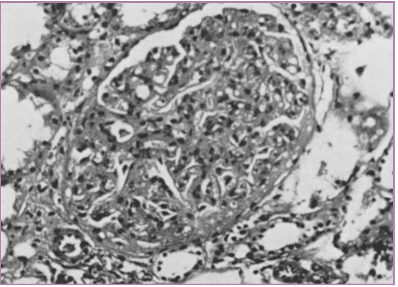
- Glomerular capillary endotheliosis (hallmark) - endothelial swelling narrows capillary lumens to near obliteration.
- Enlarged glomeruli completely filling Bowman capsule with vacuolated appearance (shown in image below).
- Occurs in 70% of primiparas vs only 14% of multiparas with preeclampsia.
- These changes resolve within 5-10 weeks after delivery.
Liver
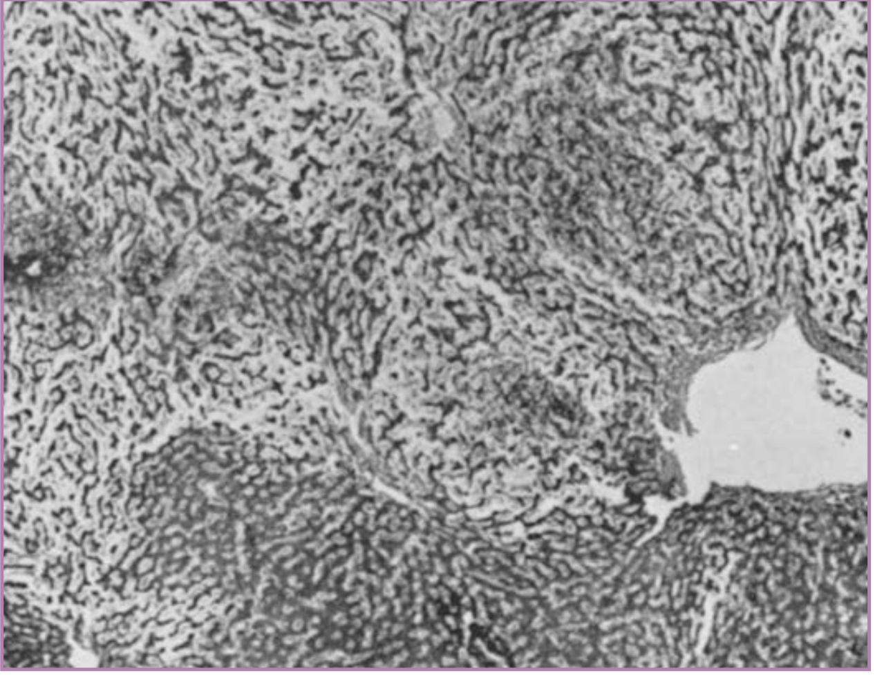
- Intense vasospasm causes hepatic infarction - small to large areas of periportal/sinusoidal necrosis extending toward portal vessels.
- HELLP syndrome (10% of severe preeclampsia): microangiopathic hemolytic anemia + elevated liver enzymes + low platelets ± DIC.
Placenta
- Multiple infarcts (far more numerous than in normal pregnancy)
- Retroplacental hemorrhage
- Ischemic villous changes (syncytial epithelial knots)
- Acute atherosis of decidual vessels (fibrinoid necrosis + lipid-laden macrophages)
Brain
- Petechial hemorrhages
- Thrombotic microangiopathy
- Vasogenic edema (parietooccipital predominance on imaging)
6. Clinical Features
Signs and Symptoms of Preeclampsia Preceding Eclampsia
- Persistent, severe frontal/occipital headache (most important warning sign)
- Visual disturbances (blurred vision, scotomata, diplopia, cortical blindness)
- Dizziness, tinnitus, drowsiness
- Oliguria (<500 mL/24 hr)
- Proteinuria (≥300 mg/24 hr or urine protein:creatinine ≥0.3)
- Rising creatinine
- Epigastric or right upper quadrant pain (due to hepatic capsule stretch/infarction)
- Nausea and vomiting
- Elevated AST/ALT
- Thrombocytopenia (<100,000/μL in HELLP)
- Microangiopathic hemolytic anemia
- Pulmonary edema (complicates 2-3% of severe preeclampsia)
The Eclamptic Seizure Itself
- Typically generalized tonic-clonic
- Usually preceded (seconds to minutes) by a prodrome: facial twitching or staring, followed by tonic phase then clonic phase
- Lasts 60-75 seconds on average
- Postictal confusion/coma follows
- May be followed by recurrent seizures or status eclampsia
- Can occur antepartum (50%), intrapartum (25%), or postpartum (25%)
7. Diagnostic Criteria
- New onset BP ≥140/90 mmHg after 20 weeks in a previously normotensive woman
- PLUS one of: proteinuria, thrombocytopenia, renal insufficiency, impaired liver function, pulmonary edema, or new-onset headache/visual symptoms
- BP monitoring (continuous)
- CBC (thrombocytopenia, hemolysis)
- Liver function tests (AST, ALT, LDH)
- Serum creatinine, uric acid
- Urinalysis/24-hr urine protein or spot PCR ratio
- Fetal assessment (CTG, ultrasound)
- CT or MRI brain: not routinely required when diagnosis is apparent, but shows posterior vasogenic edema in PRES pattern
8. Management
Immediate Priorities (the "A-B-C" approach)
- Airway - lateral decubitus to prevent aspiration; suction; oxygen
- Prevent injury during seizure - padded side rails, do NOT restrain forcefully
- IV access - establish immediately
8a. Seizure Control and Prophylaxis - Magnesium Sulfate
- Loading dose: 4-6 g IV in 100 mL, given over 20-30 minutes
- Maintenance: 2 g/hour IV infusion for at least 24 hours after last seizure or delivery (whichever is later)
- Loading dose: 4 g IV + 5 g IM each buttock
- Maintenance: 5 g IM every 4 hours
| Serum Mg (mg/dL) | Effect |
|---|---|
| 4-7 | Therapeutic (seizure prophylaxis) |
| 7-10 | Loss of patellar reflexes (early toxicity warning) |
| 10-13 | Respiratory paralysis |
| >15 | Cardiac arrest |
- Patellar reflexes (absent = approaching toxicity)
- Respiratory rate (should remain >12/min)
- Urine output (>25 mL/hr to ensure adequate excretion)
- Serum Mg levels in renal insufficiency
8b. Blood Pressure Control
| Agent | Route | Dose | Notes |
|---|---|---|---|
| Labetalol | IV | 20 mg bolus; repeat 20-80 mg q10min; max 300 mg | First-line parenteral; avoid in asthma |
| Hydralazine | IV | 5-10 mg q20min | Classic choice; may cause reflex tachycardia |
| Nifedipine | PO | 10-20 mg q20-30min (immediate release) | Oral; may enhance MgSO4 effect |
| Methyldopa | PO | 250-500 mg q8h | First-line oral; most safety data in pregnancy |
| Nicardipine | IV infusion | 5-15 mg/hr | Useful in severe hypertension |
| Nitroprusside | IV | Last resort | Risk of fetal cyanide toxicity; avoid if possible |
8c. Definitive Treatment - Delivery
- <24 weeks: Termination usually recommended due to >80% perinatal mortality and high maternal risk
- 24-34 weeks: Balance fetal benefit of prolonged gestation vs. maternal risk; expectant management with intensive monitoring may be appropriate; corticosteroids for fetal lung maturity
- 34-37 weeks: ACOG recommends delivery
- ≥37 weeks: Immediate delivery indicated
- Eclampsia: Delivery is indicated regardless of gestational age once mother is stabilized
8d. Ancillary Management
- Coagulopathy/HELLP: Replace platelets if <50,000 or active bleeding; replace coagulation factors for DIC
- Pulmonary edema: Furosemide; fluid restriction (MgSO4 infusion already adds fluid load)
- Fetal monitoring: Continuous CTG during seizure and post-seizure (transient bradycardia is common)
- Transfer: If obstetric services unavailable, stabilize and transfer to a tertiary center
9. HELLP Syndrome (Severe Variant)
- Complicates ~10% of severe preeclampsia
- Can occur without proteinuria or significant hypertension
- Diagnostic criteria: Platelets <100,000/μL + LDH >600 U/L + AST/ALT >2x upper normal
- Complications include: eclampsia (6%), placental abruption (10%), acute renal failure (5%), DIC (8%), pulmonary edema (10%), hepatic hemorrhage/rupture (rare but life-threatening)
10. Prognosis and Long-Term Outcomes
- Hypertension and proteinuria typically resolve within 1-2 weeks after delivery
- Glomerular changes resolve within 5-10 weeks postpartum
- Most cases leave no lasting sequelae if managed promptly
- Women who have had eclampsia may have subtle long-term impairment in cognitive function
- Risk of recurrence in subsequent pregnancies: previous preeclampsia is the strongest single risk factor (RR 7.19)
- Long-term cardiovascular risk is elevated (hypertension, CVD later in life)
11. Differential Diagnosis of Seizures in Pregnancy
| Condition | Key Distinguishing Features |
|---|---|
| Epilepsy | Prior seizure history; no hypertension/proteinuria |
| Thrombotic thrombocytopenic purpura (TTP) | Pentad: thrombocytopenia, hemolysis, fever, renal, neuro |
| Hyponatremia | Sodium level <120; no hypertension |
| Cerebral venous thrombosis | MRI/MRV; focal deficits; no hypertensive pattern |
| Intracranial hemorrhage | CT head; sudden onset headache |
| Meningitis/encephalitis | Fever, CSF findings |
- Creasy & Resnik's Maternal-Fetal Medicine, 9e - Chapters 45 & 48 (Preeclampsia and Eclampsia, comprehensive pathophysiology and management)
- Brenner and Rector's The Kidney - Magnesium and seizure prophylaxis, antihypertensive management
- Robbins & Kumar Basic Pathology - Pathogenesis and morphology (glomerular endotheliosis, hepatic infarction)
- Tintinalli's Emergency Medicine - Clinical management protocol
- Harrison's Principles of Internal Medicine, 22e - PRES and cerebral pathophysiology
- Sullivan M et al. Duration of postpartum MgSO4 for seizure prophylaxis. J Matern Fetal Neonatal Med. 2022. PMID 34187284 - 9 RCTs; supports 24-hour regimen.
- Diaz V et al. Alternative MgSO4 regimens for pre-eclampsia and eclampsia. Cochrane Database Syst Rev. 2023. PMID 37815037 - Standard regimen remains the evidence-based choice.
Coagulation factors to look for in eclampsia
eclampsia preeclampsia coagulation DIC HELLP
Coagulation Factors and Studies in Eclampsia
Why Coagulopathy Develops in Eclampsia
- Platelet activation and consumption at sites of endothelial damage
- Coagulation cascade activation → microthrombus formation
- Intravascular depletion of procoagulants (fibrinogen, platelets, Factor VIII)
- Fibrinolysis → fibrin degradation products (FDPs/D-dimer) accumulate
- In severe cases: frank DIC with hemorrhagic tendency
DIC is diagnosed in 10% of women with severe preeclampsia or eclampsia. Even women with mild disease may show subtle coagulation changes. (Creasy & Resnik's Maternal-Fetal Medicine, 9e)
Complete Coagulation Workup in Eclampsia
Tier 1 - MANDATORY (Send Immediately)
| Test | What It Detects | Finding in Eclampsia/DIC |
|---|---|---|
| Platelet count | Thrombocytopenia (most sensitive early marker) | ↓↓ (<100,000/μL in HELLP; <50,000 in severe DIC) |
| PT (Prothrombin Time) | Extrinsic pathway (Factors VII, X, V, II, fibrinogen) | Prolonged in overt DIC |
| aPTT (Activated Partial Thromboplastin Time) | Intrinsic pathway (Factors XII, XI, IX, VIII) | Prolonged in overt DIC |
| Fibrinogen | Primary procoagulant; consumed in DIC | ↓↓ in advanced DIC (Note: fibrinogen is elevated in normal pregnancy, so a "normal" level may actually be low relative to baseline) |
| D-dimer / FDPs (Fibrin Degradation Products) | Fibrinolysis; indicates ongoing thrombus formation and lysis | ↑↑ elevated; reflects active intravascular coagulation |
| LDH (Lactate Dehydrogenase) | Hemolysis marker + tissue ischemia | ↑ >600 IU/L in HELLP |
| AST/ALT | Hepatic involvement (microthrombi, infarction) | ↑ >2× upper limit of normal in HELLP |
| Serum bilirubin | Hemolysis (indirect) + hepatic dysfunction (direct) | ↑ ≥1.2 mg/dL in HELLP |
Tier 2 - IMPORTANT ADDITIONAL TESTS
| Test | What It Detects | Significance |
|---|---|---|
| Peripheral blood smear | Schistocytes (fragmented RBCs), burr cells | Microangiopathic hemolytic anemia - hallmark of HELLP; confirms hemolysis |
| Serum haptoglobin | Free hemoglobin binding protein; consumed in hemolysis | ↓↓ (low haptoglobin = significant hemolysis) |
| Antithrombin III (AT-III) | Natural anticoagulant; consumed in DIC | ↓ Reduced in preeclampsia even before overt DIC; one of the earliest sensitive indicators |
| Factor VIII activity:antigen ratio | Ratio falls when Factor VIII is consumed | ↓ Reduced ratio is a sensitive early marker - correlates with fetal outcome better than clinical severity |
| Serum creatinine | Renal microangiopathy/glomerular injury | ↑ >1.1 mg/dL signals severe disease |
| Uric acid | Correlates with severity; renal tubular dysfunction | ↑ Elevated - may precede other coagulation changes |
Tier 3 - IN SEVERE/REFRACTORY CASES
| Test | Indication |
|---|---|
| Thromboelastography (TEG) / ROTEM | Whole-blood viscoelastic assessment of clot formation and lysis - increasingly used in obstetric hemorrhage; guides targeted factor replacement |
| vWF (von Willebrand Factor) level | Markedly elevated in preeclampsia due to endothelial release; helps distinguish from TTP (where vWF is also elevated but ADAMTS13 is deficient) |
| ADAMTS13 activity | To differentiate eclampsia/HELLP from TTP (ADAMTS13 severely deficient <10% in TTP; normal-mildly reduced in HELLP) |
| Fibronectin | Endothelial damage marker; elevated early in preeclampsia |
| Platelet function tests | Platelet dysfunction present even when count is normal |
Specific Factor Changes Explained
Platelets - The Most Important Single Test
- Platelet consumption occurs due to endothelial activation and microthrombi formation (not platelet production failure).
- Thrombocytopenia is present in virtually all cases of HELLP and severe preeclampsia.
- Even when count appears "normal," there is increased platelet turnover (elevated mean platelet volume - MPV), altered platelet appearance, and platelet dysfunction.
- Serial platelet counts (every 6-12 hours in deteriorating patients) are essential - rapid falls signal progression.
- Class I (most severe): Platelets ≤50,000/mm³
- Class II: Platelets >50,000 and ≤100,000/mm³
- Class III: Platelets >100,000 and ≤150,000/mm³
Fibrinogen - The "Hidden" Deficiency
- Normal pregnancy doubles fibrinogen levels (baseline 400-600 mg/dL vs non-pregnant 200-400 mg/dL).
- Therefore, a fibrinogen level that appears "normal" (e.g., 300 mg/dL) may actually represent significant consumption in a pregnant woman.
- Fibrinogen <200 mg/dL in pregnancy is a marker of severe DIC and imminent hemorrhage.
- Along with platelets, fibrinogen is among the first procoagulants to fall to hemorrhagic levels in advanced DIC.
Factor VIII Activity:Antigen Ratio
- In preeclampsia, Factor VIII is activated and consumed disproportionately.
- A reduced ratio is one of the most sensitive early indicators of coagulation activation.
- Importantly, this ratio correlates better with fetal outcome than with clinical severity - making it useful for prognosis.
Antithrombin III (AT-III)
- AT-III is a key natural anticoagulant; it is consumed when thrombin is generated in excess.
- Reduced AT-III is one of the earliest detectable hematologic changes in preeclampsia, appearing before platelet count falls significantly.
- Levels fall progressively with disease severity.
- AT-III deficiency also reduces the efficacy of heparin (relevant if heparin is used).
D-dimer / FDPs
- Elevated even in mild preeclampsia; markedly elevated in overt DIC.
- FDPs impair fibrin polymerization, worsening coagulopathy.
- D-dimer elevation + thrombocytopenia + low fibrinogen = diagnostic triad of DIC.
HELLP Syndrome - Formal Diagnostic Criteria
-
Hemolysis - confirmed by ≥2 of:
- Schistocytes and burr cells on peripheral smear
- Serum bilirubin ≥1.2 mg/dL
- Low serum haptoglobin
- Severe anemia unexplained by blood loss
-
Elevated liver enzymes:
- AST or ALT ≥2× upper limit of normal
- LDH ≥2× upper limit of normal
-
Thrombocytopenia: Platelet count <100,000/mm³
- Serum AST: median 249 U/L (range 70-633)
- Serum bilirubin: median 1.5 mg/dL (range 0.5-25)
- Platelet count: median 57 × 10³/mm³
Coagulation Changes vs. Disease Spectrum
MILD PREECLAMPSIA SEVERE PREECLAMPSIA ECLAMPSIA / HELLP / DIC
| | |
↑ Platelet turnover ↓ Platelet count Platelets << 100,000
↑ Platelet activation ↑ D-dimer / FDPs ↑↑ D-dimer/FDPs
↓ AT-III (early) ↓ Fibrinogen (relative) ↓↓ Fibrinogen (<200)
↓ Factor VIII ratio Prolonged PT/aPTT (mild) Overt PT/aPTT prolonged
↓ AT-III Schistocytes on smear
↓↓ Haptoglobin
↑↑ LDH (hemolysis)
Management of Coagulopathy
| Finding | Threshold for Treatment | Intervention |
|---|---|---|
| Thrombocytopenia | <50,000 (or <100,000 with active bleeding/cesarean) | Platelet transfusion - Note: avoid at higher counts as pathology is consumption, not production failure |
| Low fibrinogen (<200 mg/dL) | Active bleeding/surgery | Cryoprecipitate (each unit raises fibrinogen ~10 mg/dL) |
| Prolonged PT/aPTT + bleeding | Present with coagulopathy | Fresh frozen plasma (FFP) - use cautiously given fluid overload risk in preeclampsia |
| DIC | Confirmed | Treat underlying cause (delivery is definitive) + replace consumed factors |
"Coagulopathy should be corrected. Caution is advised in the use of fresh frozen plasma because of the risk of volume overload in preeclampsia." - Comprehensive Clinical Nephrology, 7e
Key Distinguishing Test: Eclampsia vs. TTP
- TTP: ADAMTS13 severely deficient (<10% activity)
- HELLP/Eclampsia: ADAMTS13 normal or mildly reduced
- This distinction matters because TTP requires plasmapheresis, not delivery.
- Creasy & Resnik's Maternal-Fetal Medicine, 9e - Coagulation Changes section (DIC in 10% of severe preeclampsia; AT-III, Factor VIII ratio, platelet changes)
- Sleisenger & Fordtran's Gastrointestinal and Liver Disease - HELLP classification systems (Tennessee + Mississippi)
- Harrison's Principles of Internal Medicine, 22e - HELLP complications including coagulopathy, CVA, hepatic rupture
- Henry's Clinical Diagnosis and Management by Laboratory Methods - HELLP complications, TTP differential diagnosis
- Comprehensive Clinical Nephrology, 7e - Platelet transfusion guidance; FFP caution in preeclampsia
Infraclavicular brachial plexus block in details with usg guided with pictures
Infraclavicular Brachial Plexus Block - Detailed USG-Guided Guide
1. Anatomy Overview
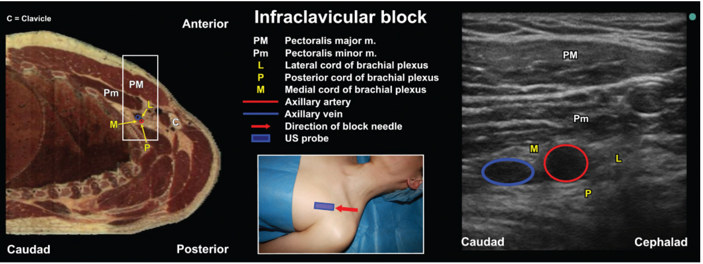
- Lateral cord - lateral/superolateral to artery (9 o'clock position)
- Posterior cord - directly posterior/deep to artery (6 o'clock position)
- Medial cord - posteromedial, between artery and vein (often at 4-5 o'clock position)
2. Indications
- Forearm, elbow, wrist, and hand surgery (primary indication)
- Surgery requiring a tourniquet at the mid-upper arm (better tolerated than axillary block)
- Situations where arm abduction is difficult or contraindicated (trauma, contractures)
- When a continuous catheter is needed for prolonged postoperative analgesia (infraclavicular region is mechanically stable for catheter fixation)
- Preferred over axillary block when musculocutaneous and axillary nerves must be included (they branch proximally and are frequently missed with axillary block)
3. Contraindications
- Absolute: Patient refusal, infection at injection site, severe coagulopathy
- Relative: Contralateral phrenic nerve palsy or pneumonectomy (phrenic nerve at this level is rarely affected, unlike interscalene), severe ipsilateral lung disease, anticoagulation (axillary approach preferred as artery is compressible)
4. Patient Positioning
- Patient supine, head turned ~45° away from operative side
- Two options for arm position:
- Arm at side with hand resting on abdomen (acceptable, especially if trauma)
- Arm abducted 90°, hand behind head (preferred) - externally rotates humeral head, retracts clavicle, straightens and superficializes the neurovascular bundle, and stretches the cords around the artery, facilitating visualization and local anesthetic spread
5. Equipment
| Item | Specification |
|---|---|
| Ultrasound probe | Linear high-frequency (7-15 MHz) for standard patients; curvilinear lower frequency (4-7 MHz) for obese patients or deep needle pass |
| Needle | 50-90 mm, 18-22 gauge insulated echogenic needle |
| Local anesthetic volume | 20-30 mL (single injection); up to 40 mL in divided doses |
| Sterile probe cover | Standard sleeve or transparent dressing |
| Nerve stimulator | Optional for combined US+NS guidance |
6. USG-Guided Technique (Step by Step)
Step 1 - Probe Placement and Scanning
- Place the linear probe in a parasagittal (paracoronal) plane, immediately medial and inferior to the coracoid process, caudad to the clavicle.
- Orient the probe so that cephalad is to the right of the screen and caudad is to the left (or vice versa per machine convention).
- Scan to obtain the best short-axis (cross-sectional) view of the axillary artery and neurovascular bundle.
Step 2 - Sonoanatomy Identification
| Layer | Structure | Appearance |
|---|---|---|
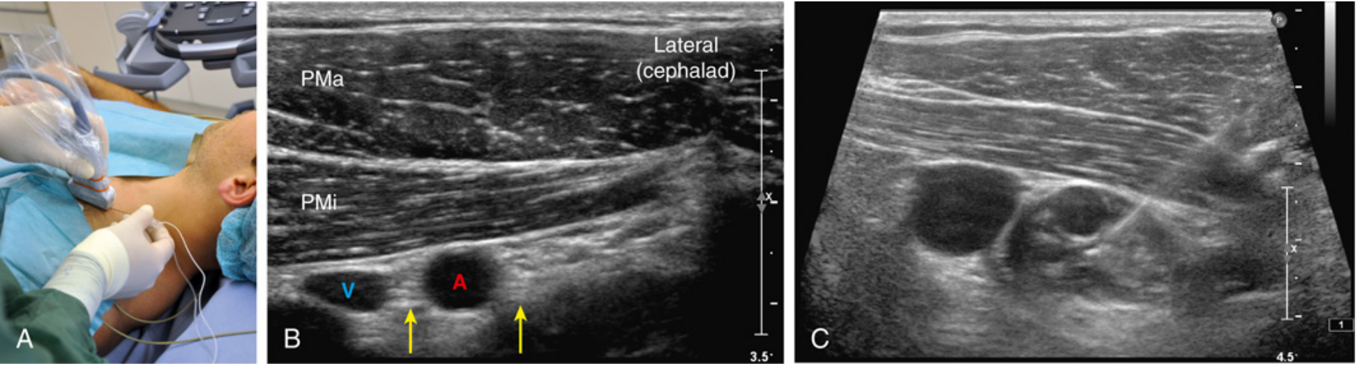
| Most superficial | Pectoralis major (PMa) | Heterogeneous muscular texture |
| Separated by hyperechoic perimysium | Pectoralis minor (PMi) | Lies deep to PMa |
| 4-5 cm deep to skin | Axillary artery (A) | Round, pulsatile, anechoic (dark), non-compressible |
| Medial to artery | Axillary vein (V) | Compressible, collapses with probe pressure |
| Adjacent to artery | Lateral cord | Hyperechoic oval, most readily visible at lateral position |
| Deep/posterior to artery | Posterior cord | May be hidden in acoustic shadow of artery |
| Between artery and vein | Medial cord | May be posterior/cephalad to artery; variable location |
Key point: There is significant individual anatomic variation in cord positions. Direct cord visualization is NOT required for a successful block - the key target is the posterior aspect of the artery.
| Proximal | Optimal Location | Distal |
|---|---|---|
| Cephalic vein | Pectoralis minor (midportion) | Subscapular artery |
| Thoracoacromial artery | Brachial plexus cords surround axillary artery | Coracobrachialis muscle |
| Chest wall and pleura | Posterior cord underneath axillary artery | Anterior/posterior circumflex arteries |
Step 3 - Needle Insertion (In-Plane Technique)
- In-plane (IP) approach is strongly preferred - allows real-time continuous needle visualization throughout its entire course.
- Insert the needle cephalad-to-caudad (i.e., from above the probe, directed posteriorly and caudally), angled 0-15° posterior to the horizontal plane.
- Insert needle at the cephalad edge of the probe to achieve a long, visible needle shaft.
- Advance through pectoralis major → pectoralis minor → toward posterior aspect of axillary artery.
- Target: Posterior to the axillary artery, adjacent to the posterior cord.
Step 4 - Local Anesthetic Injection
- Target position: Needle tip posterior to the axillary artery (6 o'clock position)
- Aspirate before each injection to rule out intravascular placement
- Inject 20-30 mL posterior to the artery in divided increments (5 mL at a time with aspiration)
- Optimal spread pattern = "U-shaped" distribution - local anesthetic wrapping around the posterior and inferior aspect of the artery, lifting it and separating the cords
- Reduction in axillary artery diameter during injection (compression from LA)
- "U-shaped" local anesthetic distribution underneath the axillary artery
- Separation of cords from the axillary artery
- "White wall" appearance of the axillary artery (free walls visible circumferentially)
- Dark layer underneath the axillary artery (long-axis view)
Substantial evidence confirms that 30-40 mL placed posterior to the axillary artery produces complete 3-cord brachial plexus block. The cords do NOT need to be individually visualized for success.
7. Local Anesthetic Choices and Doses
| Drug | Concentration | Volume | Duration |
|---|---|---|---|
| Ropivacaine | 0.5% | 20-30 mL | 12-18 hr (analgesia up to 24 hr) |
| Bupivacaine | 0.375-0.5% | 20-30 mL | 12-18 hr |
| Lidocaine (with epinephrine 1:200,000) | 1.5-2% | 20-30 mL | 3-5 hr |
| Mepivacaine | 1.5% | 20-30 mL | 4-8 hr |
- For single-injection technique: 20-25 mL posterior to artery is sufficient
- If additional injection needed anterolateral: inject 5-10 mL near lateral cord
- Total volume generally: 30-40 mL
8. Continuous Catheter Technique
- Stable anatomy (no arm movement affects position)
- Deep location protects catheter from dislodgment
- No need for arm manipulation for catheter maintenance
9. Nerve Stimulator (NS) Responses for Cord Identification
| Cord | Motor Response | Movement |
|---|---|---|
| Lateral cord | Elbow flexion (musculocutaneous), pronation | Lateral movement (pronation) |
| Medial cord | Distal flexors, finger flexion | Medial/flexion movement |
| Posterior cord | Wrist extension, finger extension (radial nerve) | Dorsal extension movement |
- First response obtained is usually musculocutaneous nerve (lateral cord) → elbow flexion
- Fifth digit (pinkie) movement is helpful: lateral = lateral cord; medial flexion = medial cord; dorsal extension = posterior cord
- Posterior cord (extension response) is the most important for complete hand anesthesia
10. Complications
| Complication | Risk Level | Comment |
|---|---|---|
| Vascular puncture (axillary artery/vein) | Most common | Deep block; frequent aspiration essential; axillary vein duplication is an anatomic variant that places an accessory vein adjacent to the lateral cord |
| Pneumothorax | Low but present | Lung pleura is close medially; lateral IP approach reduces risk significantly; higher risk with costoclavicular approach |
| Local anesthetic systemic toxicity (LAST) | Low with USG | Highly vascular area; aspirate before every injection; use incremental doses |
| Phrenic nerve block | Very rare | Much lower than interscalene block; advantage over interscalene in patients with contralateral lung pathology |
| Horner's syndrome | Very rare | Unlike interscalene/supraclavicular blocks |
| Nerve injury | Rare | Document pre-existing deficits; avoid intraneural injection |
| Block failure | 5-10% | Usually from inadequate posterior cord coverage; posterior cord under artery acoustic shadow can be missed |
11. Infraclavicular vs. Axillary Block Comparison
| Feature | Infraclavicular | Axillary |
|---|---|---|
| Depth | Deep (two overlying muscles, 4-5 cm) | Shallow |
| Onset | Slower | Faster |
| Tourniquet tolerance | Good (covers medial brachial cutaneous) | Fair |
| Catheter success | High (stable position) | Low (mobile arm) |
| Arm position required | Not required (arm can be at side) | Arm abduction needed (90°) |
| Musculocutaneous nerve | Consistently blocked | Often missed |
| Axillary nerve | Consistently blocked | Often missed |
| Phrenic nerve risk | Very rare | None |
| Pneumothorax risk | Low (lateral approach) | None |
| Compressibility if vascular | No (deep artery) | Yes (superficial artery) |
12. Clinical Pearls Summary
- Arm abduction to 90° with hand behind head is the preferred position - brings neurovascular bundle closer to surface and straightens the bundle.
- Target posterior to the artery at the 6 o'clock position - U-shaped distribution from this single injection covers all 3 cords reliably.
- Direct cord visualization not needed - focus on LA spread pattern relative to artery.
- Costoclavicular approach (cords clustered lateral to 1st part of axillary artery) offers faster onset from intra-cord spread, but carries higher pneumothorax risk due to lung proximity.
- Probes: Start with linear high-frequency; switch to curvilinear low-frequency if depth >5 cm (obese patients).
- Pre-injection D5W test dose confirms spread before committing to local anesthetic.
- For anticoagulated patients - axillary block preferred (compressible vessel); if infraclavicular is essential, use USG strictly.
- Barash, Cullen, and Stoelting's Clinical Anesthesia, 9e - Chapter 36 (Infraclavicular Block, USG technique, anatomy diagram + USG image)
- Miller's Anesthesia, 2-Volume Set, 10e - Chapter 42 (Infraclavicular Brachial Plexus Block; Sonographic landmarks table; Box 42.2 signs of success; Table 42.1 comparison with axillary block; USG images Fig 42.16)
32 yr old female post lscs twin pregnancy with headache and history of 1 seizure episode within 24 hrs and 1 reading of bp-150/90 and pr inr done within last 1 week what are the investigation do we need to eo necessarily