
What is edema. Causes and treatment of edema
Reading File
Now I'll search the medical library for edema content in parallel.
Finding Sources
Finding Sources
Excellent results. Let me now read the most comprehensive sources in parallel - Robbins Pathology (definitive pathophysiology) and Frameworks for Internal Medicine (causes/treatment).
Reading File
Reading File
Great content. Let me now read more of the Robbins Pathology section (it was truncated) and also the Goldman-Cecil Medicine section for treatment, and fetch the diagram image.
Reading File
Reading File
Loading Image
Excellent. I have rich, comprehensive content. Let me also fetch the key pathophysiology diagram from Robbins.
Loading Image
Loading Image
Reading File
I now have rich, authoritative content from multiple textbooks. Let me compile the comprehensive answer.
Edema
Definition
Edema is an abnormal accumulation of interstitial fluid within tissues. About 60% of lean body weight is water; two-thirds is intracellular and most of the remainder is interstitial fluid. When interstitial fluid accumulates beyond normal, edema results.
Fluid can also collect in body cavities:
-
Hydrothorax - pleural cavity
-
Hydropericardium - pericardial cavity
-
Hydroperitoneum (ascites) - peritoneal cavity
-
Anasarca - severe generalized edema with profound subcutaneous swelling and fluid in body cavities
-
Robbins & Kumar Basic Pathology, p. 78-79
Pathophysiology
Fluid movement across capillary walls is governed by two opposing forces:
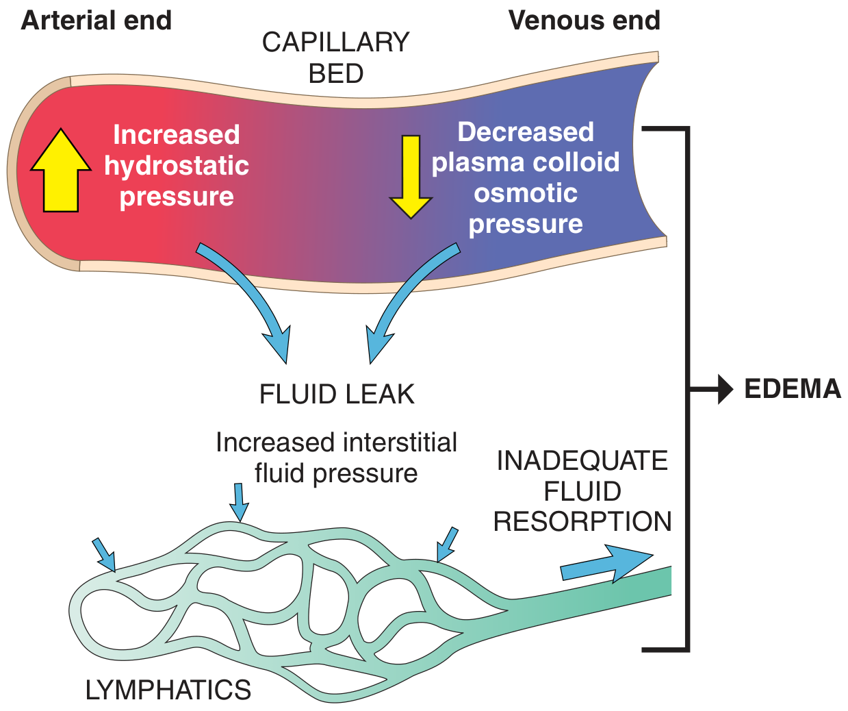
- Vascular hydrostatic pressure - pushes fluid OUT of capillaries into the interstitium
- Plasma colloid osmotic (oncotic) pressure - pulls fluid BACK into capillaries
Normally these are balanced, and the small net outflow into the interstitium is drained by lymphatics back to the circulation. Edema occurs when:
- Hydrostatic pressure increases, OR
- Colloid osmotic pressure decreases, OR
- Lymphatic drainage is blocked, OR
- Vascular permeability increases (inflammation)
Transudate (protein-poor fluid) = high hydrostatic or low oncotic pressure
Exudate (protein-rich fluid) = increased vascular permeability (inflammation)
Causes of Edema
Here is a summary diagram of peripheral edema causes:
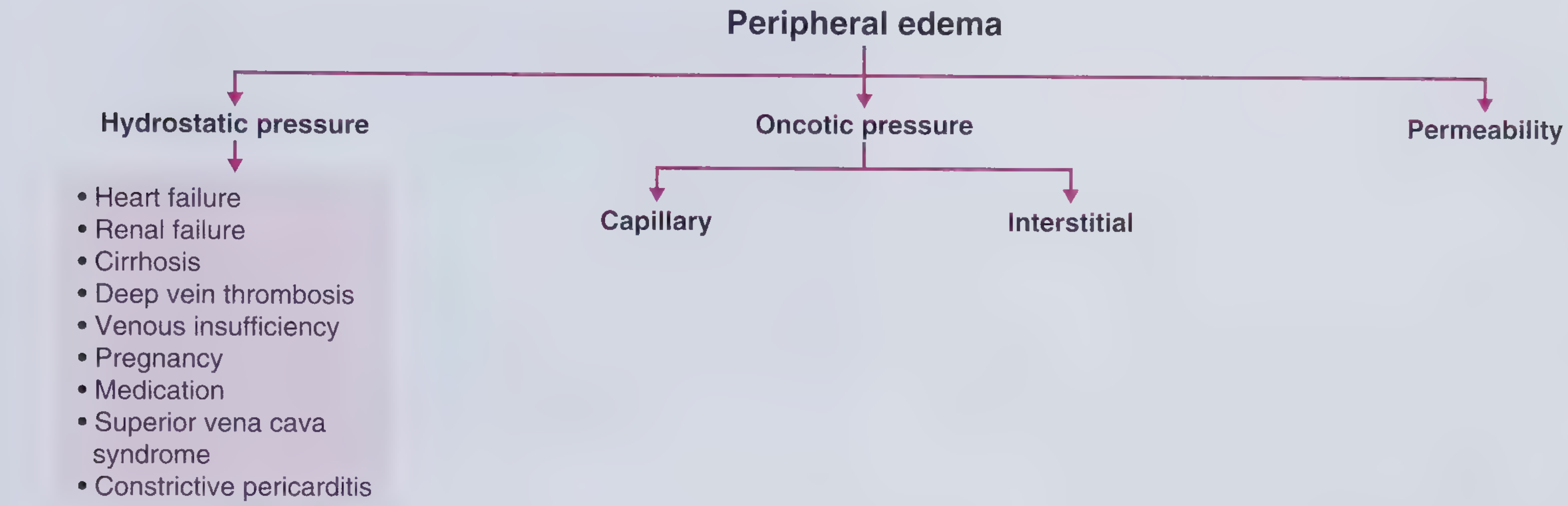
1. Increased Hydrostatic Pressure
Caused mainly by disorders that impair venous return:
| Cause | Mechanism |
|---|---|
| Congestive heart failure | Reduced cardiac output → venous pooling + renal sodium/water retention via RAAS |
| Deep vein thrombosis (DVT) | Local venous obstruction → unilateral leg edema |
| Renal failure | Sodium and water retention → increased intravascular volume |
| Liver cirrhosis | Portal hypertension → ascites and lower limb edema |
| Constrictive pericarditis | Impaired cardiac filling → elevated venous pressure |
| Venous insufficiency | Incompetent valves → chronic venous hypertension |
| Superior vena cava (SVC) syndrome | Obstruction of the SVC → face, neck, upper limb edema |
| Pregnancy | Inferior vena cava compression by gravid uterus |
| Prolonged limb dependency / inactivity | Reduced venous pump |
In congestive heart failure, a vicious cycle develops: reduced cardiac output → renal hypoperfusion → RAAS activation → Na+/water retention → increased venous pressure → worsening edema. This responds to salt restriction, diuretics, and aldosterone antagonists. - Robbins & Kumar Basic Pathology, p. 80
2. Reduced Plasma Osmotic (Oncotic) Pressure
Caused by hypoalbuminemia (plasma albumin < 2 g/dL is associated with edema):
| Cause | Mechanism |
|---|---|
| Nephrotic syndrome | Glomerular damage → albumin lost in urine |
| Liver disease (cirrhosis) | Decreased albumin synthesis |
| Malnutrition / kwashiorkor | Inadequate protein intake |
| Protein-losing enteropathy | GI losses of protein |
Low albumin leads to edema, reduced intravascular volume, renal hypoperfusion, and secondary hyperaldosteronism - which worsens edema because the primary defect (low protein) persists. - Robbins & Kumar Basic Pathology, p. 80
3. Lymphatic Obstruction (Lymphedema)
Impaired lymphatic drainage prevents resorption of interstitial fluid:
| Cause | Example |
|---|---|
| Parasitic infection | Filariasis - causes elephantiasis (massive lower limb edema) |
| Malignancy | Lymphoma obstructing lymphatics; breast cancer causing peau d'orange |
| Post-surgical / irradiation | Axillary node dissection in breast cancer → arm lymphedema |
| Iatrogenic | Radiation treatment disrupting lymphatics |
- Robbins & Kumar Basic Pathology, p. 80
4. Increased Vascular Permeability (Inflammatory Edema)
In inflammation, mediators such as histamine, bradykinin, leukotrienes, and cytokines increase capillary permeability, allowing protein-rich fluid to leak out. This produces an exudate. Examples:
- Allergic reactions / anaphylaxis
- Burns
- Sepsis / ARDS
- Infections
5. Sodium and Water Retention
Excessive salt retention increases hydrostatic pressure and dilutes plasma proteins:
- Renal failure
- Primary hyperaldosteronism
- Drugs (corticosteroids, NSAIDs, some antihypertensives)
6. Medications (Iatrogenic Edema)
Common culprits include:
- Calcium channel blockers (amlodipine)
- NSAIDs
- Corticosteroids
- Thiazolidinediones (e.g., pioglitazone)
- Hormonal therapy (estrogens)
Clinical Features
| Type | Characteristics |
|---|---|
| Heart failure | Bilateral, symmetric, dependent, pitting; gradual onset; legs primarily; may have ascites/pleural effusions |
| Renal failure | Similar to right heart failure; generalized |
| Cirrhosis | Bilateral, pitting, dependent; mainly lower limbs; ascites prominent |
| DVT | Unilateral, dependent, pitting; acute onset; associated with pain and erythema |
| SVC syndrome | Face, neck, bilateral arms |
| Lymphedema | Non-pitting (in chronic stage); involves limb; may be unilateral |
| Nephrotic syndrome | Periorbital edema (morning), then generalized; associated with proteinuria |
- Frameworks for Internal Medicine, p. 308-309
Treatment of Edema
Treatment is directed at the underlying cause plus symptomatic fluid removal:
1. Treat the Underlying Cause
- Heart failure: ACE inhibitors, ARBs, beta-blockers, cardiac resynchronization
- Renal failure: Hemodialysis or peritoneal dialysis to maintain fluid balance
- Nephrotic syndrome: Treat underlying glomerular disease; RAAS blockade; corticosteroids (where indicated)
- Cirrhosis: Treat liver disease; reduce portal hypertension
- DVT: Anticoagulation (heparin, then warfarin or DOACs)
- SVC syndrome: Treat underlying malignancy (chemotherapy, radiation, stenting)
- Lymphedema: Treat underlying infection or malignancy
2. Dietary Sodium and Fluid Restriction
- Sodium restriction is a cornerstone of edema management in heart failure, cirrhosis, and renal failure
- Diuretics work better when combined with low-sodium diet
3. Diuretics
The most widely used pharmacological treatment:
| Diuretic Class | Examples | Used in |
|---|---|---|
| Loop diuretics | Furosemide, bumetanide | Heart failure, renal failure, pulmonary edema |
| Thiazide diuretics | Hydrochlorothiazide | Mild edema, hypertension-related |
| Aldosterone antagonists | Spironolactone, eplerenone | Heart failure, cirrhosis (reduces secondary hyperaldosteronism) |
| Osmotic diuretics | Mannitol | Cerebral edema |
Note: In severe heart failure, bowel wall edema can impair oral diuretic absorption - parenteral (IV) administration is required. - Frameworks for Internal Medicine, p. 308
4. Mechanical / Physical Measures
- Leg elevation - reduces dependent edema by promoting venous return
- Compression stockings - chronic venous insufficiency, DVT prevention, lymphedema
- Compression bandaging - lymphedema management
- Manual lymphatic drainage - physiotherapy for lymphedema
5. Dialysis / Ultrafiltration
- For renal failure with hypervolemia: hemodialysis or peritoneal dialysis
- Plasma ultrafiltration can remove several liters per day in severe pulmonary edema
- Frameworks for Internal Medicine, p. 308
6. Albumin Infusion
- Used in hypoalbuminemia (nephrotic syndrome, cirrhosis) to temporarily restore oncotic pressure
- Combined with diuretics for best effect in cirrhotic ascites
7. Specific Interventions
- Paracentesis - large-volume drainage of ascites in cirrhosis
- Thoracentesis - drainage of large pleural effusions (hydrothorax)
- Surgical/endovascular - venous stenting in SVC syndrome; lymphovenous anastomosis in lymphedema
Summary Table
| Mechanism | Key Causes | Treatment Focus |
|---|---|---|
| ↑ Hydrostatic pressure | Heart failure, DVT, renal failure, cirrhosis | Diuretics, Na restriction, treat primary disease |
| ↓ Oncotic pressure | Nephrotic syndrome, cirrhosis, malnutrition | Treat underlying disease, albumin, diuretics |
| Lymphatic obstruction | Filariasis, malignancy, post-surgical | Compression, physiotherapy, treat cause |
| ↑ Vascular permeability | Inflammation, burns, sepsis, allergy | Anti-inflammatory, treat infection, manage ARDS |
| Na/H2O retention | Renal failure, aldosteronism, drugs | Dietary restriction, diuretics, dialysis |
Sources:
- Robbins & Kumar Basic Pathology (Robbins Pathology), pp. 78-80
- Frameworks for Internal Medicine, pp. 307-310
- Goldman-Cecil Medicine International Edition
- Harrison's Principles of Internal Medicine 22E
This is a shared conversation. Sign in to Orris to start your own chat.