
Frame a interesting case discussion for myxoid neoplasm in a patient presenting with nape of neck lesion
myxoid neoplasm neck soft tissue
myxoid tumor soft tissue histology microscopy
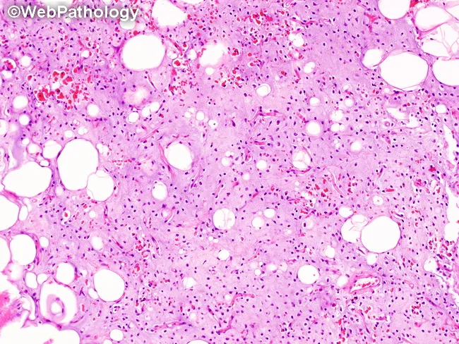
Imaging modality: Light microscopy histology of a soft tissue tumor. Specimen: formalin-fixed paraffin-embedded tissue section stained with Hematoxylin and Eosin (H&E). Anatomical context: deep soft tissue site (commonly extremities or trunk); precise origin not specified in the image. Histologic features include abundant myxoid matrix rich in acid mucopolysaccharides providing a pale, gelatinous background; scattered uniform non-adipocytic cells with spindle to stellate morphology organized in sheets; rare round cells; lipoblasts in various differentiation stages (small uni- or multi-vacuolated, signet-ring appearance) frequently located at tumor periphery; lipoblasts may be absent in some fields. Vascular pattern shows a delicate, plexiform network of thin-walled capillaries traversing the myxoid stroma, a hallmark distinguishing myxoid liposarcoma from myxoma and other myxoid neoplasms. Cytology reveals minimal atypia and low mitotic activity. The overall architecture is lobulated with myxoid septa. Diagnostic significance: this constellation—lipoblasts within a myxoid matrix and a plexiform capillary network—is characteristic of myxoid liposarcoma and supports definitive diagnosis; differential considerations include myxoid lipoma, myxoid pleomorphic sarcoma, and other liposarcoma subtypes; molecular testing for FUS-DDIT3 rearrangement can corroborate. Clinical utility: essential for surgical planning, prognosis assessment, and adjuvant therapy decisions; educational value for trainees; links to imaging and genetic studies; relevant to soft tissue sarcoma research databases.
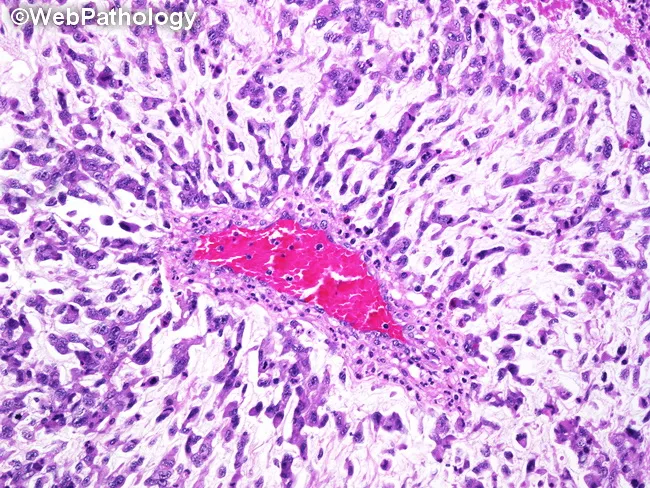
Histology image of a soft tissue mass showing features diagnostic of myxoid liposarcoma, high-grade (round cell) variant. The specimen is a tissue biopsy examined by light microscopy after Hematoxylin and Eosin staining. The architectural pattern reveals a prominent myxoid (mucoid) extracellular matrix with a delicate, arborizing capillary network radiating from vessels, a classic backdrop for myxoid adipocytic tumors. Intermixed are primitive, round to oval tumor cells arranged in cords and short trabeculae extending from the vascular core. The cells have scant cytoplasm, hyperchromatic nuclei, and mitotic activity is variably increased consistent with high-grade transformation. Occasional lipoblasts with scalloped cytoplasm may be present, highlighting adipocytic differentiation amidst the myxoid milieu. The surrounding stroma is relatively cellular in the high-grade areas, with less prominent lipogenic features compared with low-grade components. This image emphasizes the cellularity and architectural disruption associated with the round cell component, which portends a more aggressive clinical behavior and higher metastatic potential. Clinically, identification of high-grade cell populations in a predominantly myxoid liposarcoma influences prognosis and treatment planning, including surgical excision with wide margins and consideration of adjuvant chemotherapy or radiotherapy. Overall, the image exemplifies hallmarks used in pathology reports to classify liposarcoma subtype and grade for diagnostic accuracy.
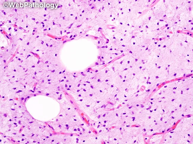
This histopathology image is a light-field microscopy view of a soft tissue tumor examined on formalin-fixed paraffin-embedded tissue stained with Hematoxylin and Eosin (H&E). The neoplasm shows an abundant myxoid (mucoid) extracellular matrix with numerous spindle, fusiform, and occasional stellate tumor cells. The cells display minimal cytologic atypia and there are no mitotic figures evident in the field, consistent with low-grade myxoid sarcoma features. A delicate, arborizing capillary network traverses the myxoid stroma and is a characteristic pattern in myxoid liposarcoma. Infrequent, non-diagnostic elements may be present, including extramedullary hematopoiesis and focal metaplasia with cartilage, bone, or smooth muscle; these incidental findings do not influence prognosis or therapeutic strategy. The lack of overt anaplasia helps distinguish this pattern from higher-grade sarcomas; however, when appreciable cellular atypia is encountered, myxofibrosarcoma or other myxoid sarcomas should be considered in the differential diagnosis. Clinically, the histology supports a myxoid liposarcoma diagnosis with favorable prognosis relative to high-grade variants, guiding surgical excision with adequate margins and potential adjuvant therapy based on location, size, and grade. Overall, the image exemplifies classic myxoid stroma with sparse, bland spindle cells and low mitotic activity, a diagnostically important pattern for soft tissue oncology and establish appropriate treatment planning.
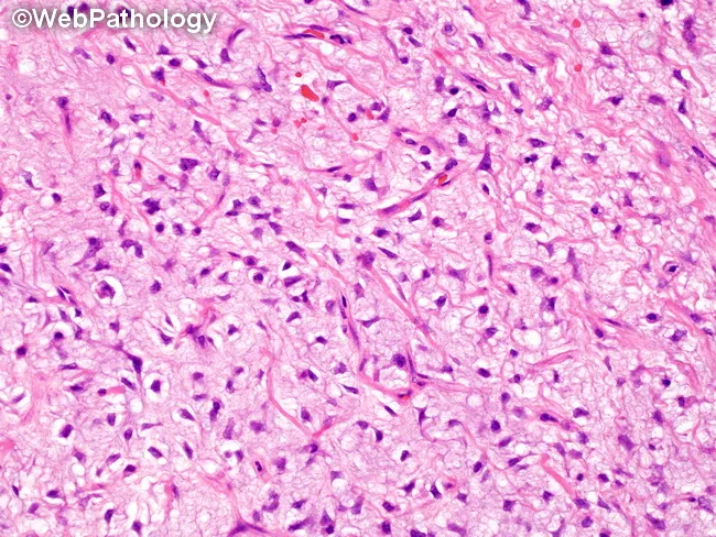
Histopathology image obtained via light microscopy of an H&E stained soft tissue specimen demonstrates the classic features of myxoid liposarcoma. The tumor is comprised of bland spindle-to-fusiform and stellate cells embedded in a abundant myxoid matrix, producing a pale basophilic, gelatinous background. The cellularity is moderate to low with uniform nuclei, minimal pleomorphism, and small inconspicuous nucleoli. A hallmark feature is the delicate arborizing/plexiform capillary network coursing through the myxoid stroma, which creates a lace-like vascular pattern. Sparse adipocytic lipoblasts may be present, often with cytoplasmic lipid vacuoles that indent nuclei. Mitotic figures are uncommon in well-differentiated areas but may be seen in higher-grade regions. The tumor margins are not well defined in this field; there may be a fibrous capsule or infiltrative growth into surrounding tissue. The combination of myxoid matrix with round-to-oval-to-spindle cells and a prominent vascular network is highly characteristic of myxoid liposarcoma and supports the diagnosis in the proper clinical context. Clinically, this histology correlates with a soft tissue sarcoma of extremities, often affecting the thigh or gluteal region, with potential for translocation t(12;16)(q13;p11) resulting in FUS-DDIT3 fusion. Diagnosis informs surgical management and adjuvant therapy decisions.
nape of neck soft tissue mass MRI imaging
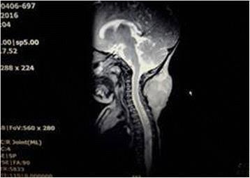
This diagnostic image is a sagittal T2-weighted contrast-enhanced magnetic resonance imaging (MRI) scan of a pediatric patient's head and neck. The image reveals a prominent, well-circumscribed soft tissue mass located in the posterior aspect of the neck, situated within the cutis and subcutaneous tissues. The mass exhibits a lobular morphology and is characterized by marked inhomogeneous hyperintensity on this T2 sequence, suggesting high vascularity or fluid content with internal septations or varying tissue density. Key anatomical landmarks visible include the cervical spine, cerebellum, and brainstem. Notably, the mass is clearly demarcated from the deeper musculoskeletal structures and does not show evidence of invasion or propagation toward the spinal canal or the prevertebral fascia. This appearance is clinically consistent with a benign vascular tumor, such as a large infantile hemangioma (capillary hemangioma), and illustrates the importance of contrast-enhanced MRI in evaluating the depth and vascular characteristics of pediatric soft tissue tumors.
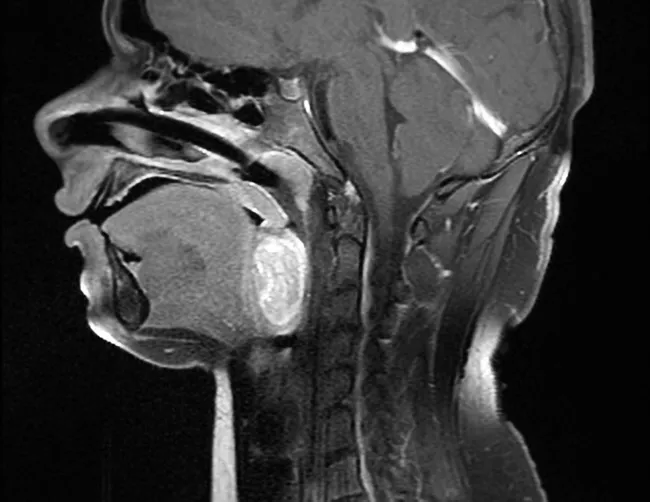
Magnetic resonance imaging (MRI) of the head and neck with gadolinium contrast demonstrates a soft-tissue mass within the oropharyngeal region anterior to the base of the tongue. The lesion is isointense to muscle on T1-weighted sequences and hyperintense on T2-weighted images, with marked, homogeneous post-contrast enhancement. The mass displays a discrete margin with the adjacent base of tongue, indicating a defined lesion abutting but separable from surrounding musculature; there is no gross invasion of the tongue musculature seen on this sagittal plane. Relative anterior orientation compared with prior radiographs suggests progression or resection planning implications. The imaging appearance supports a spindle-cell neoplasm with high vascularity and extracellular water content, consistent with monophasic synovial sarcoma in the clinical context. The findings are non-cystic, non-fatty, and show no frank calcifications on these sequences. The mass was subsequently resected, and histopathology confirmed monophasic synovial sarcoma; correlative molecular testing for SYT-SSX fusion can be pursued for definitive subtyping. Clinically, these features assist surgical planning, radiation therapy considerations, and prognosis evaluation. Differential diagnosis includes other soft-tissue sarcomas of the oropharyngeal region (rhabdomyosarcoma, fibrosarcoma, malignant peripheral nerve sheath tumor) and less likely inflammatory or infectious processes; radiologic-pathologic correlation is essential. MRI remains a critical tool for preoperative mapping and post-therapeutic surveillance in head-and-neck sarcomas.
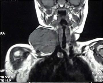
Coronal T1-weighted MRI of the neck and upper thoracic region demonstrating a large, well-circumscribed, ovoid soft tissue mass in the right lateral neck. The mass exhibits heterogeneous intermediate signal intensity. It is located in the posterior triangle and infrahyoid region, extending from the level of the mandible superiorly toward the supraclavicular fossa inferiorly. The lesion causes significant lateral displacement of the overlying soft tissues and appears to exert mass effect on adjacent structures, including the right sternocleidomastoid muscle and potentially the carotid sheath components. The trachea remains midline. This imaging is characteristic of a high-flow vascular malformation or a hypervascular neoplastic process, such as a glomangiomyoma or hemangiopericytoma-like tumor. The absence of flow voids within the bulk of the mass on this sequence, combined with its heterogeneous texture, highlights its complex tissue composition. This diagnostic image is intended for educational use in radiology and head and neck surgery, focusing on the differential diagnosis of pediatric neck masses.
myxoma intramuscular soft tissue gross pathology
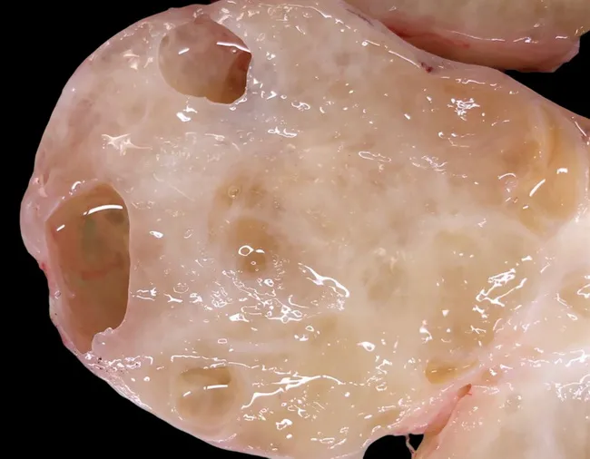
Gross pathology photograph of a sectioned thigh muscle showing an intramuscular myxoma. The specimen is a large, well-circumscribed soft tissue mass with a pink-tan, gelatinous cut surface and delicate fibrous septa. Small cystic areas contain mucoid fluid, reflecting abundant myxoid extracellular matrix. The lesion typically ranges from several centimeters up to 15 cm in greatest dimension and tends to blend with surrounding muscle rather than forming a dense capsule. On gross inspection, variability in myxoid content may produce areas that appear more solid or fibrous. The tumor is usually located within the muscle fibers of the thigh, as described in middle-aged adults, and may infiltrate adjacent muscular compartments without a true anatomic capsule. This macroscopic appearance correlates with the histologic hallmark of intramuscular myxoma: a hypocellular lesion composed of spindle to stellate cells embedded in a loose, mucopolysaccharide-rich matrix. Diagnostic significance lies in distinguishing this benign entity from myxoid sarcomas, which show higher cellularity, mitotic activity, and more complex vascular patterns. Complete surgical excision is curative in most cases, with low recurrence when margins are clear. This image illustrates the essential gross features used in initial pathologic assessment and downstream radiologic-histologic correlation.
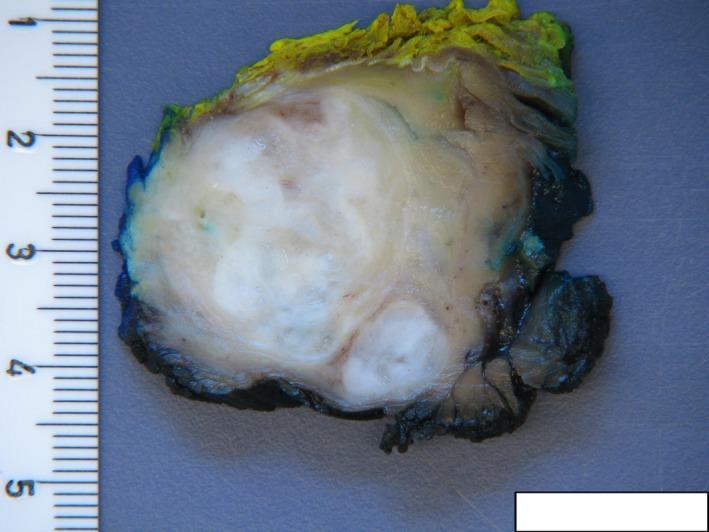
This diagnostic clinical photograph displays a gross surgical pathology specimen of an intramuscular myxoma. The image shows a cross-section of a well-circumscribed, roughly oval soft tissue mass measuring approximately 4-5 cm in diameter, as indicated by an adjacent metric ruler. The tumor exhibits a characteristic zonal architecture: a central, semi-translucent, glistening white region with a gelatinous or myxoid consistency and subtle swirling patterns, surrounded by a peripheral rim of denser, tan-to-yellowish solid tissue. The external margins of the specimen are marked with surgical inks (black, blue, and yellow) used for surgical orientation and margin assessment. The macroscopic appearance is consistent with a benign myxomatous lesion, often found within large skeletal muscles such as the deltoid. This visual is an essential educational resource for distinguishing benign myxoid tumors from more aggressive mimics like low-grade fibromyxoid sarcoma in orthopedic oncology.
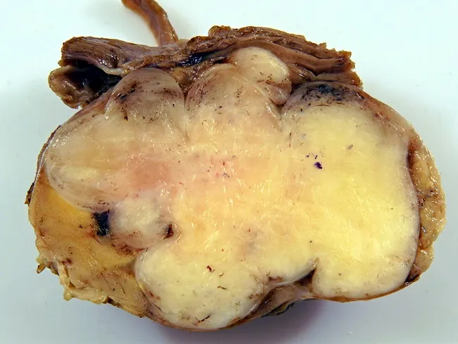
Gross-pathology view of an 8 cm intramuscular myxoma within the thigh musculature from a middle-aged man. This specimen demonstrates a circumscribed, pale, yellow-white mass with a gelatinous to mucoid cut surface in classic myxoid variants; in collagen-predominant foci the cut surface is more fibrous and tan. The lesion infiltrates adjacent skeletal muscle despite a deceptively well-defined external margin. It measures approximately 8 cm in greatest dimension and resides within a single or deep compartment of the thigh, separating but not erupting into surrounding soft tissue. Histologically, the tumor shows abundant myxoid matrix rich in acid mucopolysaccharides interlaced with small spindle or stellate cells; collagenized areas impart a fibrous texture. Although macroscopically infiltrative, such intramuscular myxomas are benign and lack overt cellular atypia or high mitotic activity. Diagnostic significance rests on distinguishing these lesions from myxoid sarcomas such as myxoid liposarcoma or myxofibrosarcoma, nodular fasciitis with myxoid change, or fibromatosis. Clinically, patients present with a slow-growing, painless mass in the proximal or mid-thigh in middle-aged adults; imaging (MRI) may show a well-defined, T2-hyperintense lesion with a myxoid halo, but histology is definitive. Complete surgical excision yields excellent prognosis; recurrence is uncommon but increases with incomplete resection. This image is a gross-pathology photograph documenting the tumor’s macroscopic appearance and its infiltration pattern.
Case Discussion: Myxoid Neoplasm Presenting as a Nape of Neck Lesion
THE CASE
PRESENTING HISTORY
PHYSICAL EXAMINATION
- Vital signs: Stable
- Local exam: A 5 x 4 cm well-defined mass at the nape of the neck, subcutaneous, with a smooth surface. The overlying skin appears normal in color, mobile over the mass. Consistency is soft to firm with a faint gelatinous "fluctuance." Non-tender. No pulsation. No bruit.
- No regional lymphadenopathy
- Neurological exam of upper limbs: Normal
- Cardiovascular exam: Normal
DIFFERENTIAL DIAGNOSIS - THE MYXOID QUIZ
| Diagnosis | Behavior | Key Features |
|---|---|---|
| Intramuscular/Subcutaneous Myxoma | Benign | Bland spindle cells in mucoid stroma; women 40-70; low T1/high T2 on MRI |
| Myxoid Liposarcoma | Malignant | Plexiform capillary network; lipoblasts; FUS-DDIT3 translocation t(12;16) |
| Myxofibrosarcoma | Malignant | Atypical spindle cells, pleomorphic multinucleated cells; poorly defined margins |
| Low-grade Fibromyxoid Sarcoma | Low-grade malignant | MUC4 positive; whorled fibrous zones + myxoid areas |
| Extraskeletal Myxoid Chondrosarcoma | Malignant | Cords of cells in abundant myxoid matrix; NR4A3 rearrangement |
| Cutaneous Myxoma | Benign | May be solitary or part of Carney Complex |
| Lipoma with myxoid change | Benign | Mature adipose component dominant |
| Ganglion Cyst (deep) | Benign | Juxta-articular, mucin-filled |
INVESTIGATION STRATEGY
Round 1: Imaging
- T1 weighted: Low signal intensity (water-like)
- T2 weighted/STIR: Markedly high signal intensity (bright as fluid)
- A "split-fat sign" or peripheral fat rind may be present
- Post-contrast: Internal enhancement distinguishes solid myxoid lesion from a pure cyst; purely peripheral enhancement in low-cellularity lesions
- Surrounding soft tissue edema-like T2 hyperintensity - a hallmark suggesting infiltrative behavior
"Myxoid tumours often resemble cysts, typically showing low SI on T1 weighted imaging and high SI on T2 weighted/fluid-sensitive sequences... A rind of fat may be seen, most prominently at the poles of the tumour." - Grainger & Allison's Diagnostic Radiology
Round 2: The Diagnostic Pivot - Core Needle Biopsy
- Hypocellular lesion with bland spindle-to-stellate cells embedded in abundant pale, gelatinous myxoid stroma
- No significant cellular atypia
- Minimal mitotic activity
- No lipoblasts identified
- No plexiform capillary network
- Alcian blue stain: positive (confirms acid mucopolysaccharides)
- Diagnosis: Intramuscular/Subcutaneous Myxoma (benign)
THE CLINICAL TEACHING TWIST: Carney Complex Alert
"Is this truly a solitary myxoma - or could this be part of a systemic syndrome?"
- Cardiac myxomas (79%) - potentially lethal due to embolism
- Cutaneous myxomas
- Spotty skin pigmentation (lentigines)
- Primary pigmented nodular adrenocortical disease (Cushing syndrome)
- Psammomatous melanotic schwannoma
- Breast myxomatosis or ductal adenoma
- GH-secreting pituitary adenoma
- Large cell calcifying Sertoli cell tumor
- Thyroid carcinoma or multiple nodules
- Andrews' Diseases of the Skin
- Echocardiography → No cardiac myxoma found
- Dermatologic exam → A few lentigines noted around the lips (previously dismissed as "freckles")
- Endocrine screen → ACTH-independent cortisol: mildly elevated (further evaluation warranted)
- PRKAR1A gene mutation testing: Sent
THE HISTOPATHOLOGY DIFFERENTIAL - How to Tell Them Apart
| Feature | Myxoma (Benign) | Myxoid Liposarcoma | Myxofibrosarcoma |
|---|---|---|---|
| Cellularity | Hypocellular | Moderate | High |
| Atypia | None / minimal | Minimal-moderate | Marked - pleomorphic cells |
| Lipoblasts | Absent | Present (peripheral) | Absent |
| Vascularity | Scant | Plexiform "chicken-wire" capillaries | Curvilinear vessels |
| Mitoses | Rare | Rare (low-grade) to present | Present |
| Margins | Well-defined | Lobulated | Poorly defined - infiltrative |
| Molecular | - | FUS-DDIT3 or EWSR1-DDIT3 | - |
| IHC | Vimentin+ | S100+, MDM2- | Vimentin+, CD34 variable |
"The diagnosis of cutaneous myxofibrosarcoma is often delayed because the tumor may appear indolent clinically and may mimic an interstitial granuloma histologically." - Andrews' Diseases of the Skin
"The differential diagnosis includes cystic masses...and myxoid soft tissue tumours including myxoid liposarcoma, myxofibrosarcoma, low-grade fibromyxoid sarcoma and extraskeletal myxoid chondrosarcoma." - Grainger & Allison's Diagnostic Radiology
THE TWIST WITHIN THE TWIST: What If It Were Malignant?
- Unusual site - thigh and retroperitoneum are most common; head and neck is rare (<3%)
- Translocation t(12;16)(q13;p11) → FUS-DDIT3 fusion (85%) or t(12;22) → EWSR1-DDIT3 (10%)
- Multifocal disease is possible - staging CT chest/abdomen/pelvis and whole-body MRI required
- Treatment: wide local excision + adjuvant radiotherapy; chemotherapy (trabectedin) for advanced disease
- Round cell transformation (>5% round cells) = high-grade behavior, worse prognosis
- Subcutaneous location is actually more common here than for other sarcomas
- Tendency for local recurrence with infiltrative margins - preoperative MRI underestimates extent
- Wide surgical margins (2 cm or more) are essential - positive margins drive recurrence
MANAGEMENT OF THE CONFIRMED BENIGN MYXOMA
- Nape of neck: avoid neurovascular structures (great occipital nerve, semispinal muscles)
- Gelatinous content can rupture intraoperatively - handle carefully to avoid implantation
- Send entire specimen for histopathology
LEARNING POINTS SUMMARY
-
Any jelly-like or "gelatinous" neck mass in a middle-aged woman should trigger a myxoid neoplasm differential - the clinical softness reflects the mucopolysaccharide-rich stroma shared across this group.
-
MRI is the best imaging modality but cannot reliably distinguish benign from malignant myxoid tumors - all are T2 bright. Tissue diagnosis is mandatory.
-
Histology is the diagnostic cornerstone: bland hypocellular stroma = myxoma; plexiform capillaries + lipoblasts = myxoid liposarcoma; pleomorphism + poorly defined margins = myxofibrosarcoma.
-
A solitary soft tissue myxoma demands screening for Carney Complex - particularly cardiac myxoma which can cause sudden death from embolism.
-
Myxofibrosarcoma mimics benign lesions clinically - delayed diagnosis is common. MRI underestimates infiltration extent. Wide margins are non-negotiable.
-
Mazabraud syndrome (soft tissue myxomas + fibrous dysplasia of bone) is another association to consider, especially if bone abnormalities are incidentally found on imaging.
-
Molecular markers matter: FUS-DDIT3 confirms myxoid liposarcoma; NR4A3 rearrangement confirms extraskeletal myxoid chondrosarcoma; PRKAR1A mutation confirms Carney Complex.
DISCUSSION QUESTIONS FOR TRAINEES
- What features on histology would make you upgrade a myxoid liposarcoma from low-grade to high-grade?
- Why does Carney Complex mandate annual echocardiography even after excision of the soft tissue myxoma?
- A patient undergoes "excision" of what appears to be a benign myxoid lesion at a peripheral center - pathology returns as myxofibrosarcoma with positive margins. What is your next step?
- Describe the Alcian blue staining pattern and its significance in soft tissue myxoid tumors.
- How does the "split-fat sign" on MRI help differentiate intramuscular from intermuscular myxoma?