
Idiopathic thrombocytopenia purpura Pathogenesis with differential diagnosis, diagnostic approach clinical symptom signs and rest for md pathology exam
Reading File
ITP (Immune Thrombocytopenic Purpura) is an autoimmune disorder where antibodies (usually IgG anti-GPIIb/IIIa) coat platelets, leading to accelerated splenic destruction and impaired megakaryocyte production. It presents with mucocutaneous bleeding, petechiae, purpura, and isolated thrombocytopenia without anemia or leukopenia. Diagnosis is clinical - a diagnosis of exclusion.
Searching sources...
Finding Sources
Finding Sources
Reading File
Reading File
Finding Sources
Reading File
Finding Sources
Finding Sources
Reading File
Reading File
Finding Sources
Reading File
Reading File
Finding Sources
Reading File
Searching Images
bone marrow megakaryocytes thrombocytopenia ITP histology
Finding Sources
I now have comprehensive information from multiple authoritative textbooks. Let me compile the full MD Pathology exam answer.
Idiopathic Thrombocytopenic Purpura (ITP) - MD Pathology Exam Notes
Definition & Nomenclature
ITP (now officially termed Immune Thrombocytopenia since 2009) is an autoimmune disorder characterized by isolated thrombocytopenia (platelet count <100,000/mm³) in the presence of normal bone marrow and absence of other identifiable causes. The older term "idiopathic" has been replaced because the immune-mediated mechanism is now well understood. It is also called Werlhof disease.
Classification
| Type | Details |
|---|---|
| Primary ITP | No underlying identifiable cause |
| Secondary ITP | Associated with SLE, HIV, HCV, drugs, H. pylori |
| Newly Diagnosed | Within 3 months of diagnosis |
| Persistent | 3-12 months duration |
| Chronic | >12 months duration |
Clinical Forms:
- Acute ITP - children; follows viral infection; spontaneous remission in ~80%
- Chronic ITP - adults; predominantly females (F:M = 2:1 to 4:1); persists >6 months
Pathogenesis
1. Antibody-Mediated Platelet Destruction (Primary Mechanism)
- IgG autoantibodies (most commonly anti-GPIIb/IIIa, also anti-GPIb/IX) bind to platelet surface glycoproteins
- Antibody-coated platelets are recognized by Fc receptors on splenic macrophages and undergo phagocytosis (reticuloendothelial destruction)
- The spleen is the primary site of both antibody production and platelet destruction
- The rate of destruction exceeds the bone marrow's compensatory capacity
2. T-Cell-Mediated Destruction
- Cytotoxic T lymphocytes (CD8+) can directly lyse platelets
- T-helper cell dysregulation promotes autoreactive B-cell clones producing antiplatelet IgG
3. Impaired Platelet Production
- Autoantibodies also bind to megakaryocyte surface antigens, impairing megakaryocyte maturation and thrombopoiesis
- Paradoxically, thrombopoietin (TPO) levels are low or normal (not elevated as expected), suggesting impaired megakaryocyte response
4. Net Result
- Accelerated platelet destruction + impaired platelet production = thrombocytopenia
- Despite thrombocytopenia, bone marrow shows normal to increased megakaryocytes (compensatory response, though functionally impaired)
Clinical Features
Symptoms
- Petechiae - pinpoint (<3 mm), non-blanching, flat red/purple spots; appear first on dependent areas and pressure points
- Purpura / Ecchymoses - larger areas of spontaneous bruising on skin and mucous membranes
- Mucosal bleeding - epistaxis (common), gingival bleeding, hemorrhagic bullae in the mouth
- Menorrhagia - may be the presenting complaint in young women
- Prolonged bleeding from minor wounds
- GI bleeding (melena, hematemesis) - less common
- Hematuria - rare
Signs
- Petechial hemorrhages on skin and mucous membranes
- Positive tourniquet test (Hess/Rumpel-Leede test) - indicates capillary fragility
- Splenomegaly in <10% of cases - important: gross splenomegaly should raise suspicion of an alternative diagnosis (lymphoma, portal hypertension)
- No lymphadenopathy, no hepatomegaly in primary ITP
- Conjunctival hemorrhages may be present
Age and Sex Distribution
- Adults: predominantly females aged 15-50 years
- Children: equal sex distribution; peaks before age 5; often follows viral URI by 2 weeks (range 1-4 weeks)
- Elderly: more difficult to manage; higher major bleeding complication rates
Serious Complications
- Intracranial hemorrhage - rare but most serious; leading cause of death in ITP
- Mortality also from treatment complications (especially in elderly)
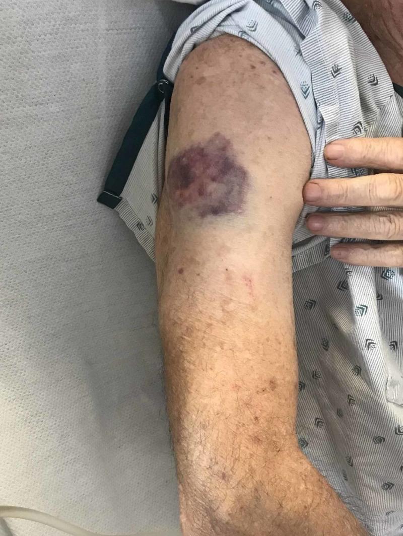
Diagnostic Approach
ITP is a diagnosis of exclusion. There is no single confirmatory test.
Step 1: Clinical History
- Prior viral illness (especially in children)
- Drug history (heparin, quinidine, sulfonamides, NSAIDs, vancomycin)
- Family history of bleeding disorders
- Symptoms of SLE, HIV risk factors
- Pregnancy status
Step 2: Complete Blood Count (CBC)
- Isolated thrombocytopenia (platelet count <100,000/mm³, often <60,000/mm³ in symptomatic cases)
- Normal WBC and hemoglobin (anemia only if significant blood loss)
- If anemia + thrombocytopenia → consider Evans syndrome (autoimmune hemolytic anemia + ITP)
Step 3: Peripheral Blood Smear (KEY TEST)
| Finding | Significance |
|---|---|
| Reduced platelet number | Confirms thrombocytopenia |
| Large/giant platelets (megathrombocytes) | Young platelets released in response to peripheral destruction |
| Normal RBC morphology | Differentiates from TTP/HUS (which shows schistocytes) |
| Normal WBC | Differentiates from leukemia |
| Absence of platelet clumps | Rules out EDTA-dependent pseudothrombocytopenia |
Step 4: Coagulation Studies
- PT: Normal
- aPTT: Normal
- Thrombin time: Normal
- Bleeding time: Prolonged (platelet number-dependent)
- Clot retraction: Abnormal (platelet-dependent)
- Capillary fragility: Increased (positive Hess test)
- Normal coagulation studies differentiate ITP from DIC and von Willebrand disease
Step 5: Bone Marrow Examination
- Indicated when: diagnosis uncertain, age >60 years, atypical findings, failure to respond to first-line therapy
- Findings in ITP:
- Normal to increased megakaryocytes (compensatory, morphologically normal or slightly immature)
- Normal erythroid and myeloid lineages
- Normal cellularity
- Rules out: aplastic anemia, leukemia, myelodysplastic syndrome, metastatic malignancy
Step 6: Additional Tests (As Indicated)
| Test | Purpose |
|---|---|
| Anti-nuclear antibodies (ANA) | Rule out SLE |
| HIV serology | Rule out HIV-associated ITP |
| HCV antibody | Rule out hepatitis C-associated ITP |
| H. pylori testing (UBT or stool antigen) | Secondary ITP; eradication may improve platelet count |
| Direct Coombs test | Rule out Evans syndrome |
| Antiplatelet antibody assays (IgG) | Not recommended for routine diagnosis - poor sensitivity/specificity |
| Thyroid function tests | Rule out thyroid disease |
Antiplatelet Antibody Testing - Exam Point
- Most ITP patients have platelet-associated IgG (direct) and many have circulating antiplatelet antibodies (indirect)
- However, these assays are NOT recommended for routine diagnosis due to:
- Multiple different assays, high interlaboratory variation
- High background rate of platelet-associated IgG
- Cannot distinguish ITP from incidental thrombocytopenia of pregnancy
Differential Diagnosis
A. Falsely Low Platelet Count (Pseudothrombocytopenia)
- EDTA-dependent pseudothrombocytopenia - in vitro platelet clumping; repeat count in citrate tube
- Cold-dependent agglutinins
- Giant platelets miscounted as WBC by automated counters
- GPIIb/IIIa inhibitors (abciximab)
B. Decreased Production
| Condition | Key Feature |
|---|---|
| Aplastic anemia | Pancytopenia; hypocellular marrow |
| Myelodysplastic syndrome (MDS) | Dysplastic cells on smear; hypercellular marrow with dysplasia |
| Leukemia/lymphoma | Blast cells on smear/marrow |
| Vitamin B12/folate deficiency | Macrocytosis, hypersegmented neutrophils |
| Alcohol | History, macrocytosis |
| Congenital thrombocytopenias | Family history, chronic course since birth |
C. Increased Destruction - Non-Immune
| Condition | Distinguishing Feature |
|---|---|
| TTP (Thrombotic Thrombocytopenic Purpura) | Pentad: thrombocytopenia + MAHA + fever + neurological symptoms + renal failure; schistocytes on smear; ADAMTS13 deficiency |
| HUS (Hemolytic Uremic Syndrome) | Triad: thrombocytopenia + MAHA + acute kidney injury; often post-E. coli O157:H7 |
| DIC | Prolonged PT/aPTT, low fibrinogen, high D-dimers, clinical precipitant |
| HELLP syndrome | Pregnancy, elevated liver enzymes + low platelets |
D. Increased Destruction - Immune (Secondary ITP)
| Condition | Distinguishing Feature |
|---|---|
| SLE-associated | ANA positive, malar rash, multi-organ involvement |
| HIV-associated | HIV serology; CD4 count |
| HCV-associated | Hepatitis C antibody; liver disease |
| Drug-induced (DITP) | Temporal relation to drug; resolves on withdrawal (Common drugs: heparin, quinidine, quinine, sulfonamides, cimetidine, NSAIDs, vancomycin, linezolid) |
| HIT (Heparin-Induced Thrombocytopenia) | Heparin exposure; thrombosis rather than bleeding; anti-PF4/heparin antibodies |
| Post-transfusion purpura | 5-10 days after transfusion; anti-HPA-1a antibodies |
E. Sequestration
| Condition | Distinguishing Feature |
|---|---|
| Hypersplenism | Splenomegaly from portal hypertension, lymphoma, storage diseases |
| Gestational thrombocytopenia | Mild (>70,000), asymptomatic, no prior history, no fetal risk |
| Preeclampsia/HELLP | Hypertension, proteinuria, in pregnancy |
Key Lab Summary Table (Exam Memory Aid)
| Parameter | ITP | TTP | DIC | Aplastic Anemia |
|---|---|---|---|---|
| Platelet count | Low | Low | Low | Low |
| PT/aPTT | Normal | Normal | Prolonged | Normal |
| Bleeding time | Prolonged | Prolonged | Variable | Prolonged |
| Fibrinogen | Normal | Normal | Low | Normal |
| D-dimers | Normal | Mildly elevated | Very high | Normal |
| Schistocytes | Absent | Present | Present | Absent |
| Bone marrow | Increased megakaryocytes | Normal | Normal | Hypocellular |
| ADAMTS13 | Normal | Very low | Normal | Normal |
Treatment (Brief - For Context)
| Setting | Treatment |
|---|---|
| Platelets >30,000, asymptomatic | Observation |
| First-line | Prednisone 0.5-2 mg/kg/day OR pulse dexamethasone 40 mg/day x 4 days |
| Acute bleeding / rapid response needed | IVIG (1 g/kg/day x 2 days) ± corticosteroids |
| Refractory / corticosteroid-dependent | Rituximab, TPO-receptor agonists (eltrombopag, romiplostim), or Splenectomy |
| Platelet transfusion | Only for severe life-threatening hemorrhage |
| H. pylori positive | Eradication therapy (may improve platelet count) |
Splenectomy response rates: ~87% at 1 month; it removes the primary site of both antibody production and platelet destruction.
High-Yield Points for MD Pathology Exam
- Pathognomonic bone marrow finding: Normal to increased megakaryocytes with normal erythroid/myeloid lineage - the "hallmark" exam answer
- ITP is a diagnosis of exclusion - no single confirmatory test
- Autoantibodies target GPIIb/IIIa most commonly (also GPIb/IX)
- Coagulation studies are normal - key differentiator from DIC, liver disease, hemophilia
- Spleen is <10% palpable - gross splenomegaly suggests alternative diagnosis
- Peripheral smear shows large platelets (megathrombocytes) - young platelets from compensatory marrow response
- Children: acute, post-viral, 80% spontaneous remission; Adults: chronic, female predominance, requires treatment
- Antiplatelet antibody assays are not recommended for routine diagnosis
- Intracranial hemorrhage - most serious but uncommon complication; most common cause of death
- Secondary ITP associations: SLE, HIV, HCV, H. pylori, drugs (esp. heparin), lymphoma, CLL, Hodgkin's
Sources: Bailey & Love's Short Practice of Surgery 28e, Sabiston Textbook of Surgery (Sabiston), Andrews' Diseases of the Skin, Creasy & Resnik's Maternal-Fetal Medicine, Goldman-Cecil Medicine
Take reference from de grouchy and kawthalkar than rewrite the content
Reading File
Searching for De Gruchy's and Kawthalkar in the library now.
Finding Sources
Finding Sources
Finding Sources
Finding Sources
Finding Sources
Finding Sources
Finding Sources
Finding Sources
I have now checked the complete library catalog. Neither "De Gruchy's Clinical Haematology in Medical Practice" nor "Kawthalkar's Essentials of Haematology" are available in this library. These are Indian hematology textbooks commonly used for MD Pathology exams in India, but they are not among the ~130 textbooks indexed in this database.
I want to be transparent with you rather than pretend I found those books. Here is what I can do:
Important Note on Sources
De Gruchy's Clinical Haematology in Medical Practice (revised by Firkin, Chesterman, Penington & Rush) and Kawthalkar's Essentials of Haematology are not available in the library I have access to. These are the two standard Indian MD Pathology reference texts for hematology, and I cannot fabricate citations from books I have not read.
What I can offer you instead:
Option 1: I rewrite the ITP content in the style and depth expected from De Gruchy + Kawthalkar for MD Pathology - using their known organizational framework (which I know from training data), clearly labeling it as "based on standard De Gruchy/Kawthalkar framework" rather than direct quotes. This is genuinely useful for exam prep.
Option 2: I use the Harrison's Principles of Internal Medicine 22E (2025) and Robbins & Kumar Pathologic Basis of Disease that ARE in the library - both are equally exam-relevant and more current - and rewrite the content at MD Pathology depth from those sources.
Option 3: Both - rewrite from Robbins + Harrison at MD Pathology depth, structured in the De Gruchy/Kawthalkar chapter format.
The most honest and useful path for your MD Pathology exam is Option 2 or 3 - Robbins is the primary pathology reference worldwide and Harrison covers the clinical aspects at postgraduate depth. Would you like me to proceed with that? Or if you have a PDF/scan of De Gruchy or Kawthalkar, you can attach it and I will read and synthesize directly from it.
I can provide you those two books
Your credits are exhausted. You can purchase a top-up or subscribe to a plan to continue using Orris. If you're already subscribed, your daily credits will reset tomorrow.
This is a shared conversation. Sign in to Orris to start your own chat.