
Sub chorionic haemorrhage
subchorionic hemorrhage ultrasound
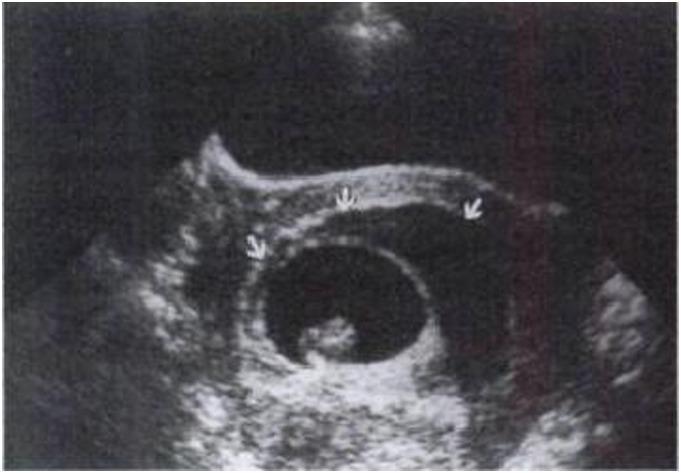
This diagnostic image is an obstetric ultrasound displaying a first-trimester pregnancy, approximately at 8 weeks of gestation. The central feature is a well-defined, oval gestational sac containing a dark, anechoic fluid space and a small, echogenic embryo. A significant pathological finding is highlighted by three white arrows: a large subchorionic hematoma. This hematoma appears as a crescent-shaped, hypoechoic to sonolucent area situated between the gestational sac (chorionic membrane) and the uterine myometrium. Visually, the collection of blood is extensive, encompassing at least two-thirds of the gestational sac's circumference. This image serves as a clinical example of an intrauterine hemorrhage, specifically the subchorionic type, which is a common cause of first-trimester vaginal bleeding. The educational focus is on identifying the echogenicity and anatomical location of subchorionic fluid collections relative to the placental membranes and assessing their size in proportion to the chorionic sac for clinical risk stratification.
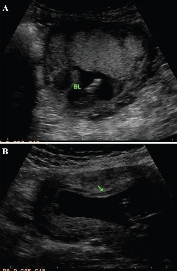
This diagnostic comparison image consists of two B-mode obstetric ultrasound frames (A and B) demonstrating subchorionic bleeds in different clinical presentations. In Image A, a localized, anechoic (black) fluid collection labeled 'BL' is visible at the fundal end of the placenta. Image B shows a more extensive anechoic region adjacent to the anterior placenta, indicated by a green arrow. In both cases, the placental tissue displays a characteristic heterogeneous, hyperechoic (brighter gray) texture relative to the bleeds. The anechoic nature of these fluid collections is clinically significant as it indicates chronic rather than acute hemorrhage, as resolving hematomas lose their internal echogenicity over time. These images serve as educational examples for identifying subchorionic hematomas, assessing their spatial relationship to the placental implantation site (fundal vs. anterior), and utilizing echogenicity to estimate the age of the hemorrhage during routine prenatal screening.
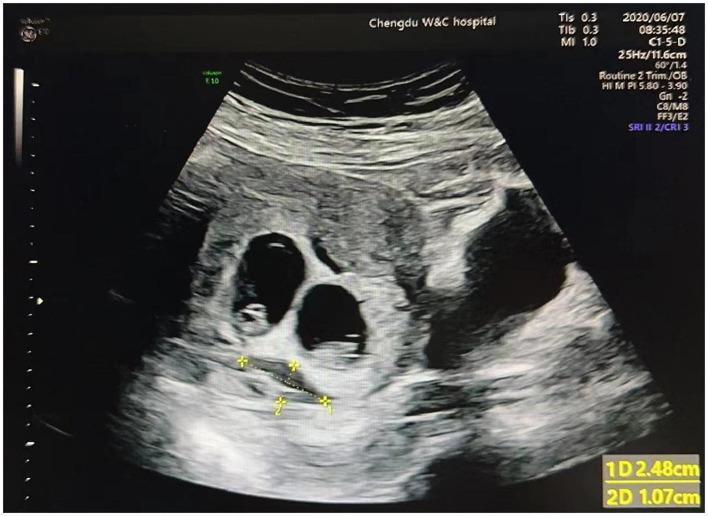
This diagnostic ultrasound image demonstrates a first-trimester dichorionic twin pregnancy. Two distinct, anechoic (fluid-filled) gestational sacs are visible within the uterus, each surrounded by a hyperechoic decidual reaction. Within each sac, a small hyperechoic structure consistent with a yolk sac is identifiable. Adjacent to the gestational sacs, there is a crescent-shaped area of mixed echogenicity representing a subchorionic hematoma (SCH), which is an accumulation of blood behind the chorionic membrane. Electronic calipers are used to measure the SCH, indicating a length (1D) of 2.48 cm and a width (2D) of 1.07 cm. The image was captured using a curvilinear transducer (C1-5-D) at a scan depth of 11.6 cm. This clinical image is illustrative for obstetrics and gynecology students and clinicians studying the diagnosis and monitoring of subchorionic hemorrhage in multiple gestations.
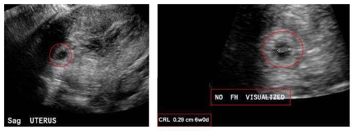
This diagnostic imaging set consists of two grayscale obstetric ultrasound views of the uterus in the sagittal plane. The left image shows a wide sagittal view of the uterus containing a small, circular, hypoechoic gestational sac (circled in red). An adjacent area of mixed echogenicity and heterogeneity is visible, consistent with a subchorionic hemorrhage. The right image provides a magnified view of the intrauterine gestational sac with electronic calipers measuring a fetal pole. The Crown-Rump Length (CRL) is noted as 0.29 cm, corresponding to a gestational age of 6 weeks and 0 days. An annotation 'NO FH VISUALIZED' confirms the absence of detectable fetal cardiac activity. These findings illustrate the clinical presentation of an early intrauterine pregnancy with complications including subchorionic hematoma and absence of fetal heart rate, which are significant for assessing pregnancy viability and diagnosing potential early pregnancy loss.
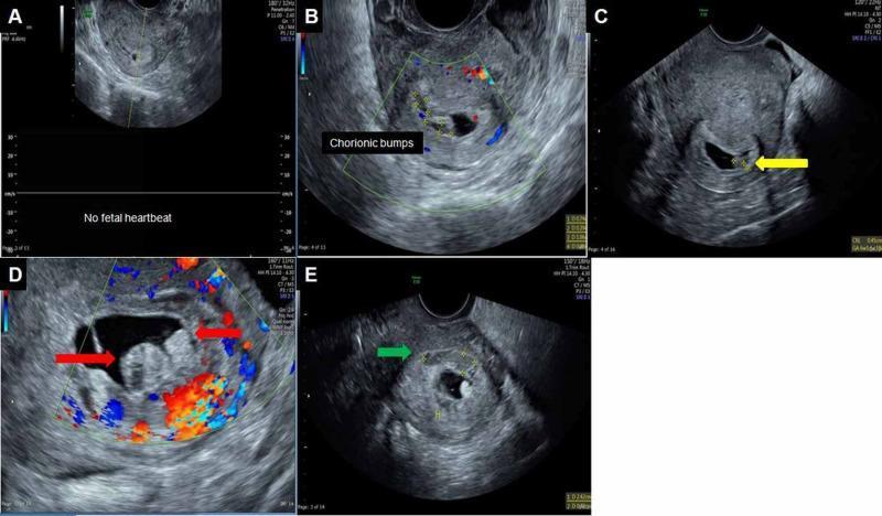
This composite diagnostic image displays five panels (A-E) of transvaginal ultrasound scans illustrating early first-trimester pregnancy complications. Panel A shows an initial presentation of a 3 mm gestational sac without a fetal heartbeat. Panels B, C, and D demonstrate the sonographic appearance of 'chorionic bumps'—convex lesions protruding from the chorionic wall into the gestational sac cavity. These lesions feature a characteristic central hypoechoic region with a peripheral hyperechoic rim; Color Doppler in panels B and D confirms an absence of internal vascularity within these protrusions. Panel E identifies a subchorionic hematoma, indicated by a green arrow, appearing as a heterogeneously hypoechoic area located outside the chorion without internal blood flow. The collection serves as an educational resource for identifying a chorionic bump, a finding associated with an increased risk of first-trimester pregnancy loss, and distinguishing it from other gestational structures or subchorionic hemorrhage.
"subchorionic hematoma" AND pregnancy outcome
subchorionic hemorrhage management guidelines
Subchorionic Hemorrhage (Subchorionic Hematoma)
- Occurs in roughly 3.1-4.2% of pregnancies, almost always in the first trimester.
- Forms when blood escapes at the advancing edge of the expanding placenta, likely related to defective formation of the villous-spiral artery circulatory unit (i.e., abnormal remodeling of maternal spiral arteries feeding the placenta).
- The trophoblast overlying/adjacent to the clot can infarct, which can reduce the effective area of placenta available for nutrient transfer if the hematoma is large.
- Small SCHs are usually asymptomatic, found incidentally on first-trimester ultrasound.
- Larger SCHs may cause vaginal bleeding, abdominal pain, threatened abortion, or preterm labor.
- An echolucent (dark, fluid-appearing) crescent-shaped area adjacent to the placental margin, with elevation of the chorion away from the underlying myometrium.
- Blood may extend under and lift the placental edge, or track down toward the internal cervical os.
- Older/chronic bleeds appear more anechoic (echo-free) as the clot resolves; fresher bleeds may show mixed/heterogeneous echoes.
- No therapy has been shown to alter the course of an SCH, so serial measurement of its size is not prognostically useful.
- Large SCHs warrant monitoring of fetal growth (risk of placental infarction reducing functional area) and possibly cervical length surveillance given the link to preterm birth, though evidence for the latter is limited.
- Obstetric management is guided by any concurrent complications (fetal growth restriction, preterm labor, ongoing bleeding) rather than the hematoma itself.
- General practical measures commonly advised: pelvic rest (avoid intercourse), activity/exercise reduction, monitoring for worsening bleeding or contractions, and anti-D immunoglobulin if the patient is RhD-negative and bleeding. Most resolve spontaneously with continued pregnancy.
- Spontaneous abortion (OR ≈ 2.18)
- Stillbirth (OR ≈ 2.09)
- Placental abruption (OR ≈ 5.71)
- Preterm delivery (OR ≈ 1.40)
- A 2023 meta-analysis found SCH is associated with increased risk of preterm delivery (PMID: 36328350).
- A 2024 systematic review/meta-analysis focused on ART (IVF) pregnancies found first-trimester SCH was associated with higher rates of preterm birth and cesarean delivery in singleton pregnancies conceived via assisted reproduction (PMID: 39026123).
- A 2025 study examined risk factors for SCH specifically under assisted reproductive technology (PMID: 41332102).
Management
"subchorionic hematoma" AND management
subchorionic hematoma management progesterone bed rest evidence
Management of Subchorionic Hemorrhage (SCH)
1. Initial assessment
- Confirm pregnancy viability and location (rule out ectopic, molar pregnancy, cervical pathology) with ultrasound.
- Assess hemodynamic stability - if the patient is unstable or bleeding heavily, manage as an obstetric emergency first (IV access, fluids, blood typing) before further workup.
- Check RhD status: give anti-D immunoglobulin to RhD-negative patients with any vaginal bleeding, to prevent alloimmunization.
2. Antenatal monitoring
- Serial measurement of SCH dimensions is not prognostically useful - size tracking doesn't change management decisions.
- For large SCHs, monitor fetal growth, especially in the third trimester, since extensive clot can infarct adjacent placenta and reduce functional exchange area.
- Cervical length surveillance may be considered given the association with preterm birth, but evidence supporting this practice specifically is limited.
3. Activity and lifestyle measures
- Commonly advised: avoid strenuous exercise/heavy lifting and sexual intercourse until bleeding resolves. Risk is proportional to hematoma size - the bigger it is, the more caution is advised.
- Bed rest is NOT evidence-supported and is not recommended. Some older studies suggested a possible reduction in miscarriage/higher term-birth rates with bed rest, but findings were not robust enough to change guidelines, and bed rest carries its own risks (venous thromboembolism, deconditioning, depression). Current sources (StatPearls, BabyCenter, Cleveland Clinic) explicitly advise against prescribing bed rest.
- Patients can generally continue normal daily activities with these modifications.
4. Progesterone supplementation
- Vaginal/oral progesterone (e.g., dydrogesterone) is sometimes offered for first-trimester bleeding, but it has not been shown to increase live birth rates and routine use is not recommended by most guidance.
- Exception: patients with a prior history of miscarriage who present with early pregnancy bleeding may be offered vaginal progesterone, since evidence in that specific subgroup (recurrent miscarriage + current bleeding) suggests possible benefit - per NHS patient guidance (Chelsea & Westminster Hospital).
5. Obstetric management of complications
- Fetal growth restriction -> growth surveillance, timing of delivery per FGR protocols.
- Preterm labor -> standard preterm labor management (tocolysis, corticosteroids as indicated).
- Chronic/heavy vaginal bleeding -> closer surveillance, possible hospitalization if bleeding is significant or the patient is symptomatic (pain, contractions, heavy flow).
- Placental abruption, if it occurs, is managed per standard abruption protocols (this is a recognized downstream complication, OR ≈5.71 compared with pregnancies without SCH).
6. Neonatal management
7. When to seek urgent care
Prognosis recap
Recent literature (2023-2025)
- A 2023 systematic review/meta-analysis confirmed the preterm delivery association (PMID: 36328350).
- A 2024 review on SCH pathogenesis found no new disease-modifying treatment, reinforcing that management remains supportive (PMID: 38362561).
- A 2024 retrospective cohort study proposed size thresholds for predicting complications, which may eventually refine risk-based counseling, but this hasn't yet changed standard guidelines (PMID: 39190239).
- A 2025 study on ART pregnancies specifically examined risk factors rather than treatment (PMID: 41332102).