
Pathogenesis of pneumonia
pneumonia pathogenesis lung inflammation diagram
community acquired pneumonia lung consolidation alveolar
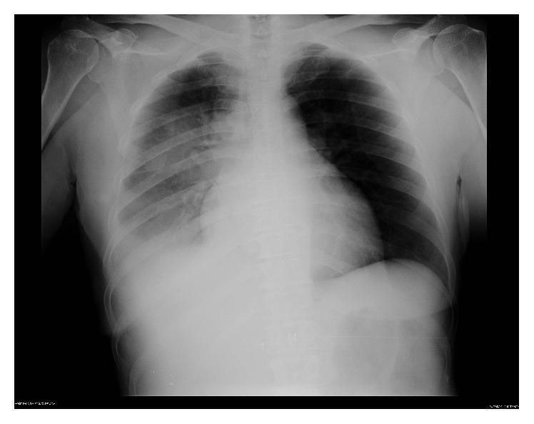
This diagnostic image is a posterior-anterior (PA) chest radiograph. The primary finding is a dense consolidation localized in the right lower lobe, characterized by increased opacity that obscures the right hemidiaphragm and portions of the heart border. Additionally, there are patchy peribronchial opacities extending into the right middle lung zone, following the bronchovascular markings. The left lung field remains relatively clear with normal vascular markings and no evidence of pleural effusion or consolidation. The cardiac silhouette appears within normal limits for size and shape. This radiographic presentation is highly characteristic of community-acquired pneumonia, specifically Legionnaires' disease (Legionella pneumophila), illustrating the progression from patchy infiltrates to denser alveolar consolidation. The image serves as an educational example for identifying unilateral focal pneumonia and distinguishing consolidation from clear lung parenchyma in an acute clinical setting.
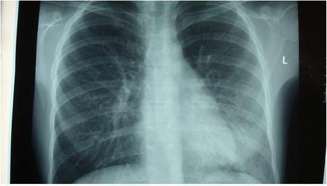
A posterior-anterior (PA) chest x-ray demonstrating localized pathology in the right lung. There is a notable area of increased opacity and consolidation in the right lower lobe, which partially obscures the right hemidiaphragm and suggests an infectious process such as pneumonia. In contrast, the left lung field appears relatively clear and well-aerated with normal vascular markings. The cardiac silhouette, mediastinal contours, and trachea appear within normal limits. The skeletal structures of the ribcage and clavicles are intact and clearly visible. This diagnostic image illustrates the typical radiographic presentation of community-acquired pneumonia, characterized by alveolar infiltration and consolidation in a specific anatomical lobe, used clinically to guide antibiotic treatment and monitor disease progression.
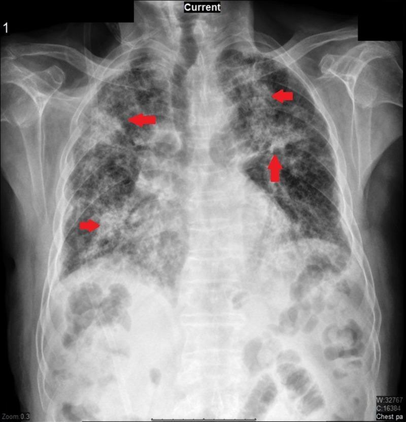
This diagnostic image is a posterior-anterior (PA) chest radiograph illustrating significant pulmonary pathology. The radiograph demonstrates diffuse, bilateral increased reticular markings and patchy opacities consistent with multi-lobular consolidation. Red arrows highlight specific areas of denser consolidation in the upper and middle lung zones. The lung parenchyma shows a mix of interstitial thickening and alveolar filling, which clinically correlates with infectious or inflammatory processes such as community-acquired pneumonia or an exacerbation of underlying interstitial lung disease (ILD). Key anatomical landmarks including the clavicles, ribs, and mediastinal silhouette are visible, though the diaphragmatic borders are partially obscured by the extensive pulmonary opacification. The image is a primary educational resource for recognizing the radiological presentation of multi-lobar pneumonia and diffuse lung disease in a clinical setting.
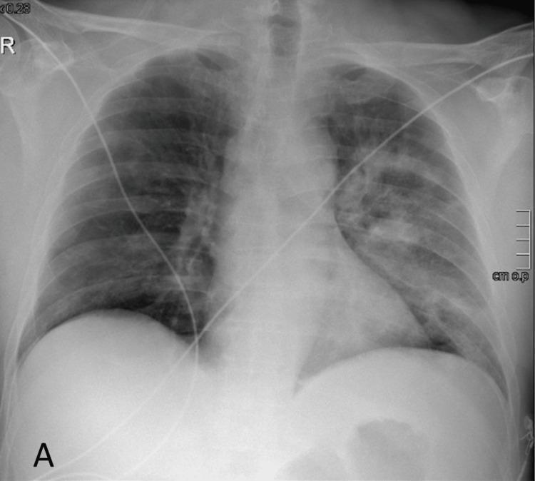
This diagnostic image is an anteroposterior (AP) chest X-ray of an adult male. The primary finding is the presence of patchy, ill-defined alveolar opacities and infiltrates concentrated in the left mid-to-upper lung zones, consistent with lobar or multifocal pneumonia. These infiltrates represent consolidation or inflammatory fluid within the alveolar spaces. The right lung field appears relatively clear, although some mild interstitial markings are visible. The cardiac silhouette and mediastinal contours are within normal limits, with no evidence of cardiomegaly or pleural effusion. External medical equipment is present, including monitoring leads and tubing crossing the thoracic field. The bony structures, including the ribs and clavicles, show no acute fractures or abnormalities. This radiograph demonstrates typical findings of community-acquired pneumonia (CAP) in a clinical setting of septic shock or acute respiratory infection, illustrating the radiographic hallmarks of consolidation and inflammatory lung disease for medical educational indexing.
Pathogenesis of Pneumonia
Definition
1. Routes of Microbial Entry
| Route | Details |
|---|---|
| Microaspiration | The most common route - silent aspiration of oropharyngeal organisms into the lower airways |
| Inhalation | Direct inhalation of aerosolized particles (e.g., Legionella, TB, influenza) |
| Macroaspiration | Large-volume aspiration of gastric or oropharyngeal contents (aspiration pneumonia, ~5-15% of CAP) |
| Hematogenous spread | From a distant infected site (e.g., right-sided endocarditis, line sepsis) |
| Direct mucosal dispersion | Contiguous spread from an adjacent structure |
- Harrison's Principles of Internal Medicine, 22E
2. Host Defense Mechanisms (and How They Fail)
- Mechanical barriers: nasal hairs, turbinates, the branching tracheobronchial tree, and the gag/cough reflexes
- Mucociliary clearance: the ciliated epithelium propels particles and pathogens upward
- Innate immunity: alveolar macrophages are the first-line phagocytes; they clear small inocula without triggering overt inflammation
- Adaptive immunity: antibodies (especially secretory IgA and opsonizing IgG) and T-cell responses
3. The Modern Model: Lung Microbiota Disruption
- Microbial entry into the lungs
- Microbial elimination (by local defenses)
- Regional growth conditions (pH, oxygen tension, temperature)
"An inflammatory event resulting in epithelial and/or endothelial injury results in the release of cytokines, chemokines, and catecholamines, some of which may selectively promote the growth of certain bacteria, such as Streptococcus pneumoniae and Pseudomonas aeruginosa. This cycle of inflammation, enhanced nutrient availability, and release of potential bacterial growth factors may result in a positive feedback loop that further accelerates inflammation and the growth of particular bacteria, which may then become dominant."
- Harrison's Principles of Internal Medicine, 22E
4. Inflammatory Cascade
- Alveolar macrophage activation - the first responders; they attempt to phagocytose and kill bacteria, and release pro-inflammatory cytokines
- Cytokine/chemokine release:
- IL-6 and TNF → systemic fever, acute-phase response
- IL-8 and G-CSF → recruitment of large numbers of neutrophils to the alveoli
- Neutrophil influx - the dominant effector cells; they release proteases, reactive oxygen species, and antimicrobial peptides
- Alveolar capillary leak - mediators released by macrophages and neutrophils damage the alveolar-capillary membrane, causing exudation of protein-rich fluid into the alveolar space
- Bacterial toxins - many pathogens (e.g., S. pneumoniae pneumolysin, S. aureus toxins) further amplify the inflammatory response
- Impaired hypoxic vasoconstriction - some bacterial pathogens interfere with the normal vasoconstrictive response to hypoxic alveoli, worsening ventilation-perfusion (V/Q) mismatch and causing severe hypoxemia
- Harrison's Principles of Internal Medicine, 22E
5. Pathological Stages (Classic Lobar Pneumonia)
| Stage | Timing | Pathological Features |
|---|---|---|
| 1. Edema | Early | Proteinaceous exudate + bacteria in alveoli; alveolar capillary congestion |
| 2. Red hepatization | ~Day 1-3 | Alveolar spaces flooded with erythrocytes, neutrophils, and fibrin; lung appears red and liver-like |
| 3. Gray hepatization | ~Day 3-8 | Erythrocytes lysed; neutrophils predominate; abundant fibrin; bacteria disappear; corresponds with infection containment |
| 4. Resolution | Days to weeks | Macrophages dominate; debris from neutrophils, bacteria, and fibrin is cleared; normal architecture restored |
6. Gas Exchange Impairment
- Intrapulmonary shunt: consolidated alveoli are perfused but not ventilated → deoxygenated blood bypasses gas exchange
- Low V/Q regions: partially filled alveoli have reduced ventilation relative to perfusion
7. Viral Pneumonia Pathogenesis
- Initial infection of ciliated respiratory mucosal epithelial cells → widespread cellular destruction
- Tracheobronchial involvement: tracheitis, bronchitis, bronchiolitis; loss of ciliated epithelium; submucosal hemorrhage and edema
- Alveolar involvement: neutrophils and mononuclear cells mixed with fibrin and edema fluid; intra-alveolar hemorrhage; hyaline membranes lining alveolar ducts
- Bacterial superinfection: viral disruption of the epithelial barrier and mucociliary clearance, plus the type I interferon response downregulating antibacterial defenses, markedly increases susceptibility to secondary bacterial pneumonia (classically appearing 2-14 days after initial viral illness)
- Fishman's Pulmonary Diseases and Disorders
8. Summary Diagram (Conceptual)
Microbial Entry (aspiration / inhalation / hematogenous)
        ↓
Overwhelm of local defenses (mucociliary, macrophages, IgA)
        ↓
Alveolar epithelial/endothelial injury
        ↓
Cytokine/chemokine release (IL-6, TNF, IL-8, G-CSF)
        ↓
Neutrophil recruitment → Alveolar exudate (edema stage)
        ↓
Consolidation: Red → Gray hepatization
        ↓
V/Q mismatch → Intrapulmonary shunt → Hypoxemia
        ↓
Resolution (macrophage clearance) OR
Complications (abscess, empyema, bacteremia, ARDS)