
Elderly male patient known case of heart block with pace maker implantation presented with acute onset SOB with AKI and ALI to rule out ADHF or Acute on CKD evaluation and management
Reading File
Now let me run parallel searches across the library for all key topics in this complex case.
Finding Sources
Finding Sources
Finding Sources
Finding Sources
Reading File
Reading File
Finding Sources
Finding Sources
Reading File
Finding Sources
Finding Sources
Reading File
Reading File
Finding Sources
Finding Sources
Searching PubMed
acute decompensated heart failure cardiorenal syndrome management
I now have sufficient high-quality data from Brenner & Rector's The Kidney, Harrison's, Braunwald's, and Fuster's Heart textbooks, plus PubMed 2024-2026 evidence. Let me compose the comprehensive clinical response.
Elderly Male with Pacemaker: Acute SOB + AKI + ALI - ADHF vs Acute-on-CKD Evaluation & Management
Clinical Context
This is a high-complexity, multi-system presentation in an elderly male with:
- Known complete heart block with permanent pacemaker (PPM)
- Acute onset dyspnea / shortness of breath (SOB)
- Acute Kidney Injury (AKI)
- Acute Lung Injury (ALI) / pulmonary edema
- Primary differential: ADHF (Cardiorenal Syndrome type 1) vs Acute-on-CKD with fluid overload
Step 1: Initial Rapid Assessment (First 30-60 minutes)
A. Hemodynamic Profile - "Wet vs Dry / Cold vs Warm"
Use the Stevenson classification at the bedside:
| Wet (congested) | Dry (euvolemic) | |
|---|---|---|
| Cold (hypoperfused) | Cold & Wet = Cardiogenic shock | Cold & Dry = Hypovolemia/low output |
| Warm (perfused) | Warm & Wet = Most common ADHF | Warm & Dry = Compensated HF |
- Most ADHF presentations are Warm & Wet (elevated filling pressures, adequate perfusion)
- Cold & Wet = most severe - requires urgent inotropic support
B. Rapid Bedside Assessment
| Feature | Supports ADHF | Supports Acute-on-CKD |
|---|---|---|
| Orthopnea / PND | Yes | Less specific |
| Bilateral crackles | Yes (pulmonary edema) | Possible if fluid overload |
| S3 gallop | Strongly supports | Absent |
| JVP elevated | Yes | Yes (fluid overload) |
| Pitting edema | Yes | Yes |
| Oliguria | Present in both | Prominent |
| Weight gain (acute) | Typically >2-3 kg | Variable |
| Prior HF history | Key context | Look for CKD baseline |
| Pacemaker | May indicate CMP | Cor pulmonale possible |
Step 2: Pacemaker-Specific Considerations
This patient's pacemaker history adds critical considerations:
-
Pacemaker interrogation is mandatory - Check for:
- Mode switch events (indicating AF burden)
- Rate-responsive function (may mask tachycardia)
- Pacing threshold changes (electrode dislodgement or lead failure)
- Impedance trends (lead insulation breach)
- Stored electrograms - episodes of fast AF, VT
-
Pacemaker-Induced Cardiomyopathy (PICM):
- Occurs in ~20% of patients with chronic RV pacing
- Causes dyssynchrony - LVEF drops, can precipitate ADHF
- Look for LBBB morphology on ECG/paced beats
- If pacing burden >40% over years, suspect PICM
-
Pacemaker syndrome (in VVI devices):
- VA conduction causes retrograde P-waves, symptoms of low output
-
Device infection - Consider if fever + sepsis contribution to AKI
-
ECG: Will show paced rhythm; compare with prior ECG for changes in morphology
Step 3: Key Investigations
A. Immediate (Bedside/Emergency)
| Test | Purpose |
|---|---|
| ECG | Paced rhythm, AF burden, ischemic changes, QRS duration |
| Chest X-ray (PA/AP) | Pulmonary edema (bat-wing, Kerley B), cardiomegaly, pleural effusion, device lead position |
| Pulse oximetry / ABG | Hypoxia severity, respiratory acidosis (ALI), metabolic acidosis (AKI) |
| Bedside echo (POCUS) | LV function, LVEF, IVC diameter (volume status), pericardial effusion, lung B-lines |
| BP, HR, UO monitoring | Hemodynamic profile |
B. Laboratory - Priority
| Investigation | Significance |
|---|---|
| Serum creatinine + eGFR | Confirm AKI; compare with baseline |
| BUN/Creatinine ratio | >20:1 suggests prerenal; <10:1 intrinsic renal |
| Urine output (catheterize) | Oliguria < 0.5 mL/kg/hr = AKI criterion |
| Serum electrolytes (Na, K, Cl, HCO3) | Hyponatremia in advanced HF; hyperkalemia in AKI |
| ABG | Type of respiratory failure; metabolic acidosis |
| BNP or NT-proBNP | Key diagnostic anchor for ADHF |
| Troponin (hsTnI/T) | Rule out ACS precipitant; also elevated in ADHF from wall stress |
| CBC | Anemia (worsens HF); infection (AKI precipitant) |
| LFTs, albumin | Hepatic congestion (congestive hepatopathy) |
| Urine analysis + microscopy | Casts (ATN vs prerenal); proteinuria; specific gravity |
| Urine Na, FENa | FENa <1% = prerenal; >2% = ATN |
| Procalcitonin / CRP | Infectious trigger for decompensation |
| Serum lactate | Cardiogenic shock / hypoperfusion |
| Phosphorus, Uric acid, Magnesium | AKI metabolic complications |
C. BNP/NT-proBNP Interpretation
- BNP >100 pg/mL or NT-proBNP >300 pg/mL = Supports HF diagnosis (Class I ACC/AHA recommendation)
- NT-proBNP age-adjusted cut-offs: <75 yrs: >125 pg/mL; >75 yrs: >450 pg/mL (higher with AKI/CKD - interpret cautiously)
- BNP <35 or NT-proBNP <125: Makes ADHF unlikely
- Caveat: Both are elevated in CKD independent of HF - use relative levels and clinical correlation
Note: "BNP and NT-proBNP are similar for diagnosis of AHF, but NT-proBNP is superior in predicting clinical outcome." - Fuster and Hurst's The Heart, 15th Ed.
D. Echocardiography (Formal, urgent)
- LVEF: HFrEF (<40%) vs HFpEF (≥50%)
- RWMA - Regional wall motion abnormalities (ACS precipitant)
- Diastolic function - E/e' ratio - elevated in HFpEF
- RV function - Cor pulmonale from chronic lung disease
- Pericardial effusion - Tamponade causing low output
- Lead position - Right ventricular lead position
- Valvular disease - TR, MR (functional) worsening
Step 4: Cardiorenal Syndrome Classification
This patient almost certainly has Cardiorenal Syndrome (CRS). Classification guides management:
| CRS Type | Name | Mechanism in This Patient |
|---|---|---|
| Type 1 | Acute cardiorenal | ADHF → AKI (most likely if primary cardiac event) |
| Type 2 | Chronic cardiorenal | Pacemaker CMP → chronic HF → CKD progression |
| Type 4 | Chronic renocardiac | Prior CKD → HFPEF/HFREF (common in elderly) |
| Type 5 | Systemic | Sepsis, contrast, drugs causing both simultaneously |
"At least five subtypes of cardiorenal syndrome have been proposed. Worsening renal function in heart failure is not the same as acute kidney injury (AKI) in heart failure." - Brenner & Rector's The Kidney
Key pathophysiology to understand:
- Reduced cardiac output → Reduced renal blood flow (RBF) → Prerenal AKI
- Elevated CVP → Elevated renal venous pressure → Reduced GFR (even without reduced RBF) - this is often underappreciated
- RAAS/SNS activation → Vasoconstriction → Oliguria
"Higher CVP was an important predictor of worsening renal function in acute HF" - Brenner & Rector's The Kidney
Step 5: ADHF vs Acute-on-CKD - Differentiating
| Feature | ADHF (CRS Type 1) | Acute-on-CKD |
|---|---|---|
| BNP/NT-proBNP | Markedly elevated | Elevated but may be chronic |
| Echo: LVEF | Low in HFrEF; diastolic dysfunction in HFpEF | Usually normal unless concurrent HF |
| Response to IV diuresis | Good diuresis, SOB improves | Variable; may worsen if volume depleted |
| Urine sediment | Usually benign; granular casts if ATN develops | Granular/pigmented casts, proteinuria |
| FENa | Often <1% (prerenal from low CO) | >2% if ATN superimposed |
| Prior creatinine baseline | Critical to establish | History of CKD, prior Cr values |
| Precipitating factor | New AF, missed meds, salt excess, ischemia | NSAIDs, contrast, infection, hypovolemia |
Step 6: Management
A. Immediate Stabilization
- Positioning: Head of bed 45°, legs dependent (reduces preload, eases breathing)
- Oxygen: Target SpO2 92-96%; avoid hyperoxia
- Non-invasive ventilation (NIV/BiPAP): If SpO2 <90% or respiratory distress despite O2 - reduces intubation, reduces preload, improves oxygenation
- Reserve intubation for failure of NIV
- IV access + monitoring: Continuous ECG, BP q15-30 min, urine output hourly
- Hold nephrotoxins: NSAIDs, aminoglycosides, contrast (if avoidable), ACE inhibitors/ARBs temporarily if K >5.5 or creatinine rising rapidly
B. Decongestion - Core of ADHF Management
"After initial stabilization, the mainstay of treatment is vasodilator and diuretic therapy." - Brenner & Rector's The Kidney
IV Loop Diuretics (First-line):
| Drug | Starting Dose | Notes |
|---|---|---|
| Furosemide IV | 40-80 mg bolus (2.5x oral equivalent if already on oral) | Erratic bioavailability in HF; prefer IV |
| Torsemide | 10-20 mg IV | Longer acting, more predictable; 50% reduction in re-hospitalization vs furosemide |
| Bumetanide | 0.5-1 mg IV | Reliable bioavailability |
- Target urine output: 100-200 mL/hour in first few hours
- Diuretic resistance (common in CRS): Furosemide response is blunted in HF due to rightward/downward shift of dose-response curve
- Add thiazide (metolazone 2.5-5 mg PO 30 min before loop diuretic) for sequential nephron blockade
- Increase IV dose; consider continuous infusion
- Consider aldosterone antagonist addition
- Monitor: Creatinine, BUN, electrolytes q8-12h; tolerate creatinine rise up to ~0.3 mg/dL if congestion is improving ("aggressive decongestion associated with improved survival" even when creatinine worsens - Brenner & Rector)
IV Vasodilators (if BP allows, SBP >100 mmHg):
- IV Nitroglycerin: Start at 5-10 mcg/min, titrate up - reduces preload rapidly; useful for acute pulmonary edema
- Nitroprusside: For severe hypertensive ADHF
- Nesiritide: Recombinant BNP; vasodilator + mild natriuretic effect; not shown to improve mortality
C. Pacemaker Management in ADHF
- Urgent pacemaker interrogation
- If PICM suspected (LVEF drop + high RV pacing burden):
- Upgrade to CRT-D (Cardiac Resynchronization Therapy with Defibrillator) - this is the key intervention
- Reduces dyssynchrony, improves LVEF, reduces HF hospitalizations
- If pacemaker-dependent and hemodynamically unstable: Ensure programming is appropriate (rate-response on, avoid very slow programmed rates)
- Check for device-related infection as AKI precipitant
D. Managing AKI in the Context of ADHF
- Establish if prerenal vs intrinsic AKI:
- FENa <1% + response to decongestion = prerenal (most common in ADHF)
- Rising creatinine despite adequate decongestion = intrinsic/ATN
- Do NOT reflexively hold diuretics - aggressive decongestion improves renal perfusion by reducing CVP
- RAAS inhibitors: Hold ACEi/ARB temporarily if K >5.5 or creatinine rising >50% from baseline; restart at lower dose once stable
- Monitor for hyperkalemia - ECG changes (peaked T waves, wide QRS) - requires urgent treatment
- Dialysis indications: Refractory fluid overload, hyperkalemia unresponsive to medical management, severe acidosis (pH <7.1), uremic symptoms
- Ultrafiltration: An option for diuretic-resistant fluid overload - recent meta-analysis (2024) shows better clinical decongestion than IV loop diuretics but no mortality benefit; higher risk of adverse events - reserve for refractory cases
E. Precipitant Identification and Treatment
Search for and treat the trigger:
- New AF (very common) - Rate control; anticoagulation with DOAC (dose-adjust for eGFR)
- ACS - Troponin elevation, RWMA on echo; urgent PCI if STEMI/NSTEMI
- Infection/sepsis - Procalcitonin, cultures, antibiotics
- Medication non-compliance - History critical
- Dietary salt indiscretion - Common trigger
- Drug culprits (NSAIDs, verapamil, diltiazem, contrast) - Review medication list
F. Pacemaker Patient - Special Drug Considerations
| Drug | Consideration |
|---|---|
| Beta-blockers | Continue if tolerated (unless cold/wet profile); improve LVEF in HFrEF |
| ACEi/ARB/ARNI | Hold temporarily in acute AKI; restart when creatinine stabilizes |
| Spironolactone/eplerenone | Hold if K >5.0 or creatinine >2.5 mg/dL (CRS risk) |
| Digoxin | Renal clearance - toxic in AKI; check levels; avoid or reduce dose |
| DOACs | Dose-reduce per eGFR for AF anticoagulation |
| IV contrast | Avoid if possible; if needed, pre/post hydrate |
G. Monitoring & Goals of Therapy
- Daily weight (target 0.5-1 kg/day weight loss while decongesting)
- Strict fluid balance - aim for negative balance initially
- Creatinine/electrolytes q12h
- BNP/NT-proBNP at 24-48h - trends guide decongestion adequacy
- Repeat echo at 48-72h - track LVEF, IVC, diastolic function
- Pacemaker check formal report
Step 7: ADHF vs Acute-on-CKD - Summary Algorithm
Elderly male + PPM + Acute SOB + AKI + ALI
        ↓
1. ABG, SpO2, ECG, CXR, POCUS within 15 min
2. BNP/NT-proBNP + Troponin + RFT + Urine analysis
        ↓
BNP >300 + Echo: EF↓ or DD + Bilateral B-lines (POCUS)?
        ↓YES                    ↓NO
    ADHF (CRS)          Acute-on-CKD (consider infection,
    Diurese + vasodilate  NSAIDs, obstruction, ATN)
        ↓
Pacemaker interrogation
        ↓
PICM present? → CRT upgrade
AF present? → Rate control + anticoagulation
ACS? → PCI
        ↓
Daily monitoring: Weight, Cr, K+, BNP trend
Hold nephrotoxins; adjust diuretics for response
Key Points Summary
- Classify CRS type - this patient likely has CRS Type 1 or 2 (cardiac primary, renal secondary)
- Pacemaker interrogation is non-negotiable - must rule out PICM, device arrhythmia, and lead issues
- BNP/NT-proBNP is the diagnostic anchor for ADHF; interpret in context of CKD (falsely elevated)
- Aggressive IV diuresis is the cornerstone of ADHF management - do not be deterred by mild creatinine rise
- Elevated CVP harms the kidney via venous congestion - decongestion itself improves renal function
- Hold nephrotoxins and dose-adjust renally cleared drugs (digoxin, DOACs, spironolactone)
- NIV/BiPAP for ALI/respiratory failure - reduces intubation need
- CRT upgrade if PICM confirmed - most impactful intervention for ADHF from pacemaker-induced dyssynchrony
- Recent (2026) meta-analysis on ARNi in ADHF supports angiotensin-neprilysin inhibition post-stabilization
Sources: Brenner & Rector's The Kidney, 2-Volume Set | Fuster and Hurst's The Heart, 15th Ed. | Goldman-Cecil Medicine | Murray & Nadel's Respiratory Medicine | PubMed 2024-2026
Elderly male patient known case of heart block with pacemaker implantation presented with acute onset SOB to rule out ADHF and with AKI ?Acute on CKD with ALI ... evaluation and management
Reading File
Finding Sources
Finding Sources
Finding Sources
Finding Sources
Reading File
Reading File
Finding Sources
Finding Sources
Reading File
Finding Sources
Finding Sources
Reading File
Reading File
Searching PubMed
acute decompensated heart failure AKI cardiorenal treatment
Searching Images
cardiorenal syndrome heart failure AKI classification diagram
Searching Images
loop diuretic dose response curve heart failure CKD
Loading Image
Here is the full structured clinical write-up, integrating Brenner & Rector's The Kidney, Fuster & Hurst's The Heart, Murray & Nadel's Respiratory Medicine, Harrison's, and 2024-2026 PubMed evidence.
Elderly Male + Pacemaker (Heart Block) | Acute SOB | AKI | ALI
Rule Out: ADHF vs Acute-on-CKD
OVERVIEW OF THE CLINICAL PROBLEM
This presentation sits at the intersection of three simultaneous organ system crises - cardiac, renal, and pulmonary - in a patient whose baseline is already modified by complete heart block and chronic pacemaker dependency. The central unifying diagnosis is almost certainly Cardiorenal Syndrome (CRS), most likely Type 1 (Acute Cardiorenal): acute cardiac decompensation driving AKI, with the ALI representing cardiogenic pulmonary edema. However, Acute-on-CKD with volume overload must be formally excluded.
"The interplay between heart and kidney is key to the pathophysiologic processes that occur in HF and end-stage renal disease, and the synergistic failure of both organs together has been termed cardiorenal syndrome." - Brenner & Rector's The Kidney
PART 1: SYSTEMATIC EVALUATION
A. Immediate Bedside Assessment (First 15 Minutes)
1. Hemodynamic Profile - Stevenson Classification
Every ADHF patient must be classified on two axes:
| WET (congested: JVP↑, edema, crackles, orthopnea) | DRY (euvolemic) | |
|---|---|---|
| WARM (perfused: warm peripheries, good capillary refill) | Warm & Wet - Most common ADHF | Compensated HF |
| COLD (hypoperfused: cool extremities, narrow pulse pressure, altered sensorium) | Cold & Wet - Cardiogenic shock | Cold & Dry - Hypovolemia / Excess diuresis |
- Warm & Wet: IV diuretics + vasodilators
- Cold & Wet: Inotropes + cautious diuresis (this is the dangerous group)
2. POCUS (Point-of-Care Ultrasound) - Do This Immediately
| Window | What to Look For |
|---|---|
| Subcostal / Parasternal | LV systolic function (eyeball EF), RV dilatation, pericardial effusion |
| IVC | Dilated, non-collapsing IVC (>2.1 cm, <50% collapse) = elevated CVP / volume overload |
| Lung (bilateral) | B-lines (>3 per zone bilaterally) = pulmonary edema, supports ADHF |
| Pleural | Effusion (confirms congestion) |
B. History - Key Points in This Patient
| Question | Clinical Significance |
|---|---|
| Prior heart failure diagnosis, LVEF known? | Baseline for comparison |
| Usual diuretic regimen - last dose taken? | Medication non-compliance = most common trigger |
| Recent weight gain (>2 kg in 48h)? | Confirms fluid accumulation |
| Orthopnea, PND, ankle swelling trend? | Confirms chronic congestion |
| Pacemaker type and date of implant? | Duration of RV pacing (PICM risk) |
| Last pacemaker check? | Device malfunction, AF burden |
| Prior creatinine values? | Essential to define AKI vs Acute-on-CKD |
| Nephrotoxin exposure (NSAIDs, contrast, antibiotics)? | Intrinsic AKI trigger |
| Fluid intake / dietary salt? | Dietary excess as trigger |
| Fever, dysuria, productive cough? | Infection as ADHF trigger |
C. Physical Examination - Targeted Findings
| System | ADHF Features | Acute-on-CKD Features |
|---|---|---|
| JVP | Markedly elevated (>4 cm above sternal angle) | Elevated if fluid overloaded |
| Chest | Bilateral crackles (basal or diffuse); wheeze ("cardiac asthma") | May have crackles if fluid-overloaded |
| Heart | S3 gallop (volume overload), displaced apex | May have pericardial rub |
| Abdomen | Hepatomegaly, hepatojugular reflux, ascites | Less specific |
| Legs | Pitting bilateral edema, sacral edema (bedridden) | Edema if nephrotic |
| Skin | Cool, mottled (if cold/wet); or warm and diaphoretic | Pallor (anemia of CKD) |
| Periphery | Narrow pulse pressure (low output) | Broad spectrum |
| Pacemaker site | Check for pocket warmth/swelling/discharge (device infection) | - |
D. Investigations - Priority Ladder
STAT (Arrive in ≤30 min)
| Test | Rationale |
|---|---|
| ECG (12-lead) | Paced rhythm analysis; underlying rhythm; ischemic changes; QRS duration; compare with prior |
| Chest X-ray (PA/AP) | Cardiomegaly; vascular redistribution; Kerley B lines; bilateral infiltrates; pleural effusion; pacemaker lead positions |
| ABG | Type of respiratory failure (Type I = hypoxic = pulmonary edema vs Type II = hypercapnic = COPD); acidosis pattern |
| SpO2 continuous | Oxygenation status |
| Serum Creatinine + BUN + eGFR | AKI confirmation; BUN/Cr ratio |
| Serum Electrolytes | Sodium (dilutional hyponatremia = poor prognosis); Potassium (hyperkalemia = emergency) |
| Blood glucose | Diabetic ketoacidosis can mimic |
| CBC | Anemia (high-output HF contribution); leucocytosis (infection trigger) |
| BNP or NT-proBNP | Diagnostic anchor for ADHF |
| Troponin (hsTnI/T) | Rule out ACS; also elevated from myocardial wall stress in ADHF |
| Serum Lactate | Tissue hypoperfusion / cardiogenic shock marker |
URGENT (2-4 hours)
| Test | Rationale |
|---|---|
| Urine analysis + microscopy | Casts: granular/muddy-brown = ATN; RBC casts = GN; proteinuria = nephrotic; sp. gravity |
| Urine sodium + FENa | FENa <1% = prerenal (functional); FENa >2% = intrinsic (ATN) |
| LFTs, albumin, bilirubin | Hepatic congestion (congestive hepatopathy); hypoalbuminemia worsens edema |
| Procalcitonin / CRP | Infection as decompensation trigger |
| Uric acid, phosphorus, magnesium | AKI metabolic complications |
| Thyroid function (TSH) | Hypothyroidism precipitating HF |
| 2D Echo (formal) | LVEF, diastolic function, valves, RV, IVC, pericardium |
| Pacemaker interrogation | MANDATORY - see below |
INTERPRETIVE GUIDE - NT-proBNP / BNP
"When clinical signs and symptoms strongly support ADHF, the diagnosis can be made with high probability even in the absence of additional testing. For dyspneic patients with intermediate probability, the addition of serum natriuretic peptide testing is warranted." - Murray & Nadel's Respiratory Medicine
| Result | Interpretation |
|---|---|
| NT-proBNP >900 pg/mL (patient age ≥50) | Strongly supports ADHF |
| NT-proBNP <300 pg/mL | NPV 99% - ADHF highly unlikely |
| NT-proBNP 300-900 pg/mL | Grey zone - integrate with clinical/echo |
| BNP >100 pg/mL | Supports HF (Class I ACC/AHA) |
| Caution in CKD | Both markers elevated independently of HF in CKD - use higher thresholds and trend values, not single readings |
| Very high NT-proBNP (>3000) | Also seen in ARDS - does not always mean cardiogenic |
"BNP and NT-proBNP are similar for diagnosis of AHF, but NT-proBNP is superior in predicting clinical outcome." - Fuster & Hurst's The Heart, 15th Ed.
E. Pacemaker-Specific Evaluation (Non-Negotiable)
Mandatory pacemaker interrogation - request device clinic/cardiac device nurse:
| Parameter to Check | Clinical Significance |
|---|---|
| Pacing mode | VVI/VVIR/DDD/DDDR - relevant for hemodynamics |
| Pacing threshold | Elevated = lead dislodgement or exit block |
| Lead impedance | Out of range = insulation failure or fracture |
| Sensing function | Under/oversensing causing inappropriate inhibition |
| Pacing burden (% RV pacing) | >40% chronic RV pacing = risk for Pacemaker-Induced Cardiomyopathy (PICM) |
| Mode switch episodes | Indicates AF burden (may be trigger for decompensation) |
| Stored electrograms | VT/AF episodes? Fast AF = most common treatable trigger |
| Battery status | End-of-life device = pacing failure |
Pacemaker-Induced Cardiomyopathy (PICM):
- Occurs in ~20% of patients with chronic RV-only pacing
- Mechanism: RV pacing creates left bundle branch block (LBBB) morphology → dyssynchronous LV contraction → LVEF drop over months-years
- Suspect when: LVEF <50%, high cumulative RV pacing burden, no other structural cause of HF
- ECG: Wide paced QRS with LBBB pattern
- Treatment: Upgrade to CRT (Cardiac Resynchronization Therapy) - resynchronizes LV contraction, dramatically improves LVEF
F. Key Differentiating Features: ADHF vs Acute-on-CKD
| Feature | Favours ADHF (CRS Type 1) | Favours Acute-on-CKD |
|---|---|---|
| NT-proBNP | Acutely markedly elevated | Chronically elevated - look for delta change |
| Echo: LVEF | Reduced (HFrEF) or diastolic dysfunction (HFpEF) | Usually preserved unless co-existing HF |
| POCUS lung B-lines | Bilateral, diffuse | Absent unless concurrent pulmonary edema |
| IVC | Plethoric (full, non-collapsing) | May be normal or plethoric |
| S3 gallop | Present | Absent |
| Urine specific gravity | High (>1.020) | Low in CKD (isosthenuria ~1.010) |
| FENa | <1% (low CO → prerenal) | >2% if intrinsic AKI (ATN) |
| Urine casts | Hyaline/few granular initially | Granular/pigmented = ATN |
| BUN:Creatinine ratio | >20:1 (prerenal) | <10:1 (intrinsic) |
| Response to IV diuresis | Diuresis → dyspnea improves | Variable; may worsen if volume depleted |
| Prior CKD history | Note baseline Cr | Cr chronically elevated from prior records |
| Precipitant | AF, ACS, missed meds, dietary salt | NSAIDs, contrast, infection, obstruction, hypovolemia |
| CXR | Cardiomegaly + pulmonary vascular congestion + effusions | Clear fields unless concurrent overload |
PART 2: PATHOPHYSIOLOGY (Understanding the CRS)
Cardiorenal Syndrome Classification (Ronco et al, per Brenner & Rector):
| Type | Name | This Patient |
|---|---|---|
| 1 | Acute Cardiorenal | ADHF → AKI (most likely: acute decompensation reduces CO + elevates CVP → AKI) |
| 2 | Chronic Cardiorenal | Pacemaker CMP → chronic HF → progressive CKD |
| 3 | Acute Renocardiac | AKI → acute HF (less likely primary here) |
| 4 | Chronic Renocardiac | Pre-existing CKD → drives HF (very common in elderly) |
| 5 | Systemic | Sepsis/shock causing both simultaneously |
Two Pathways to AKI in ADHF:
- Reduced Cardiac Output pathway: Low CO → reduced renal blood flow → prerenal AKI → FENa <1%
- Elevated CVP / Venous Congestion pathway (often underappreciated):
- High CVP → elevated renal venous pressure → reduces glomerular perfusion pressure (MAP - Renal Venous Pressure)
- Also: elevated renal interstitial pressure → raised Bowman capsule pressure → reduced GFR
- This is RBF-independent AKI - the kidney is being strangled from the venous side
"Higher CVP was an important predictor of worsening renal function in acute HF, whereas CVP was also the strongest independent factor associated with eGFR in a large heterogeneous cardiovascular population." - Brenner & Rector's The Kidney
This explains why aggressive decongestion (reducing CVP by diuresis) actually improves renal function even as creatinine transiently rises.
PART 3: MANAGEMENT
A. Immediate Stabilization (First Hour)
| Intervention | Detail |
|---|---|
| Positioning | Sit upright (45-90°), legs dependent - reduces preload, eases breathing |
| Oxygen | Target SpO2 92-96%; avoid hyperoxia (causes vasoconstriction) |
| NIV / BiPAP | If SpO2 <90% despite O2, or moderate-severe respiratory distress - reduces work of breathing, reduces preload, reduces intubation need; start: IPAP 10-12, EPAP 5-6, titrate |
| IV access | Two large-bore lines; avoid subclavian (pacemaker on ipsilateral side) |
| Monitoring | Continuous ECG, BP q15-30min, hourly urine output via catheter |
| Intubation | Reserve for NIV failure, apnoea, GCS <8 |
| Hold nephrotoxins | Stop NSAIDs, aminoglycosides, contrast agents, withhold ACEi/ARB if K >5.5 or creatinine rapidly rising |
B. Decongestion - The Core Treatment
"After initial stabilization, the mainstay of treatment is vasodilator and diuretic therapy." - Brenner & Rector's The Kidney
Step 1 - IV Loop Diuretics (First-Line)
| Drug | Dose | Notes |
|---|---|---|
| Furosemide IV | 40-80 mg bolus; if on oral furosemide, give 2.5× the daily oral dose as IV | Bioavailability erratic in HF; IV preferred |
| Torsemide IV | 10-20 mg | Longer acting, more predictable; associated with 50% reduction in HF re-hospitalization vs furosemide |
| Bumetanide IV | 0.5-1 mg | Reliable absorption; useful alternative |
- Target: Urine output 100-200 mL/hour in first 1-3 hours
- Dose titration: Re-assess at 1-2h; if UO <50 mL/hr, double the dose
- Continuous infusion vs bolus: No significant difference shown in DOSE trial; bolus dosing is acceptable
- Monitor: Creatinine, BUN, K+, Na+ q8-12h
Why this patient's HF diuretic response is blunted (illustrated below):
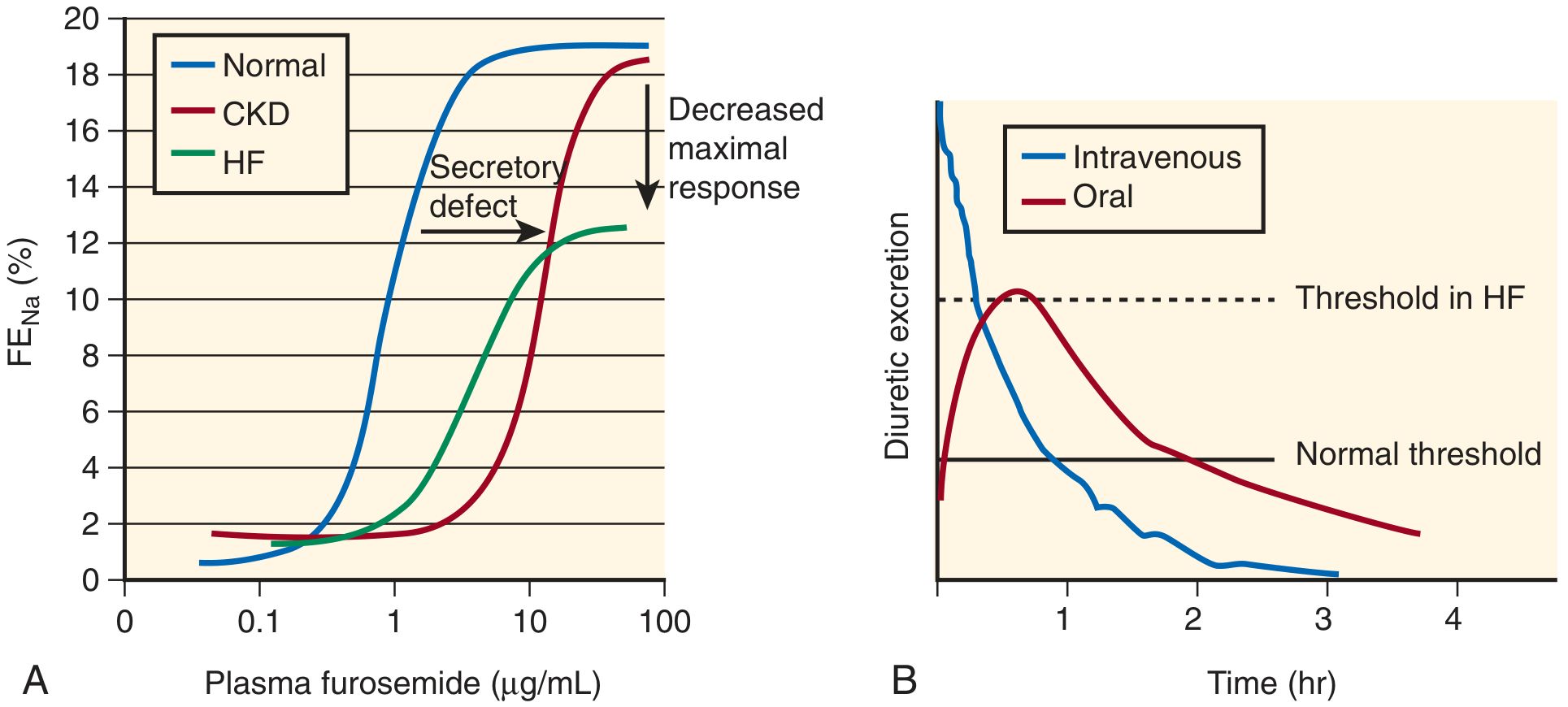
CKD = rightward shift (impaired tubular secretion). HF = rightward AND downward shift (secretory defect + reduced maximal response). This patient has BOTH - diuretic resistance is expected and doses must be escalated.
Step 2 - Managing Diuretic Resistance
When diuresis is inadequate despite adequate IV loop diuretic doses:
- Sequential nephron blockade: Add oral metolazone 2.5-5 mg PO 30-60 min before IV loop diuretic - blocks distal tubule and synergizes; monitor K+ closely
- Switch to torsemide or bumetanide if on furosemide (more predictable kinetics)
- Continuous IV infusion of loop diuretic
- Add low-dose aldosterone antagonist (spironolactone 12.5-25 mg if K+ <5.0)
Step 3 - IV Vasodilators (if SBP >100 mmHg)
| Drug | Role |
|---|---|
| IV Nitroglycerin | 5-10 mcg/min, titrate to 200 mcg/min; primarily reduces preload; first-line in acute pulmonary edema; fast onset |
| Sodium nitroprusside | Hypertensive ADHF with high afterload; requires arterial line; risk of cyanide toxicity with prolonged use in renal failure |
| Nesiritide | Recombinant BNP; vasodilator + mild natriuresis; no mortality benefit shown; use as adjunct |
C. Pacemaker-Specific Management
- Formal device interrogation (same day)
- If PICM identified (LVEF drop + high RV pacing burden + LBBB morphology):
- Upgrade to CRT-D (Cardiac Resynchronization Therapy - Defibrillator): restores LV dyssynchrony, dramatically improves LVEF, reduces HF hospitalizations and mortality
- This is the single most impactful intervention for pacing-induced HF
- If new AF detected on interrogation:
- Rate control: IV metoprolol or amiodarone (if LVEF low, avoid non-dihydropyridine CCBs)
- Anticoagulation: DOAC - dose-adjust for eGFR (apixaban preferred in renal impairment)
- If ACS triggered decompensation: Urgent cardiology review, PCI if indicated
- Rule out device pocket infection - if suspected, blood cultures, wound swab; device explant may be needed
- Do NOT place central line or pacemaker lead on same side as existing device without cardiology guidance
D. Managing AKI in the Context of ADHF
Classify AKI type first:
| Finding | Interpretation | Action |
|---|---|---|
| FENa <1% + responds to diuresis | Prerenal (low CO / high CVP) | Diurese, improve cardiac output |
| FENa >2% + rising Cr despite diuresis | Intrinsic AKI (ATN) | Supportive; review nephrotoxins |
| Urine eosinophils / interstitial casts | AIN (drug-induced) | Stop culprit drug |
| Proteinuria >1 g/day | Glomerular disease (consider renal biopsy if persistent) | Nephrology consult |
Key principles:
- Do NOT reflexively hold diuretics when creatinine rises: aggressive decongestion reduces CVP, improves renal perfusion, and improves survival even with transient creatinine rise
- Tolerate creatinine rise up to ~0.3-0.5 mg/dL if congestion is clearing and urine output is maintained
- RAAS inhibitors (ACEi/ARB): hold temporarily if K+ >5.5 or creatinine rises >50% from baseline; restart at lower dose once stable - do NOT permanently stop
- Avoid hyperkalemia - monitor K+ q6-8h; give calcium gluconate for ECG changes; insulin-dextrose for acute K+ >6.5
- Indications for urgent dialysis/CRRT:
- Refractory hyperkalemia (K+ >6.5 unresponsive to medical treatment)
- Refractory pulmonary edema (no response to IV diuretics + ultrafiltration)
- Severe metabolic acidosis (pH <7.1)
- Uremic encephalopathy or pericarditis
Ultrafiltration (if diuretic-resistant):
- Better clinical decongestion than IV loop diuretics per 2024 meta-analysis (PMID 37469222)
- Does NOT improve re-hospitalization or mortality
- Higher risk of adverse events (catheter-related)
- Reserve for truly diuretic-refractory cases
E. ALI Management (Cardiogenic Pulmonary Edema)
This patient's ALI is almost certainly hydrostatic / cardiogenic pulmonary edema (elevated PCWP causing fluid transudation into alveoli), not ARDS-type permeability edema. The distinction matters:
| Cardiogenic Pulmonary Edema | ARDS / Non-Cardiogenic ALI | |
|---|---|---|
| Mechanism | Elevated PCWP (>18 mmHg) | Increased alveolar-capillary permeability |
| CXR | Central / perihilar ("bat-wing"), cardiomegaly, effusions | Bilateral peripheral infiltrates, normal heart size |
| NT-proBNP | Markedly elevated | Can also be elevated (>3000) - overlap exists |
| Echo | Low EF or diastolic dysfunction | Usually preserved EF |
| Protein in BAL fluid | Low (transudate) | High (exudate) |
| Response to diuresis | Rapid improvement | Poor response |
Management of cardiogenic pulmonary edema:
- Upright positioning + NIV/BiPAP
- IV loop diuretics (reduce pulmonary venous pressure rapidly)
- IV nitrates (immediate preload reduction - venodilation precedes diuresis)
- If hypotensive + pulmonary edema = cardiogenic shock → dobutamine or milrinone (cautious inotropic support)
F. Inotropes - When and Which
Reserved for Cold & Wet profile (cardiogenic shock with congestion):
| Drug | Mechanism | Use |
|---|---|---|
| Dobutamine | β1-agonist; ↑CO, ↓filling pressures | First-line positive inotrope |
| Milrinone | PDE3 inhibitor; ↑CO, vasodilation | Preferred if on beta-blockers; caution - causes hypotension |
| Dopamine (low dose) | No proven renal-protective benefit in DOSE trial; avoid for renal indications alone | |
| Vasopressors (noradrenaline) | For distributive shock component (sepsis) if MAP <65 |
"IV inotropes dobutamine and milrinone are reserved for situations in which ADHF is complicated by unresponsiveness to standard therapies, diminished peripheral perfusion, end-organ dysfunction, and/or hypotension (low-output syndrome)." - Brenner & Rector's The Kidney
G. Drug Adjustments in This Patient (Elderly + AKI + PPM)
| Drug | Consideration |
|---|---|
| ACEi / ARB | Hold if K+ >5.5 or Cr rising acutely; restart at 50% dose when stable |
| ARNI (Sacubitril-Valsartan) | New 2026 meta-analysis (PMID 40689605) supports use in ADHF post-stabilization - superior to ACEi alone |
| Beta-blockers | Continue at reduced dose if tolerated (Warm & Wet); hold if Cold & Wet |
| Spironolactone / Eplerenone | Hold if K+ >5.0 or eGFR <30 |
| Digoxin | Renally cleared - toxic in AKI; check serum level; hold or dose-reduce |
| DOACs | Dose-reduce per eGFR (apixaban 2.5 mg BD if ≥2 criteria: age ≥80, weight ≤60 kg, Cr ≥1.5); avoid rivaroxaban if eGFR <30 |
| NSAIDs | Absolute contraindication - worsen HF and AKI |
| IV contrast | Avoid; if mandatory, use iso-osmolar contrast + pre/post IV hydration (isotonic saline 1 mL/kg/hr 3-12h before) |
| Aminoglycosides | Avoid; if essential, extended-interval dosing + drug levels |
H. Monitoring Goals During Admission
| Parameter | Target / Frequency |
|---|---|
| Daily weight | 0.5-1 kg/day weight loss during decongestion |
| Fluid balance | Negative balance (-1 to -2 L/day) |
| Urine output | >0.5 mL/kg/hr; >30 mL/hr minimum |
| Serum K+ | q6-8h during active diuresis; target 4.0-5.0 mEq/L |
| Creatinine / BUN | q12h; tolerate mild rise during decongestion |
| BNP/NT-proBNP | Trending down = adequate decongestion; static or rising = reassess |
| SpO2 | >92-96% on minimal O2 |
| ECG | Daily - watch for QT prolongation (electrolyte shifts), new AF |
| Pacemaker check | Formal report within 24-48h of admission |
DECISION ALGORITHM
ELDERLY MALE + PACEMAKER + ACUTE SOB + AKI + ALI
              ↓
    ┌─────────────────────────────────┐
    │  IMMEDIATE (0-15 min)           │
    │  ABG, ECG, CXR, POCUS           │
    │  Sit up, O2/BiPAP               │
    └─────────────────────────────────┘
              ↓
    NT-proBNP + Troponin + BNP
    + Creatinine (compare baseline)
    + Echo (LVEF, IVC, B-lines)
              ↓
    ┌──────────────────────┬──────────────────────────┐
    │  ADHF (CRS Type 1)   │  Acute-on-CKD primary    │
    │  BNP high, EF low,   │  Prior CKD, normal echo, │
    │  B-lines+, IVC full  │  urinary casts, nephrotoxin│
    └──────────────────────┴──────────────────────────┘
              ↓
    PACEMAKER INTERROGATION
              ↓
    ┌──────────────────────────────────┐
    │ PICM? → CRT-D upgrade            │
    │ New AF? → Rate control + DOAC    │
    │ ACS? → Cardiology / PCI          │
    │ Sepsis? → Cultures + antibiotics │
    └──────────────────────────────────┘
              ↓
    IV DIURETICS + IV NITRATES
    (Warm & Wet: diurese + vasodilate)
    (Cold & Wet: inotrope + cautious diuresis)
              ↓
    Daily: Weight, Cr, K+, NT-proBNP trend
    Restart RAAS/BBlocker when stable
    Transition to oral diuretics when euvolemic
SUMMARY OF KEY PRINCIPLES
- Classify hemodynamic profile first (Warm/Cold × Wet/Dry) - guides initial therapy
- Pacemaker interrogation is non-negotiable - PICM, AF, lead failure must be excluded
- NT-proBNP is the diagnostic anchor for ADHF; interpret with caution in CKD (falsely elevated) - trend matters more than a single value
- Elevated CVP damages the kidney from the venous side - decongestion is renoprotective even when creatinine transiently rises
- IV torsemide or bumetanide preferred over furosemide in HF+CKD (more predictable kinetics, less diuretic resistance)
- Sequential nephron blockade (metolazone + loop diuretic) for diuretic-resistant cases
- NIV/BiPAP for cardiogenic ALI - reduces intubation rates
- Hold nephrotoxins (NSAIDs, contrast, aminoglycosides); temporarily hold RAAS inhibitors if AKI is worsening
- Dialysis/CRRT if refractory hyperkalemia, fluid overload, acidosis, or uremia
- Post-stabilization: restart ACEi/ARB/ARNI (sacubitril-valsartan now supported by 2026 meta-analysis, PMID 40689605), beta-blocker, MRA; consider CRT-D if PICM; DOAC if AF
Sources: Brenner & Rector's The Kidney (2-Vol Set) | Fuster & Hurst's The Heart, 15th Ed. | Murray & Nadel's Respiratory Medicine | Harrison's Internal Medicine 22E | Goldman-Cecil Medicine | PubMed Meta-Analyses 2024-2026
This is a shared conversation. Sign in to Orris to start your own chat.