
analyse_media
Reading File
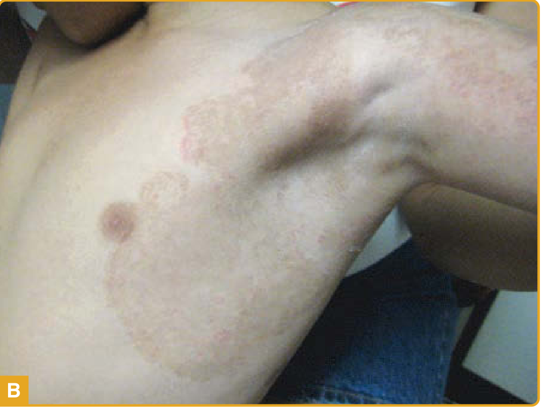
The image shows a well-demarcated, oval plaque on the lateral torso of what appears to be a child, with a central hyperpigmented/grayish area, a collarette of peeling scale at the border, and reddish-brown edges - strongly suggestive of a resolving ringworm (tinea corporis) or possibly a resolving burn/impetigo lesion.
Searching sources...
Finding Sources
Finding Sources
Searching Images
tinea corporis ringworm annular scaly plaque child skin
Reading File
Finding Sources
Loading Image
Reading File
Clinical Assessment
Most likely diagnosis: Tinea Corporis (Ringworm)
The lesion in the image is highly consistent with tinea corporis - a superficial dermatophyte infection of glabrous (non-hairy) skin. Here is the breakdown:
What the Image Shows
| Feature | Observation |
|---|---|
| Shape | Well-demarcated, oval/annular plaque |
| Center | Hyperpigmented, grayish-brown, dry - showing central clearing/resolution |
| Border | Raised, reddish-brown erythematous rim |
| Surface | Classic "collarette of scale" - peeling epidermal tissue at the periphery |
| Location | Lateral torso/flank, above waistline |
| Patient | Appears to be a child |
This matches textbook description almost exactly: "The classic presentation is that of an annular ('ringworm'-like) or serpiginous plaque with scale across the entire active erythematous border. The border advances centrifugally. The center of the plaque is usually scaly but may exhibit complete clearance." - Fitzpatrick's Dermatology
Here is a textbook reference image of tinea corporis for comparison:
Why Tinea Corporis?
- Annular morphology with central clearing and an active, scaly, raised border - the hallmark
- Collarette of peeling scale at the periphery (desquamating epidermal edge)
- Location on glabrous skin (torso/flank) - by definition tinea corporis is dermatophytosis of glabrous skin excluding palms, soles, and groin
- Child patient - children commonly acquire zoophilic strains (e.g., Microsporum canis from dogs/cats); common organisms also include Trichophyton rubrum and T. tonsurans
- The central hyperpigmentation with dry, slightly shiny surface suggests the lesion is in a resolving or partially treated phase
Differential Diagnosis to Consider
| Category | Conditions |
|---|---|
| Most Likely | Tinea corporis, nummular eczema, erythema annulare centrifugum |
| Consider | Psoriasis (plaque), contact/atopic dermatitis, pityriasis rosea |
| Rule Out | Subacute cutaneous lupus (in adults), secondary syphilis, mycosis fungoides |
Recommended Management
Confirm with: KOH preparation (potassium hydroxide scraping of the scaly border) - will show fungal hyphae. Wood's lamp may fluoresce with M. canis.
For an isolated plaque (topical antifungal - first line):
- Terbinafine 1% cream - apply twice daily for 1-2 weeks (allylamine - fungicidal)
- Clotrimazole 1% cream - apply twice daily for 2-4 weeks (imidazole)
- Miconazole 2% cream - twice daily for 2-4 weeks
- Extend 1-2 cm beyond the visible border; treat for at least 1-2 weeks after clinical resolution
Oral antifungals (if widespread, inflamed, or unresponsive to topicals):
- Terbinafine 250 mg/day x 2-4 weeks
- Griseofulvin (especially in children, particularly for M. canis)
- Itraconazole or fluconazole as alternatives
(Source: Fitzpatrick's Dermatology; Goldman-Cecil Medicine; Andrews' Diseases of the Skin)
Important Notes
- Avoid applying topical corticosteroids alone - this can suppress inflammation and mask the infection ("tinea incognito"), worsening the underlying fungal infection
- Keep the skin dry; avoid occlusive clothing
- Check household pets or close contacts for infection sources (zoophilic transmission in children)
- If no improvement after 2-4 weeks of topical treatment, refer to dermatology
Disclaimer: This is a clinical image analysis for educational purposes. A definitive diagnosis requires in-person examination, KOH microscopy, and/or fungal culture. Please consult a healthcare provider for treatment decisions.
This is a shared conversation. Sign in to Orris to start your own chat.