
Toxic shock syndrome
toxic shock syndrome rash clinical presentation
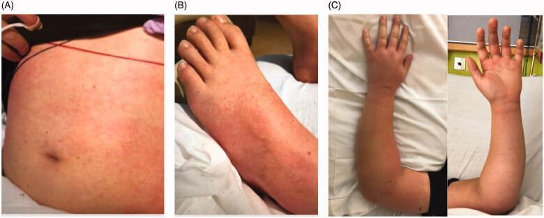
This composite clinical photograph displays the dermatological and musculoskeletal presentation of a pediatric patient with systemic illness, likely Toxic Shock Syndrome secondary to Group A Streptococcus infection. Panel A shows the abdomen/trunk with a diffuse, erythematous macular rash consisting of non-elevated, blanching red spots. Panel B illustrates similar macular findings on the dorsal surface of the foot and lower leg. Panel C provides two views (dorsal and volar) of the left upper extremity, demonstrating significant localized edema (swelling) of the forearm, wrist, and hand. The skin of the forearm appears erythematous and tense, though no overt skin breaks or cellulitis are visible at the time of presentation. This combination of findings—a diffuse scarlatiniform-like rash and localized extremity swelling—is clinically significant for identifying early signs of invasive soft tissue infection and systemic inflammatory response.
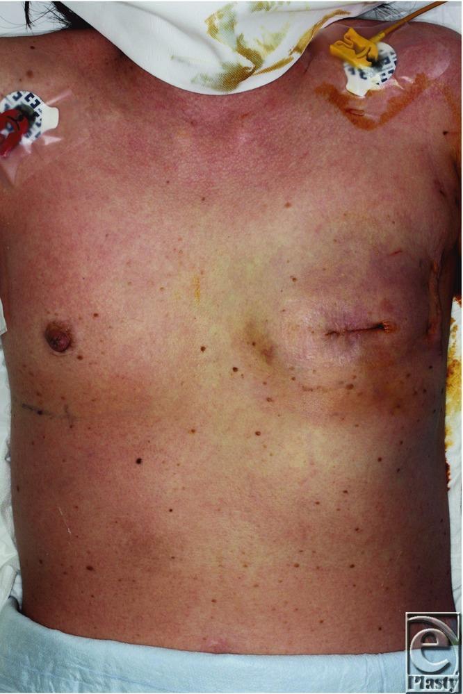
Clinical photograph of a 54-year-old female patient's anterior torso demonstrating a diffuse, erythematous macular rash on the chest and abdomen. The skin exhibits confluent redness, characteristic of a systemic inflammatory or toxic response. Anatomically, the left breast region shows evidence of recent surgery (mastectomy/reconstruction) with a visible surgical incision and suture lines. On the right and left infraclavicular regions, central venous access devices or monitoring lines are present, secured with adhesive dressings. Notably, the right-sided insertion site shows peri-catheter reddish-brown staining. Multiple small, benign-appearing pigmented nevi are scattered across the trunk. This visual presentation, in the context of postoperative fever and hypotension, is clinically representative of Toxic Shock Syndrome (TSS) following breast surgery and tissue expander placement. The image serves as an educational example of early-onset postoperative TSS manifestations, emphasizing the appearance of the classic diffuse scarlatiniform eruption.
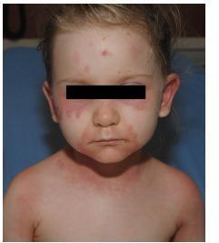
This clinical photograph shows the facial and upper chest region of a pediatric patient presenting with an extensive, symmetrically distributed dermatological eruption. The primary visual findings include a confluent, erythematous, and macular rash with an edematous appearance. The distribution is prominently periorbital (involving the infraorbital and malar areas) and perioral, with extension onto the cheeks and chin. Scattered erythematous macules are also visible on the forehead. The rash continues inferiorly onto the neck and upper chest, where it appears more diffuse and confluent. The skin in the affected areas shows signs of mild swelling without evident deep ulceration or mucosal involvement in the visible oral cavity. This clinical presentation is characteristic of systemic inflammatory or infectious processes such as Staphylococcal Scalded Skin Syndrome (SSSS), early Stevens-Johnson Syndrome (SJS), or toxic shock syndrome, where characteristic periorificial accentuation and superficial skin desquamation are common educational hallmarks.
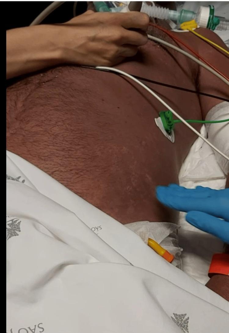
Clinical photograph of a patient's torso in a critical care setting, demonstrating an exuberant, diffuse erythematous rash. The skin exhibits a widespread, uniform reddish discoloration across the chest and abdomen. A notable feature is the blanching nature of the erythema, where the redness temporarily disappears under digital pressure, as demonstrated by the presence of a gloved hand and lighter patches in the lower left abdominal region. The rash appears macular and confluent without obvious vesicles, papules, or petechiae. Medical monitoring equipment, including ECG leads and tubing, is visible on the patient, indicating an acute clinical presentation. This visual manifestation is associated with streptococcal shock or toxic shock syndrome (TSS), representing a systemic inflammatory response. The image is used to teach medical students and residents about the dermatologic signs of sepsis and the physical examination technique of blanching in capillary-refill assessment.
"toxic shock syndrome"[MeSH Terms] AND management
Toxic Shock Syndrome (TSS)
Definition and Overview
- Rosen's Emergency Medicine, p. 2603
- Dermatology 2-Volume Set 5e, p. 1522
Etiology
| Feature | Staphylococcal TSS | Streptococcal TSS |
|---|---|---|
| Organism | Staphylococcus aureus | Group A Streptococcus pyogenes |
| Typical patient | Young (15-35 yrs), healthy | Young (20-50 yrs), healthy |
| Diffuse erythroderma | Very common | Less common |
| Soft tissue infection | Rare | Common (necrotizing fasciitis) |
| Blood culture positive | <15% | >50% |
| Mortality | <3% | 30-60% |
| Key predisposing factors | Tampon, surgical packing, abscess | Lacerations, bites, bruises, varicella |
Pathophysiology
- They bind directly to MHC class II molecules on antigen-presenting cells AND to the V-beta region of T-cell receptors
- This activates up to 20-30% of all T cells simultaneously (vs. <0.01% in a normal immune response)
- Massive release of cytokines: TNF-alpha, interleukins (IL-1, IL-2, IL-6)
- This cytokine storm drives systemic vasculitis, capillary leak, hypotension, and multisystem organ failure
- Rosen's Emergency Medicine, p. 2603-2604
Risk Factors
- Use of superabsorbent tampons
- Postoperative wound infections / nasal packing / surgical mesh
- Postpartum period
- Burns, dialysis catheters
- Common bacterial infections
- Influenza A or varicella infection
- Diabetes mellitus, HIV, chronic cardiac/pulmonary disease
- Ethanol abuse
- Cancer
- NSAID use (may mask symptoms rather than directly predispose)
Clinical Features
Diagnostic Criteria
Staphylococcal TSS (CDC Case Definition - Revised)
- Fever: Temperature >38.9°C
- Rash: Diffuse macular erythroderma
- Desquamation: 1-2 weeks after onset, especially palms and soles
- Hypotension: Systolic BP <90 mmHg (adults) or <5th percentile for age (children <16 yr)
- Multisystem involvement - 3 or more of:
- GI: Vomiting or diarrhea at onset
- Muscular: Severe myalgia or CK ≥2x upper limit of normal
- Mucous membranes: Vaginal, oropharyngeal, or conjunctival hyperemia
- Renal: BUN or creatinine ≥2x ULN or pyuria without UTI
- Hepatic: Bilirubin, AST, ALT ≥2x ULN
- Hematologic: Platelets <100,000/mm³
- CNS: Disorientation or altered consciousness without focal neurologic signs (when fever and hypotension absent)
- Negative cultures for other causes (blood, throat, CSF); negative serology for RMSF, leptospirosis, measles
Streptococcal TSS (CDC Case Definition)
- Isolation of group A streptococcus from a sterile site (DEFINITE) or non-sterile site (PROBABLE)
- PLUS Hypotension (SBP ≤90 mmHg)
- PLUS 2 or more of: renal impairment, coagulopathy/DIC, liver impairment, ARDS, generalized erythematous rash ± desquamation, soft tissue necrosis (necrotizing fasciitis, myositis, gangrene)
Differential Diagnosis
- Kawasaki disease (especially in children)
- Scarlet fever
- Staphylococcal scalded skin syndrome (SSSS)
- Early toxic epidermal necrolysis (TEN)
- Rocky Mountain spotted fever
- Leptospirosis
- Meningococcemia
Treatment
1. Aggressive Resuscitation
- IV fluid replacement - large volumes for septic shock physiology
- Vasopressors (norepinephrine first-line) for refractory hypotension
- Ventilatory support as needed (ARDS is common)
2. Source Control
- Remove the focus immediately: tampon, nasal packing, surgical mesh, barrier contraceptives
- Drain abscesses; surgical debridement for necrotizing fasciitis (streptococcal TSS)
3. Antibiotics (empirical, cover both Staph and Strep including MRSA)
- Clindamycin - preferred adjunct because it suppresses toxin/protein synthesis (anti-ribosomal), reducing ongoing cytokine drive
- Vancomycin - for MRSA coverage
- Linezolid - alternative; also suppresses protein synthesis
- Beta-lactamase-resistant penicillins (nafcillin) or first-generation cephalosporins for MSSA
- Broad-spectrum options: piperacillin-tazobactam, imipenem/meropenem
4. Adjuncts
- IV Immunoglobulin (IVIg) - neutralizes circulating superantigen toxins; used in severe/refractory cases
- Low-dose corticosteroids - considered in severe shock unresponsive to antibiotics and vasopressors
Prognosis and Complications
- Decreased renal function
- Prolonged weakness and fatigue
- Protracted myalgias
- Vocal cord paralysis, upper extremity paresthesias
- Carpal tunnel syndrome
- Arthralgias
- Amenorrhea
- Gangrene (in streptococcal TSS)
- Telogen effluvium (hair loss) in severe cases