
Introduction, morphology, lifecycle, pathogenesis, lab diagonos, about cyclospora
Reading File
Now I'll search the medical library database for Cyclospora content, running parallel queries:
Finding Sources
Finding Sources
Excellent results. Now let me read the key sections in parallel:
Reading File
Reading File
Reading File
Let me read more from the Sleisenger book (diagnosis section) and the Tietz lab textbook in parallel:
Reading File
Reading File
Reading File
Excellent content gathered. Let me also read the Tietz lab book's full microscopy/lab diagnosis section and fetch the comparison image:
Reading File
Loading Image
I now have comprehensive information from multiple authoritative textbooks. Here is the full, detailed answer:
Cyclospora cayetanensis - A Comprehensive Overview
1. Introduction
Cyclospora cayetanensis is an obligate intracellular coccidian protozoan parasite belonging to the phylum Apicomplexa. It is the only species of the genus Cyclospora known to infect humans.
-
Taxonomy: Related to Cryptosporidium parvum, Cystoisospora spp., Toxoplasma gondii, and Eimeria. Ribosomal DNA phylogenetic analysis shows it is most closely related to Eimeria.
-
Historical Discovery: First reported as a cause of human disease by Ashford in 1979, in Papua New Guinea, as an "unnamed coccidian parasite." It came to wider attention in the 1980s-1990s as a cause of protracted diarrhea in AIDS patients and in immunocompetent travelers. In 1993, Ortega et al. at Cayetano Heredia University in Peru formally classified it as Cyclospora cayetanensis, confirmed by electron microscopy.
-
Public Health Significance: A nationally notifiable disease in the United States. Major foodborne outbreaks continue to be reported annually, linked to produce imported from Latin America (raspberries, mesclun lettuce, basil, pre-prepared salads).
-
Hosts: Humans appear to be the primary (and possibly only) host. Cyclospora oocysts have been isolated from chickens, but animal reservoirs are poorly understood.
-
Medical Microbiology 9e, p. 809
-
Sleisenger and Fordtran's Gastrointestinal and Liver Disease, p. 2256
2. Morphology
Oocysts (the diagnostic stage)
| Feature | Cyclospora | Cryptosporidium | Cystoisospora |
|---|---|---|---|
| Shape | Spherical | Spherical | Elliptical |
| Size | 8-10 μm | 4-7 μm (smaller) | 15-30 μm (much larger) |
| Sporulation state when shed | Unsporulated | Fully sporulated (immediately infectious) | Unsporulated |
| Sporocysts per oocyst | 2 | None (4 naked sporozoites) | 2 |
| Sporozoites per sporocyst | 2 | - | 4 |
| Total sporozoites | 4 | 4 | 8 |
-
The oocysts of Cyclospora are spherical, ~8-10 μm, approximately twice the size of Cryptosporidium oocysts (4-5 μm) - this size difference is the key morphological distinguishing feature.
-
Each mature oocyst contains two sporocysts, each containing two sporozoites. Sporozoites contain a membrane-bound nucleus and micronemes (characteristic of Sporozoans).
-
Oocysts contain visible internal granules.
-
When passed in stool, the oocysts are unsporulated (non-infectious morula stage).
-
On modified acid-fast stain: round, slightly wrinkled, deep pink (variable staining) structures, with some appearing as unstained "ghost" cells (variable acid-fastness is a key feature differentiating it from Cryptosporidium, which stains uniformly).
-
Autofluorescence: Striking blue-green autofluorescence under UV fluorescence microscopy - this initially caused confusion with cyanobacteria (blue-green algae), hence the organism's early names like "cyanobacterium-like bodies" or "CLB."
-
Medical Microbiology 9e, p. 809; Sherris & Ryan's Medical Microbiology, p. 1738; Tietz Textbook of Laboratory Medicine, 7th Edition, p. 3573
3. Life Cycle
The life cycle is entirely within a single host (monoxenous), similar to Cryptosporidium, but with a critical environmental maturation step.
Step-by-Step:
- Excretion: Unsporulated (non-infectious) oocysts are shed in the feces of infected humans.
- Environmental Sporulation: Outside the host, sporulation requires 1-2 weeks at temperatures of 22-32°C. This produces mature oocysts containing 2 sporocysts, each with 2 sporozoites. This delay is why direct person-to-person fecal-oral transmission does NOT occur (unlike Cryptosporidium).
- Ingestion: Humans are infected by consuming sporulated oocysts in fecally contaminated food or water.
- Excystation: In the small bowel (primarily jejunum), sporulated oocysts excyst, releasing sporozoites.
- Asexual Multiplication (Merogony):
- Sporozoites invade epithelial cells of the small intestine and reside in cytoplasmic vacuoles.
- They develop into Type I merozoites, which produce Type II merozoites.
- Sexual Multiplication (Gametogony):
- Type II merozoites differentiate into microgametocytes (male) and macrogametocytes (female).
- A microgamete fertilizes a macrogamete to form a zygote.
- Oocyst Formation (Sporogony):
- The zygote develops into an unsporulated thick-walled oocyst, which is released into the intestinal lumen and shed in stool.
- The cycle restarts in the environment.
- Medical Microbiology 9e, p. 809; Tietz Textbook of Laboratory Medicine, p. 3573; Jawetz Melnick & Adelberg's Medical Microbiology 28e, p. 879
4. Pathogenesis
Target Site
The organism primarily infects the upper small bowel (jejunum), residing within cytoplasmic vacuoles of jejunal epithelial cells.
Histopathological Changes
- Villous blunting/atrophy (shortening of intestinal villi)
- Crypt hyperplasia
- Diffuse edema of the lamina propria
- Inflammatory cell infiltrate in the lamina propria (mild)
- These changes lead to a malabsorptive diarrhea pattern.
Key Pathogenic Features
- Incubation period: ~1 week (7-14 days after ingestion of sporulated oocysts)
- Mechanism of diarrhea: Villous atrophy and mucosal damage impair absorptive surface area. Inflammatory changes disrupt normal epithelial function.
- The exact molecular mechanisms are less well-studied than for Cryptosporidium, but appear similar.
- Unlike Cryptosporidium, which is immediately infectious when shed, Cyclospora cannot auto-infect the same host or cause hyperinfection - this limits its severity in most immunocompetent patients.
- In immunocompromised patients (HIV/AIDS): Infection can be prolonged, relapsing, and associated with acalculous cholecystitis and significant weight loss (up to 10% of body weight). Rare dissemination to the biliary tract and possibly respiratory tract has been reported.
Clinical Features
-
Profuse, "explosive" watery diarrhea - the hallmark symptom
-
Abdominal cramps and bloating
-
Nausea, anorexia
-
Fatigue and malaise (often more severe than with Cryptosporidium)
-
Flatulence
-
Low-grade fever
-
Weight loss
-
Duration: 1-3 weeks in immunocompetent individuals; can last months in immunocompromised
-
In endemic regions: may be asymptomatic (acquired immunity with repeated exposure); symptomatic disease more common in children and travelers from non-endemic areas.
-
Sleisenger and Fordtran's, p. 2256-2257; Jawetz Melnick, p. 884; Tietz, p. 3573
5. Laboratory Diagnosis
A. Microscopy (Gold Standard)
Modified Acid-Fast Stain (Kinyoun or Ziehl-Neelsen):
- Oocysts appear as round, slightly wrinkled, deep pink structures (8-10 μm)
- Staining is variable - some oocysts stain intensely red-pink, others appear as unstained "ghost" cells or lightly staining forms
- This variability is characteristic and helps distinguish Cyclospora from Cryptosporidium (which stains uniformly)
- Internal granules are commonly visible
- Must measure size using a well-calibrated ocular micrometer to differentiate from Cryptosporidium (4-5 μm)
Below: Side-by-side acid-fast stain comparison - Cryptosporidium (left) vs. Cyclospora (right, larger oocysts):
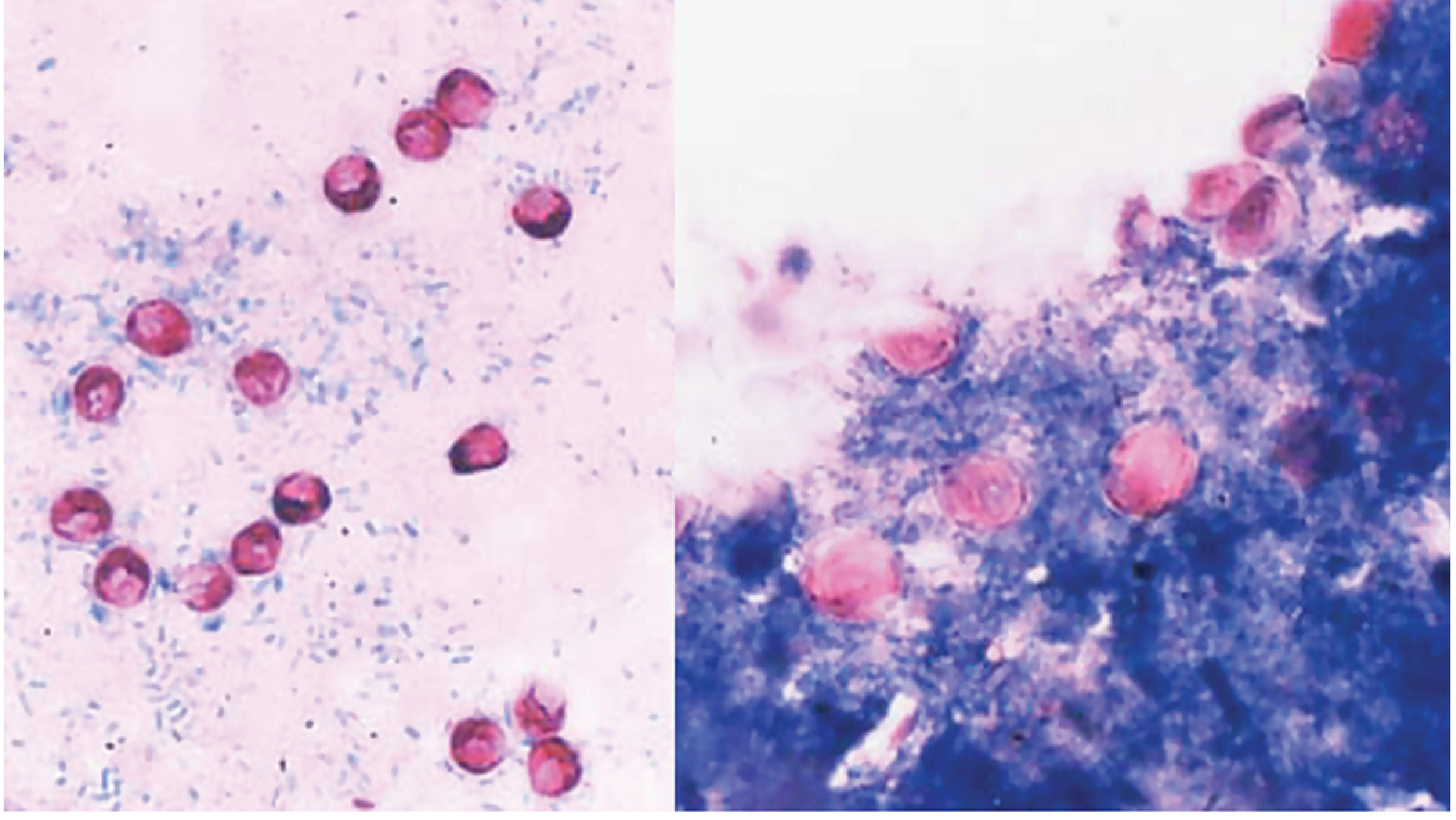
Cyclospora oocysts (right panel) are larger (~8-10 μm) with variable pink staining and internal granules. Cryptosporidium (left) stains more uniformly and is smaller.
B. Fluorescence Microscopy (UV Autofluorescence)
- Cyclospora oocysts exhibit striking blue-green autofluorescence under UV light
- This is a highly useful, specific property that aids rapid screening
- Does not require special staining reagents
C. Wet Preparation
- Unstained oocysts may be visible as refractile, wrinkled spheres on direct wet preparation, but sensitivity is low
D. Molecular Diagnostics (NAAT)
- PCR-based assays: Increased sensitivity compared to microscopy
- BioFire GI Panel: Commercially available multiplex PCR panel that targets C. cayetanensis, with reported sensitivity and specificity of 100%. This panel demonstrated utility in the 2013 US outbreak, identifying cases 1 week earlier than conventional microscopy in a validation study.
E. Histology
- C. cayetanensis may be seen within enterocyte cytoplasm on standard H&E sections of small intestinal biopsy. Both asexual and sexual developmental stages may be visible.
Important Laboratory Considerations
-
Routine stool O&P (ova and parasite) exam will miss Cyclospora - specific requests for Cyclospora detection are required
-
Multiple stool samples should be examined (oocysts may be shed intermittently)
-
Size measurement is essential to differentiate from Cryptosporidium
-
Cyclospora responds to TMP-SMX, while Cryptosporidium does not - making correct identification clinically important
-
Tietz Textbook of Laboratory Medicine, p. 3573-3574; Henry's Clinical Diagnosis and Management by Laboratory Methods; Jawetz Melnick, p. 886
6. Treatment & Prevention (Brief)
| Aspect | Detail |
|---|---|
| Drug of Choice | Trimethoprim-Sulfamethoxazole (TMP-SMX) 160/800 mg twice daily for 7 days |
| HIV/AIDS patients | Extended treatment + maintenance therapy (TMP-SMX 3x/week) to prevent relapse |
| Alternatives | Ciprofloxacin, Nitazoxanide |
| Key point | Unlike Cryptosporidium, Cyclospora is readily treatable even in immunocompromised patients |
| Prevention | Avoid contaminated water/produce; boiling water; chlorination alone is insufficient (chlorine-resistant) |
Summary Table: Cyclospora vs. Cryptosporidium
| Feature | Cyclospora cayetanensis | Cryptosporidium parvum |
|---|---|---|
| Oocyst size | 8-10 μm | 4-5 μm |
| Sporulation state when shed | Unsporulated | Fully sporulated |
| Person-to-person transmission | No | Yes (common) |
| Acid-fast staining | Variable (+ and -) | Uniform + |
| Autofluorescence | Blue-green under UV | Present |
| Treatment | TMP-SMX (effective) | No effective treatment |
| HIV/AIDS disease | Prolonged, relapsing | Fulminating |
Sources: Medical Microbiology 9e; Sleisenger & Fordtran's GI and Liver Disease; Jawetz Melnick & Adelberg's Medical Microbiology 28e; Sherris & Ryan's Medical Microbiology, 8th ed.; Tietz Textbook of Laboratory Medicine, 7th ed.
This is a shared conversation. Sign in to Orris to start your own chat.