
anticoagulents (warferrin)
Reading File
Warfarin is a vitamin K antagonist (VKA) used for anticoagulation - it inhibits the synthesis of clotting factors II, VII, IX, and X by blocking vitamin K epoxide reductase. It's monitored via the INR, with typical therapeutic range of 2.0-3.0.
Searching sources...
Finding Sources
Finding Sources
Searching Images
warfarin mechanism of action vitamin K clotting factors
Good - I have multiple excellent sources. Let me now read the detailed content from key textbooks.
Reading File
Reading File
Reading File
Excellent content retrieved. Let me now fetch the mechanism diagram and read the sections on dosing, monitoring, adverse effects, and drug interactions.
Loading Image
Reading File
Reading File
Now let me read the dosing, monitoring, drug interactions, and reversal sections from Harrison's:
Reading File
I now have comprehensive information. Let me compile the complete answer.
Warfarin (Anticoagulant) - Complete Overview
1. Classification & Background
Warfarin is a vitamin K antagonist (VKA) - a water-soluble coumarin derivative originally developed as a rodenticide that became one of the most widely prescribed oral anticoagulants in the world. It is a racemic mixture of R- and S-enantiomers, with S-warfarin being approximately 5x more pharmacologically active than the R-form.
2. Mechanism of Action
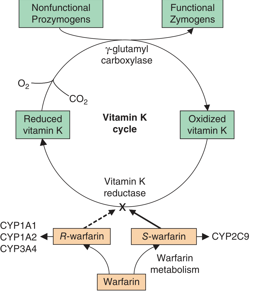
Figure: Warfarin blocks Vitamin K epoxide reductase, preventing recycling of vitamin K. This stops gamma-carboxylation of clotting factors, producing nonfunctional prozymogens instead of functional zymogens.
The mechanism involves the Vitamin K cycle:
- Vitamin K (from diet) is reduced to vitamin K hydroquinone (active form) by vitamin K reductase
- Vitamin K hydroquinone acts as a cofactor for gamma-glutamyl carboxylase, which adds a carboxyl group to glutamic acid residues on clotting factors (gamma-carboxylation)
- This gamma-carboxyglutamic acid modification is essential - it allows calcium-dependent binding of clotting factors to anionic phospholipid surfaces (the site of coagulation)
- During this process, vitamin K hydroquinone is oxidized to vitamin K epoxide, which is recycled back to reduced vitamin K by vitamin K epoxide reductase (VKOR)
Warfarin blocks VKOR (encoded by the VKORC1 gene), preventing regeneration of active vitamin K. This depletes the cofactor needed for gamma-carboxylation, resulting in the synthesis of clotting factors with little or no biological activity (nonfunctional prozymogens).
Factors affected: II (prothrombin), VII, IX, X - and anticoagulant proteins C and S are also reduced.
Key point: The onset of action is delayed (72-96 hours) because warfarin does not neutralize already-circulating clotting factors. It must wait for the existing active factors to be cleared. The antithrombotic effect specifically requires depletion of factor X (t½ = 24 h) and prothrombin (t½ = 72 h).
3. Pharmacokinetics
| Parameter | Detail |
|---|---|
| Bioavailability | ~100% (rapidly and almost completely absorbed orally) |
| Peak blood levels | ~90 minutes after oral administration |
| Plasma half-life | 36-42 hours (racemic mixture) |
| Protein binding | >97% bound to albumin (only free fraction is active) |
| Metabolism | Liver - CYP2C9 metabolizes S-warfarin; R-warfarin via CYP1A1, CYP1A2, CYP3A4 |
| Excretion | Urine and feces (as inactive glucuronide conjugates) |
Warfarin crosses the placenta and can cause fetal abnormalities. It does NOT cross into CSF or breast milk significantly because of its high albumin binding.
4. Therapeutic Uses
- Atrial fibrillation - stroke prevention
- DVT and PE - treatment and prevention of recurrence
- Prosthetic heart valves (mechanical valves require lifelong anticoagulation)
- Venous thromboembolism prophylaxis post-orthopedic surgery
- Protein C/S deficiency and antiphospholipid syndrome
5. Monitoring: INR
Warfarin has a narrow therapeutic window and requires frequent INR (International Normalized Ratio) monitoring.
| Indication | Target INR |
|---|---|
| Most indications (AF, DVT, PE) | 2.0 - 3.0 |
| Mechanical heart valves (high risk) | 2.5 - 3.5 |
- Stable patients: INR every 3-4 weeks
- Any new medication added: more frequent monitoring required
- The INR reflects the extrinsic pathway (factors II, VII, X)
6. Dosing
- Typical starting dose: 2-5 mg/day orally (individual variation is large)
- Bridging therapy: Because of the delayed onset, patients with established thrombosis or high thromboembolic risk require concomitant parenteral anticoagulation (heparin, LMWH, or fondaparinux) for at least 5 days until INR is therapeutic
- Doses are adjusted based on INR response, genetics, and interacting drugs
7. Genetics and Pharmacogenomics
Two key gene polymorphisms significantly influence warfarin dosing:
CYP2C9 variants (affect warfarin metabolism):
- CYP2C9*2 and 2C93 encode enzymes with reduced activity
- ~25% of Caucasians carry at least one variant allele
- Heterozygous carriers need 20-30% dose reduction; homozygous carriers need 50-70% reduction
- Variant carriers have ~2x higher bleeding risk
VKORC1 variants (affect warfarin sensitivity):
- Account for ~30% of warfarin dose variability
- Higher prevalence in Asians > Caucasians > African Americans
- A/A haplotype carriers require the lowest doses
8. Drug Interactions
Warfarin has numerous drug interactions that can potentiate or reduce its effect:
Drugs that INCREASE INR (increase bleeding risk):
- Broad-spectrum antibiotics (reduce gut vitamin K-producing bacteria)
- Fluconazole, metronidazole, amiodarone (CYP2C9 inhibitors)
- Aspirin / NSAIDs (additive bleeding risk + displace warfarin from albumin)
- Sulfonamides (displace warfarin from albumin binding site)
Drugs that DECREASE INR (reduce anticoagulant effect):
- Rifampicin, carbamazepine, phenytoin (CYP inducers)
- Cholestyramine (reduces warfarin absorption)
Food interactions:
- Vitamin K-rich foods (green leafy vegetables, broccoli, spinach) reduce warfarin efficacy
- Grapefruit can inhibit CYP metabolism
- Patients should be counseled to maintain consistent dietary vitamin K intake rather than eliminate it
9. Adverse Effects
| Effect | Details |
|---|---|
| Bleeding (major) | Most common - ranges from minor (epistaxis, hematuria) to life-threatening (intracranial, GI, retroperitoneal) |
| Skin necrosis | Rare, occurs 2-5 days after initiation; seen in protein C/S deficiency; thrombi in microvasculature of fatty tissues (thighs, buttocks, breasts, toes) |
| Purple toe syndrome | Rare; painful blue-tinged discoloration from cholesterol emboli |
| Teratogenicity | Warfarin is contraindicated in pregnancy - crosses placenta, causes fetal warfarin syndrome (nasal hypoplasia, stippled epiphyses, CNS defects) |
10. Management of Elevated INR / Bleeding
| INR | Action |
|---|---|
| INR 3.5-10, no bleeding | Withhold warfarin; monitor; allow INR to return to range |
| INR >10, no bleeding | Oral vitamin K 2.5-5 mg |
| Serious/life-threatening bleeding | IV vitamin K 5-10 mg (slow infusion) + 4-factor prothrombin complex concentrate (4F-PCC) - faster than fresh frozen plasma (FFP) |
Note: High-dose vitamin K (5-10 mg) may cause temporary resistance to warfarin when it is restarted.
11. Reversal Agent
- Vitamin K (phytomenadione) is the primary antidote
- Oral vitamin K takes ~24 hours to work (requires degradation of existing clotting factors, then resynthesis)
- IV vitamin K acts faster (~6-12 hours)
- 4-factor PCC (factors II, VII, IX, X) provides immediate reversal for emergencies
- FFP can also be used if PCC is unavailable
12. Special Situations
Protein C/S deficiency: Initiating warfarin can cause paradoxical hypercoagulable state early (protein C/S fall faster than pro-coagulant factors), leading to skin necrosis. Always overlap with parenteral anticoagulation, start at low doses.
Pregnancy: Warfarin is contraindicated throughout pregnancy. Use LMWH instead. Women with mechanical heart valves on warfarin require careful individualized management.
Perioperative management: Warfarin should be bridged with LMWH for high-risk patients undergoing surgery.
13. Comparison: Warfarin vs DOACs
| Feature | Warfarin | DOACs (dabigatran, rivaroxaban, apixaban) |
|---|---|---|
| Target | Factors II, VII, IX, X (via vitamin K) | Thrombin (dabigatran) or Factor Xa |
| Monitoring | Required (INR) | Not routinely required |
| Food interactions | Significant | Minimal |
| Onset | Delayed (days) | Rapid (hours) |
| Reversal | Vitamin K, PCC | Idarucizumab (dabigatran), andexanet alfa (Xa inhibitors) |
| Mechanical valves | Yes (preferred) | Not approved |
| Cost | Low | Higher |
Sources:
- Harrison's Principles of Internal Medicine, 22nd Ed. (2025), Chapter 123, p. 994-996
- Braunwald's Heart Disease, Chapter 95
- Lippincott Illustrated Reviews: Pharmacology, Chapter 13, p. 458-461
- Goodman & Gilman's Pharmacological Basis of Therapeutics
hematinic
Hematinics are agents that promote blood formation - primarily iron, vitamin B12, and folic acid. They are used to treat various types of anemia by supplying essential nutrients for red blood cell synthesis and hemoglobin production.
Searching sources...
Finding Sources
Finding Sources
Searching Images
iron absorption mechanism hematinic anemia treatment
Finding Sources
Finding Sources
Finding Sources
Reading File
Reading File
Reading File
Reading File
Reading File
Finding Sources
Reading File
Reading File
Reading File
Reading File
Reading File
Reading File
Reading File
I now have comprehensive information to compile a thorough answer on hematinics.
Finding Sources
Searching Images
iron absorption duodenum enterocyte DMT1 ferroportin hepcidin
I have all the information needed. Here is the comprehensive answer:
Hematinics - Complete Overview
Hematinics are agents that promote the formation of blood by providing the essential building blocks required for erythropoiesis (red blood cell production) and hemoglobin synthesis. They correct nutritional or functional deficiencies that cause anemia.
Classification of Hematinics
| Category | Agents |
|---|---|
| Iron preparations | Ferrous sulfate, ferrous fumarate, ferrous gluconate, ferric carboxymaltose, iron dextran, iron sucrose |
| Vitamin B12 (Cobalamin) | Cyanocobalamin, hydroxocobalamin, methylcobalamin |
| Folic acid | Folic acid (pteroylglutamic acid), folinic acid (leucovorin) |
| Erythropoiesis-stimulating agents (ESAs) | Epoetin alfa, darbepoetin alfa |
I. IRON
Iron Metabolism and Physiology
Total body iron is approximately 2.5 g in women and 3.5 g in men:
- ~80% is in hemoglobin, myoglobin, and iron-containing enzymes (catalase, cytochromes)
- ~15-20% is in storage form as ferritin and hemosiderin (primarily in macrophages of liver, spleen, bone marrow)
- Transport form: iron is carried in plasma bound to transferrin (normally ~33% saturated)
Normal serum values:
- Serum iron: ~120 µg/dL (men), ~100 µg/dL (women)
- Total iron-binding capacity (TIBC): 300-350 µg/dL
Iron Absorption
Dietary iron exists as heme iron (from meat/poultry - ~20% absorbed) and non-heme iron (vegetables - only 1-2% absorbed). Absorption occurs in the duodenum:
- Ferric iron (Fe³⁺) is reduced to ferrous iron (Fe²⁺) by duodenal cytochrome B (a ferric reductase)
- Fe²⁺ enters the enterocyte via DMT-1 (divalent metal transporter-1) on the apical membrane
- Iron exits the enterocyte into plasma via ferroportin on the basolateral membrane
- Iron is re-oxidized to Fe³⁺ by hephaestin/ceruloplasmin before binding transferrin
Hepcidin - the master regulator of iron homeostasis:
- A small peptide secreted by the liver
- Negatively regulates ferroportin - when hepcidin is high, ferroportin is degraded, trapping iron in enterocytes and macrophages
- High in inflammation (IL-6 stimulus) → explains anemia of chronic disease
- Low in iron deficiency and high erythroid demand (erythroferrone from erythroblasts suppresses hepcidin)
Iron Deficiency Anemia
Causes:
- Chronic blood loss (most common in developed countries - GI: peptic ulcer, colon cancer; or menorrhagia)
- Poor dietary intake (most common in developing countries)
- Increased demand (pregnancy, infancy)
- Malabsorption (celiac disease, gastrectomy)
Stages of iron deficiency:
- Depletion of iron stores → falling serum ferritin, absent bone marrow iron
- Fall in serum iron + rise in TIBC (transferrin rises to compensate)
- Microcytic hypochromic anemia
Lab findings:
- Low serum ferritin (most sensitive early marker)
- Low serum iron, high TIBC, low transferrin saturation
- Peripheral smear: microcytic, hypochromic RBCs
- Thrombocytosis (platelet count often elevated)
Clinical features: Weakness, fatigue, pallor, glossitis, angular stomatitis, koilonychia (spoon nails), pica (craving non-food items such as dirt)
Treatment: Oral Iron
The preferred first-line treatment is ferrous sulfate (cost-effective, well-absorbed):
- Ferrous salts (Fe²⁺) are absorbed ~3x better than ferric salts (Fe³⁺)
- Ferrous sulfate: 325 mg tablet = 65 mg elemental iron; typical dose 150-200 mg elemental iron/day
- Other ferrous salts (fumarate, gluconate, succinate) are absorbed to approximately the same extent - the dose is based on elemental iron content
Oral iron delivers at most 40-60 mg of iron/day to the erythroid marrow with tolerable doses.
Enhancers of iron absorption:
- Vitamin C (ascorbic acid) - reduces Fe³⁺ to Fe²⁺
- Acidic environment (take on empty stomach if tolerated)
- Heme iron
Inhibitors of absorption:
- Phytates (grains, bran), tannins (tea, coffee)
- Calcium, antacids, proton pump inhibitors
- Tetracyclines, fluoroquinolones (form chelates)
Monitoring response:
- Reticulocytosis expected within 4-7 days
- Hemoglobin rise of 2 g/dL or more within 4 weeks = adequate response
- Treatment should continue for 3-6 months after normalization to replenish stores
Adverse effects: GI disturbances (nausea, constipation, dark stools), dose-related
Parenteral Iron
Used when oral iron is insufficient or not tolerated:
- Ferric carboxymaltose, iron sucrose, low-molecular-weight iron dextran, ferumoxytol
- Indicated for: malabsorption, chronic blood loss exceeding oral absorption capacity, non-compliance, IBD
- Risk of anaphylaxis (lower with newer preparations); always have resuscitation available
- Can deliver complete iron deficit in 1-2 infusions
II. VITAMIN B12 (Cobalamin)
Biochemical Roles
Vitamin B12 exists as two active coenzymes intracellularly:
1. Methylcobalamin - cofactor for methionine synthase:
- Converts homocysteine → methionine (using methyltetrahydrofolate as methyl donor)
- This reaction is critical for folate recycling: it regenerates tetrahydrofolate (THF) for DNA synthesis
- Lack of B12 "traps" folate as methylTHF (the methyl-trap hypothesis), explaining why B12 deficiency causes megaloblastic anemia
2. Deoxyadenosylcobalamin - cofactor for methylmalonyl-CoA mutase:
- Converts methylmalonyl-CoA → succinyl-CoA
- Deficiency → accumulation of methylmalonic acid (MMA) - a diagnostic marker for B12 deficiency
Absorption of Vitamin B12
- Dietary B12 is released from food protein by gastric acid and pepsin
- B12 binds haptocorrin (R-protein) in stomach
- In duodenum, pancreatic enzymes degrade haptocorrin
- B12 binds intrinsic factor (IF) secreted by gastric parietal cells
- B12-IF complex is absorbed in the terminal ileum via cubilin receptors
Deficiency - Causes
- Pernicious anemia (PA) - autoimmune destruction of gastric parietal cells → no IF → B12 malabsorption (most common cause in adults >40 yrs; 90% have anti-parietal cell antibodies, 60% anti-IF antibodies)
- Total or partial gastrectomy
- Terminal ileum disease (Crohn's, ileal resection)
- Strict vegan diet (no animal products)
- Medications: metformin, PPIs, nitrous oxide (oxidizes B12)
- Bacterial overgrowth, tapeworm (Diphyllobothrium latum)
- Pancreatic insufficiency
Clinical Features of B12 Deficiency
- Hematological: Macrocytic (megaloblastic) anemia, hypersegmented neutrophils (>5 lobes), macroovalocytes, pancytopenia
- Neurological (unique to B12, not folate):
- Subacute combined degeneration of the spinal cord (posterior columns + lateral corticospinal tracts)
- Peripheral neuropathy, paresthesias, ataxia, cognitive impairment
- Neurological damage may be irreversible if not treated promptly
- Other: Glossitis, jaundice, hyperpigmentation of skin creases
Diagnosis
- Low serum B12 (<200 pg/mL)
- Elevated methylmalonic acid (MMA) - most sensitive and specific
- Elevated homocysteine (also elevated in folate deficiency)
- Anti-IF antibodies (specific for pernicious anemia)
Key distinction: MMA is elevated only in B12 deficiency; homocysteine is elevated in BOTH B12 and folate deficiency.
Treatment
Pernicious anemia / severe malabsorption:
- IM cyanocobalamin 1000 µg/day x 7 days, then 1000 µg/week x 4 weeks, then 1000 µg/month for life
Maintenance / non-malabsorption causes:
- High-dose oral cyanocobalamin 1000-2000 µg/day (passive absorption bypasses need for IF)
Response: Reticulocytosis within 1 week; Hgb normalizes over 6-9 weeks. Neurological recovery is slower and may be incomplete.
Warning: Do NOT treat B12 deficiency with folic acid alone - it will correct the anemia but allow neurological damage to progress undetected.
III. FOLIC ACID
Biochemical Roles
Folic acid (pteroylglutamic acid) is converted to tetrahydrofolate (THF) after absorption. Key roles:
- Thymidylate synthesis: 5,10-methyleneTHF donates a methylene group to dUMP → dTMP (rate-limiting step in DNA synthesis)
- Purine synthesis: formyl-THF donates carbon atoms to the purine ring
- Methionine synthesis: with B12 as cofactor (homocysteine → methionine)
Sources and Requirements
- Rich dietary sources: fresh green vegetables (spinach, broccoli), liver, yeast, citrus fruits
- Daily requirement: ~200 µg/day (400 µg/day in pregnancy)
- Body stores: ~3-5 months (depleted much faster than B12, which lasts 3-5 years)
- Cooking destroys up to 90% of dietary folate
Deficiency - Causes
- Malnutrition (alcoholism, poverty, elderly, psychiatric patients)
- Pregnancy/lactation (3-4x increased daily requirements)
- Malabsorption (celiac disease, bariatric surgery)
- Drugs that block folate: methotrexate, trimethoprim, pyrimethamine, phenytoin, sulfasalazine, ethanol
- Increased demand: hemolytic anemia (sickle cell, thalassemia), rapid cell turnover
Clinical Features
- Macrocytic megaloblastic anemia (identical to B12 deficiency on blood film)
- Hypersegmented neutrophils
- Glossitis
- No neurological involvement (this is the key distinguishing feature from B12 deficiency)
- Neural tube defects in the fetus (if mother deficient in early pregnancy)
Treatment
- Folic acid 1 mg/day PO (5 mg/day in malabsorption)
- Duration: until deficiency corrected (usually 4 months)
- Prevention of neural tube defects: 400-800 µg/day before conception and during first trimester
- High-risk groups (hemolytic anemia, dialysis): ongoing supplementation
IV. ERYTHROPOIESIS-STIMULATING AGENTS (ESAs)
These are biologic hematinics used when the problem is insufficient erythropoietin production.
| Drug | Features |
|---|---|
| Epoetin alfa | Recombinant human EPO; SC or IV; 3x/week |
| Darbepoetin alfa | Long-acting ESA; weekly or biweekly dosing |
Mechanism: Bind erythropoietin receptor on committed erythroid progenitors in bone marrow → promote survival, proliferation, and differentiation into RBCs.
Indications:
- Anemia of chronic kidney disease (CKD)
- Chemotherapy-induced anemia
- Anemia in HIV patients on zidovudine
- Low-grade myelodysplastic syndrome
Key points:
- Always ensure adequate iron stores before/during ESA therapy (functional iron deficiency is a common cause of ESA resistance)
- Target hemoglobin: < 11 g/dL - treatment to higher levels increases risk of cardiovascular events and stroke
- Common adverse effects: hypertension, thromboembolic events, bone pain (with G-CSF)
Summary Comparison Table
| Feature | Iron | Vitamin B12 | Folic Acid |
|---|---|---|---|
| Anemia type | Microcytic, hypochromic | Macrocytic, megaloblastic | Macrocytic, megaloblastic |
| Neurological features | None | Yes (subacute combined degeneration) | None |
| Diagnostic marker | Low ferritin, low TIBC saturation | Elevated MMA + homocysteine | Elevated homocysteine only |
| Main absorption site | Duodenum | Terminal ileum (needs IF) | Upper small intestine |
| Body stores duration | Months | 3-5 years | 3-5 months |
| First-line treatment | Ferrous sulfate PO | Cyanocobalamin IM or PO | Folic acid 1 mg/day PO |
Sources:
- Goodman & Gilman's Pharmacological Basis of Therapeutics, Chapter 45
- Robbins & Kumar Basic Pathology, Chapter 10
- The Washington Manual of Medical Therapeutics
- Harrison's Principles of Internal Medicine, 22nd Ed.
This is a shared conversation. Sign in to Orris to start your own chat.