
Recess of eisler
recess of Eisler anatomy elbow joint
"recess of Eisler" OR "Eisler recess" anatomy definition
recess of Eisler orbital anatomy lateral canthus
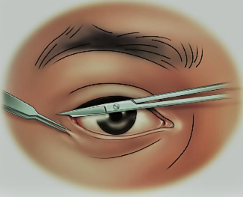
A medical illustration depicting the surgical procedure for a lateral canthotomy on a right human eye. The diagram shows the periorbital anatomy including the eyebrow, upper and lower eyelids, and the globe with a visible iris and sclera. Two primary surgical instruments are shown: a scalpel performing an initial incision at the lateral canthus (the outer corner of the eye) and iris scissors positioned to lengthen the incision. This procedure is clinically indicated for the emergency management of orbital compartment syndrome to rapidly reduce intraocular pressure and prevent optic nerve ischemia. The educational focus is on the correct anatomical placement of instruments relative to the lateral canthal tendon to achieve ocular decompression.
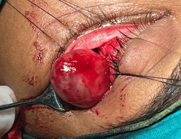
This clinical intraoperative photograph documents a surgical excision of an orbital mass via a lateral orbitotomy approach on the left eye. The image shows a well-defined, spherical, bulbous mass being mobilized from the intraconal compartment. The tumor displays a smooth, encapsulated surface with a reddish-pink, vascular appearance and areas of pale discoloration. Surgical exposure is achieved through an incision at the lateral canthus (lateral canthotomy), with black traction sutures clearly visible holding the eyelid and orbital margins apart to maintain surgical access. A surgical instrument is seen supporting and elevating the mass from the retrobulbar space, displacing surrounding orbital fat and tissues. This procedure is indicated for the management of retrobulbar tumors, such as orbital schwannomas, which often present with axial proptosis. The photograph provides educational insight into orbital surgical anatomy, the presentation of intraconal tumors, and the technique for lateral orbital decompression and tumor delivery.
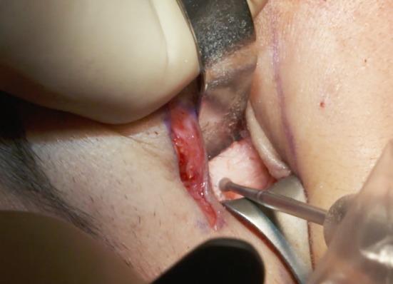
This clinical photograph captures an intraoperative view of a surgical procedure involving the lateral orbital rim. A surgical incision has been made through the skin and orbicularis muscle near the lateral canthus, with the surrounding skin marked by purple surgical ink. The surgical field is exposed using a metal retractor to provide visualization of the underlying bone. A surgical drill with a small round burr is being used to create a burr hole (identified as hole n°4) in the lateral orbital rim, just below the level of the lateral canthus. The procedure illustrates a key step in a concentric malar lift or canthopexy, where bone anchoring points are established for suture fixation. The image demonstrates the precise handling of specialized surgical instruments and the subperiosteal exposure required for mid-face rejuvenation and lower eyelid stabilization.
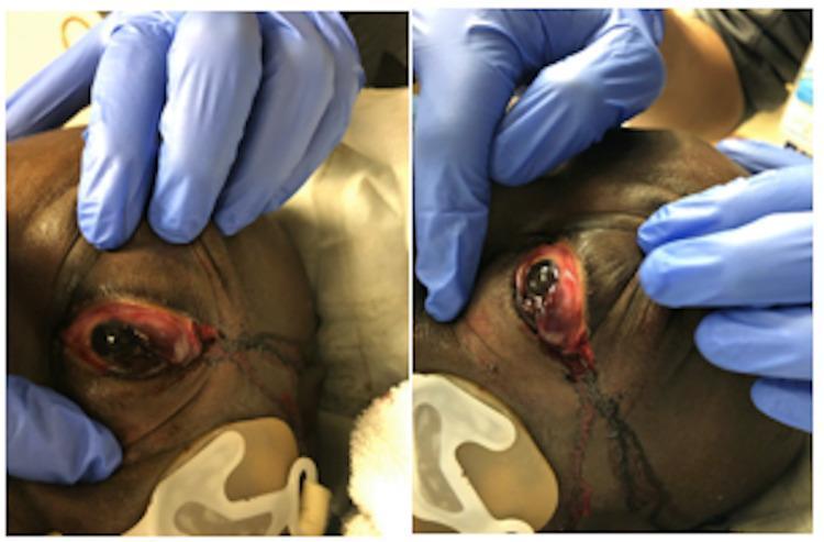
Clinical photographs in frontal (A) and lateral (B) views demonstrating the post-procedural appearance of a lateral canthotomy and inferior cantholysis performed for orbital compartment syndrome. The images show a 57-year-old male with significant proptosis and chemosis of the left eye. In view A, the surgical incision at the lateral canthus is visible, with marked erythema and edema of the periorbital tissues. In view B, the lateral profile emphasizes the degree of globe protrusion (proptosis) and reveals surgical trauma with minor bleeding and tissue disruption at the lateral commissure where the canthal tendons were incised. Blue-gloved hands are seen retracting the upper and lower eyelids to expose the ocular surface and surgical site. This visual documentation serves to illustrate the emergency surgical management required to reduce intraocular pressure and prevent permanent vision loss in cases of retrobulbar pressure buildup.
Recess of Eisler (Premarginal Recess of Eisler)
What It Is
Anatomical Location and Formation
- A few millimeters lateral to the zygomaticomaxillary suture, the septum's line of attachment leaves the orbital rim and lies several millimeters away from it on the facial (anterior) surface of the zygomatic bone.
- This separation between the septum and the zygomatic bone creates a small pocket - the premarginal recess of Eisler - which is filled with fat.
- The septum then returns to the lateral orbital rim just below the level of Whitnall's ligament.
Key Relations
| Structure | Relationship |
|---|---|
| Lateral palpebral ligament (lateral canthal tendon, LCT) | Lies deep to the orbital septum; the septum and LCT are separated by Eisler's pocket |
| Whitnall's tubercle | Just above the recess; the LCT inserts here |
| Orbital septum | Forms the roof/anterior wall of the recess |
| Zygomatic bone | Forms the posterior boundary |
| Fat | Fills the recess |
Clinical and Surgical Significance
-
Blepharoplasty and lower eyelid surgery: Eisler's pocket is a recognizable fat compartment. The inferolateral septum's offset insertion means the lateral fat pad can slightly spill over the orbital rim, contributing to lower eyelid fullness. Disrupting this recess improperly during surgery can cause aesthetic complications.
-
Lacrimal system surgery and orbital dissection: The recess is encountered during surgical approaches to the lateral orbit and must be recognized to avoid inadvertent tissue damage.
-
Aging changes: As the orbital septum weakens with age, the Eisler fat pocket can become more prominent, contributing to lateral lower eyelid fat prolapse and a puffy appearance.
-
Canthoplasty / canthopexy: Understanding the relationship of the orbital septum, lateral canthal tendon, and Eisler's pocket is important when performing lateral canthal procedures, as the recess marks a key transition zone in the anatomy.
Summary
Blow in and blow out fractures of orbit. Explain with diagram also.
blowout fracture orbit mechanism diagram orbital floor
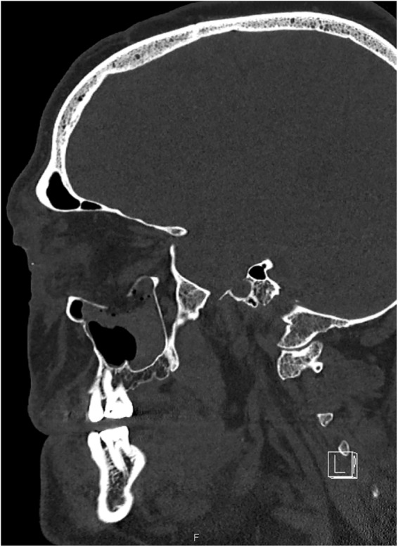
**Imaging Modality:** Computed Tomography (CT), bone window setting. **Anatomical Region:** Midface and neurocranium, sagittal reformatted view. **Observed Pathology:** Extended orbital floor blowout fracture. The image displays a significant disruption and inferior displacement of the bony orbital floor into the underlying maxillary sinus. There is an associated herniation of orbital soft tissue (extraconal fat and potentially the inferior rectus muscle) into the superior aspect of the maxillary antrum. **Characteristic Visual Features:** - **Osseous Findings:** Comminuted fracture of the floor of the orbit with a large bony defect. - **Soft Tissue Findings:** "Teardrop sign" or trapdoor-type appearance where orbital contents prolapse through the fracture site. - **Secondary Features:** Partial opacification of the maxillary sinus, likely representing hemosinus or displaced soft tissue. The anterior and posterior walls of the maxillary sinus appear grossly intact in this plane, localizing the primary trauma to the orbital interface. **Diagnostic Features:** These findings are characteristic of a high-energy "blowout" mechanism, frequently resulting in globe displacement or entrapment of extraocular muscles. The visualization of the defect in the sagittal plane confirms the anteroposterior extent of the orbital floor involvement.
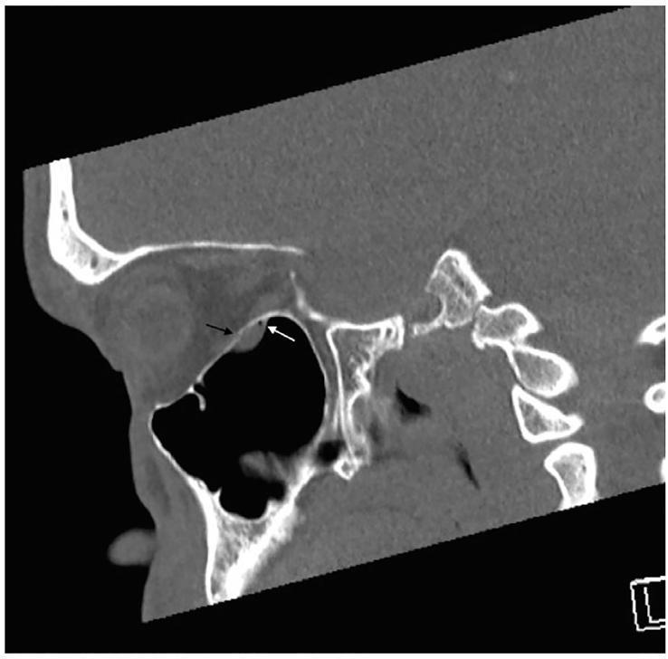
This diagnostic image is a sagittal non-contrast CT scan of the orbit and midface presented in a bone window. The image demonstrates a subtle blowout fracture of the orbital floor. Key findings include a focal bony defect (indicated by a black arrow) in the orbital floor, representing a 'trapdoor' type fracture. Immediately inferior to this defect, a soft tissue density is visible prolapsing into the superior aspect of the maxillary sinus. A small focus of orbital emphysema (intraorbital air), highlighted by a white arrow, is situated adjacent to this herniated soft tissue. The appearance is highly suggestive of orbital fat and inferior rectus muscle entrapment, which often presents clinically with vertical diplopia and restricted ocular motility. This educational visual emphasizes the importance of identifying subtle radiological signs of orbital trauma, particularly when gross fractures are absent, to prevent permanent muscle ischemia or gaze restriction.
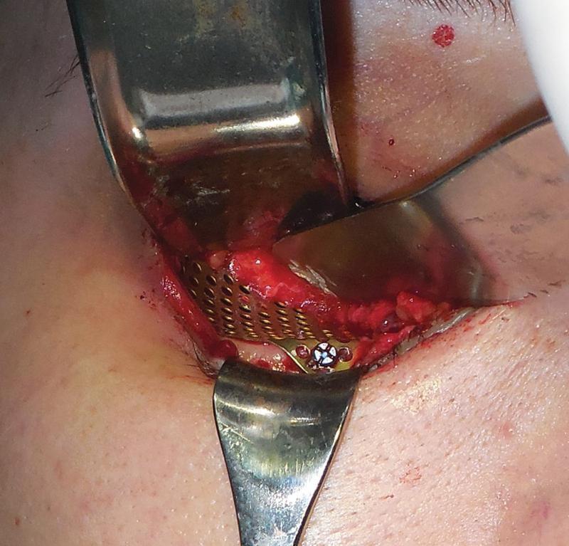
This clinical photograph captures an intraoperative view of an orbital floor reconstruction following a pure blowout fracture. The surgical site is accessed through a transconjunctival approach, with metallic retractors providing exposure of the inferior orbital rim and cavity. Centrally, a titanium mesh implant with a regular perforated grid pattern is visible, precisely contoured to reconstruct the orbital floor boundary. The mesh is rigidly secured to the bone using a micro-screw, visible with a cross-slotted head. The surrounding area shows exposed soft tissue and minor surgical hemorrhage consistent with an active procedure. This image illustrates the surgical management of orbital trauma and the use of internal fixation materials to prevent enophthalmos and muscle entrapment by restoring anatomical volume and structural integrity to the orbit.
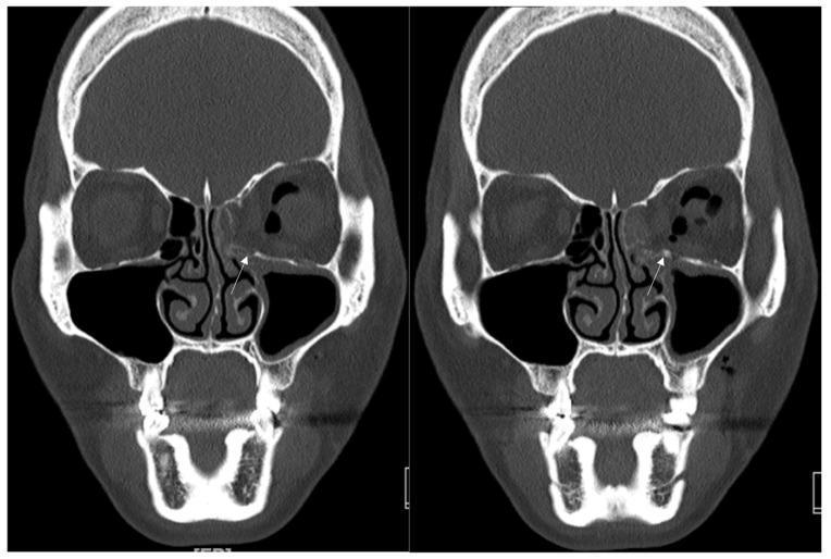
This diagnostic image consists of two side-by-side coronal slices from a facial computerized tomography (CT) scan (bone window) demonstrating surgical reconstruction of an orbital blowout fracture. The left orbit (viewer's right) exhibits a fracture involving the inferomedial wall. A hyperdense, radio-opaque implant, identified as a titanium-reinforced porous polyethylene (TR-PPE) plate, is seen positioned along the left medial orbital floor and lower medial wall to reconstruct the defect. A small, focal, high-density point (marked by a white arrow) indicates a single titanium screw used to fixate the plate to the inferior orbital rim. Anatomical landmarks include the bilateral maxillary sinuses, ethmoid air cells, and the nasal cavity. The left orbit shows some soft tissue herniation and postoperative changes compared to the intact right orbit. This image illustrates maxillofacial trauma management and the use of alloplastic materials in orbital floor reconstruction to restore orbital volume and prevent enophthalmos.
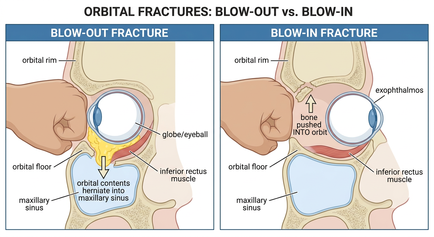
Medical anatomy diagram showing blow-out and blow-in orbital fractures side by side. Left panel labeled "BLOW-OUT FRACTURE": A sagittal cross-section of the orbit showing a large blunt object (fist/ball) hitting the eye from the front, the orbital rim remains intact, the orbital floor fractures downward into the maxillary sinus, orbital fat and inferior rectus muscle herniate down through the floor defect into the maxillary antrum, arrow pointing downward labeled "orbital contents herniate into maxillary sinus". Right panel labeled "BLOW-IN FRACTURE": Same view showing direct trauma to the orbital rim, bone fragment displaced inward and upward into the orbit, arrow pointing upward into the orbit labeled "bone pushed INTO orbit", globe shows exophthalmos (pushed forward). Both panels clearly labeled with: orbital rim, orbital floor, maxillary sinus, inferior rectus muscle, globe/eyeball. Clean educational medical illustration style with clear labels.
Blow-Out and Blow-In Fractures of the Orbit
Mechanism Diagram
1. Blow-Out Fracture
Definition
Mechanism
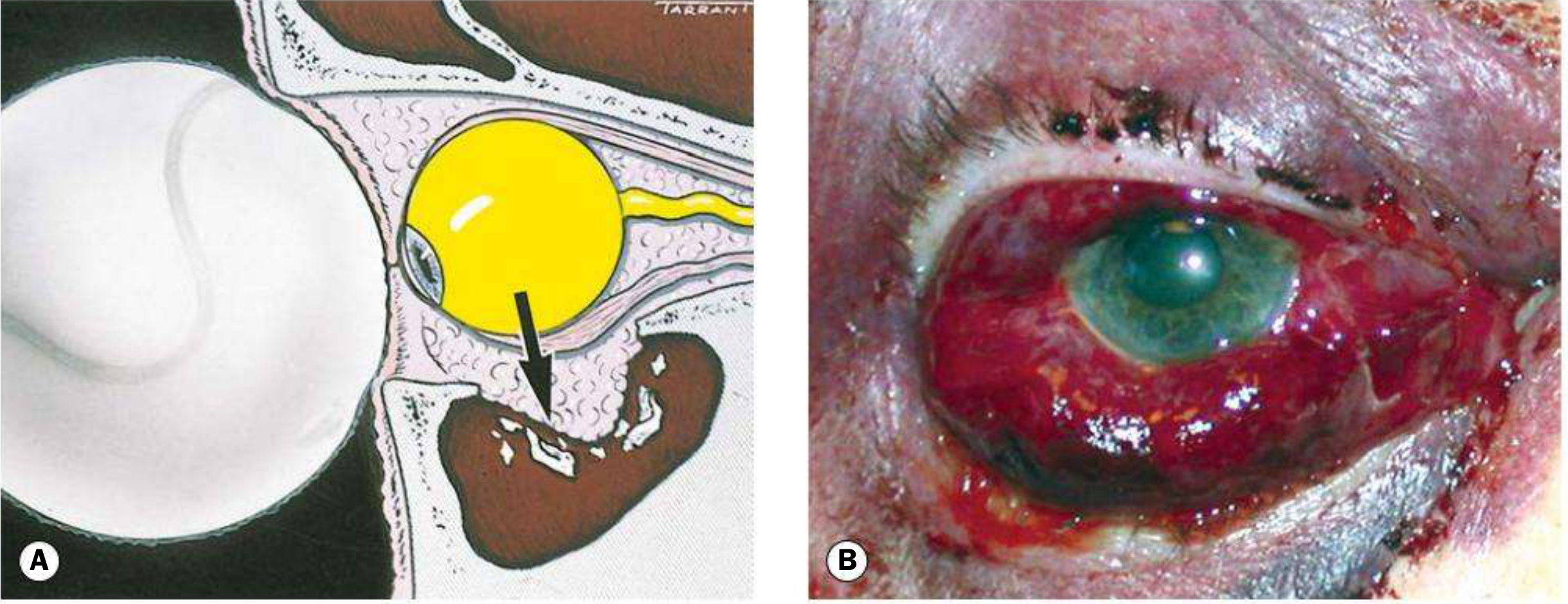
- A blunt object larger than the orbital aperture (e.g., fist, tennis ball, cricket ball) strikes the eyeball directly.
- The eyeball is displaced posteriorly.
- This causes a sudden rise in intraorbital pressure.
- The pressure is transmitted to the weakest orbital wall - the posteromedial floor (thin bone of the maxillary roof, medial to the infraorbital neurovascular bundle).
- The floor fractures downward ("blows out") into the maxillary antrum.
- The orbital rim remains intact because the force is transmitted hydraulically, not directly to the rim.
- Direct trauma to the orbital rim causes a shock wave that buckles the thinner orbital floor at its weakest point.
Site of Fracture
- Most common: posteromedial orbital floor (maxillary bone), medial to the infraorbital canal
- The floor is the thinnest and shortest orbital wall (equilateral triangle shape)
- Less commonly: medial wall (lamina papyracea of ethmoid) may also fracture
Clinical Features
- Pain, blurred vision
- Binocular vertical/oblique diplopia (double vision)
- Eyelid edema and ecchymosis (periorbital bruising)
- Subconjunctival hemorrhage
- Subcutaneous or orbital emphysema (crackling on palpation - air from maxillary sinus entering orbit)
- Enophthalmos - sinking of the globe due to increased orbital volume (may be masked initially by edema; becomes apparent after 2-4 weeks)
- Globe ptosis (hypoglobus)
- Numbness of lower eyelid, cheek, side of nose, upper lip, upper teeth and gums
- Caused by fracture involving the infraorbital canal
- Hemorrhage and edema tightening orbital septa - usually improves as swelling resolves
- Mechanical entrapment of inferior rectus or inferior oblique muscle (or fat and connective tissue) within the fracture - most significant
- Direct injury to extraocular muscle
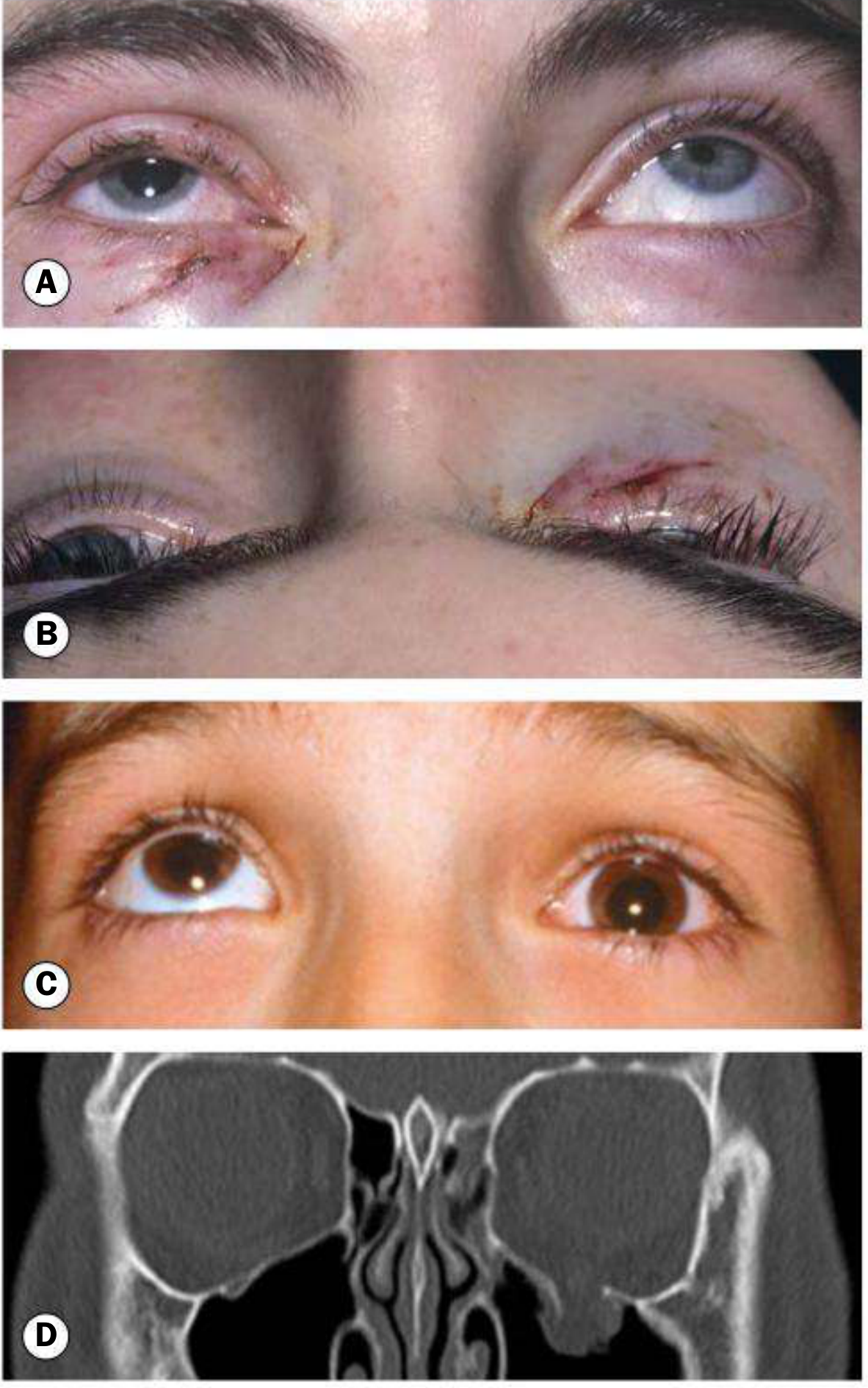
Special Variant: "White-Eye" Blow-Out Fracture (Trapdoor Fracture)
- Seen almost exclusively in children (<18 years) due to greater bone elasticity (greenstick fracture pattern)
- The orbital floor fractures and immediately springs back ("trapdoor"), trapping the inferior rectus muscle between the fracture edges
- Externally, the eye appears surprisingly normal - minimal or no subconjunctival hemorrhage (hence "white eye")
- CT may show minimal or no obvious fracture
- However, the patient has:
- Severe restriction of upgaze
- Oculocardiac reflex: vagal triad of bradycardia, nausea, vomiting (sometimes syncope) due to traction on the entrapped muscle
- This is a surgical emergency - irreversible ischemic damage to the muscle can occur within hours → permanent restrictive strabismus and fibrosis
Imaging
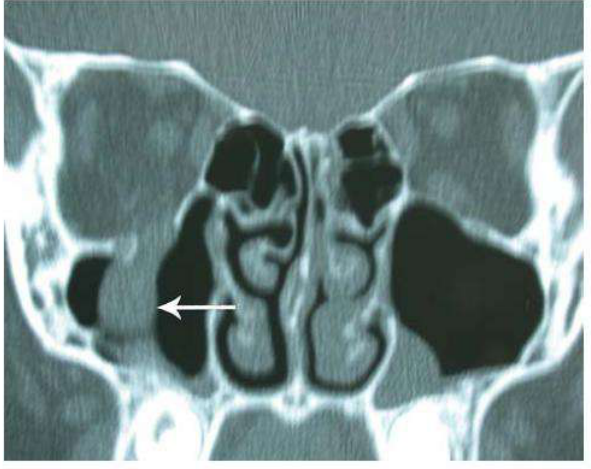
- Shows floor defect
- "Teardrop sign" - soft tissue (fat/muscle) prolapsing through the defect into the maxillary antrum, creating a teardrop-shaped opacity
2. Blow-In Fracture
Definition
| Feature | Blow-Out | Blow-In |
|---|---|---|
| Mechanism | Indirect - globe compressed, floor fractures outward | Direct trauma to orbital rim - bone pushed inward |
| Orbital volume | Increases (floor displaced downward) | Decreases (bone pushed into orbit) |
| Globe position | Enophthalmos (globe sinks in) | Exophthalmos / proptosis (globe pushed out) |
| Orbital rim | Intact | Usually involved |
| Contents | Herniate into sinus | Compressed/restricted |
Mechanism
- Direct, high-energy blow to the orbital rim itself
- The bone fragment is driven inward into the orbit
- This reduces the orbital volume, pushing the globe anteriorly
Clinical Features
- Exophthalmos/proptosis (globe pushed forward) - the hallmark
- Orbital contents are compressed and restricted rather than prolapsing
- Vision may be compromised by pressure on the optic nerve or globe
- Restricted eye movements due to compression
Comparison Summary Table
| Feature | Blow-Out Fracture | Blow-In Fracture |
|---|---|---|
| Cause | Blunt trauma > orbital aperture diameter | Direct blow to orbital rim |
| Rim | Intact | Fractured/involved |
| Floor | Displaced downward/outward | Displaced inward into orbit |
| Orbital volume | Increased | Decreased |
| Globe | Enophthalmos | Exophthalmos |
| Diplopia | Yes (upgaze/downgaze) | Yes (compression) |
| Infraorbital numbness | Common | Less typical |
| Trapdoor variant | Yes (children) | No |
| Key complication | Muscle entrapment, enophthalmos | Optic nerve compression, proptosis |
Treatment of Blow-Out Fracture
- Oral antibiotics (risk of sinus contamination)
- Ice packs, nasal decongestants
- Patient must NOT blow their nose (forces infected sinus air into orbit, risks orbital cellulitis)
- Systemic steroids if severe orbital edema compresses the optic nerve
- Persistent diplopia in primary or reading position not improving
- Enophthalmos >2 mm
- Fracture involving >50% of orbital floor
- Entrapment of orbital contents on imaging
- White-eye trapdoor fracture in a child - urgent/emergency repair
- Transconjunctival or subciliary incision
- Periosteum elevated from orbital floor
- Entrapped orbital contents freed
- Bony defect repaired with synthetic implant (titanium mesh or porous polyethylene plate)