
Knee joint anatomy long question Post graduate answer
knee joint anatomy diagram ligaments menisci bones
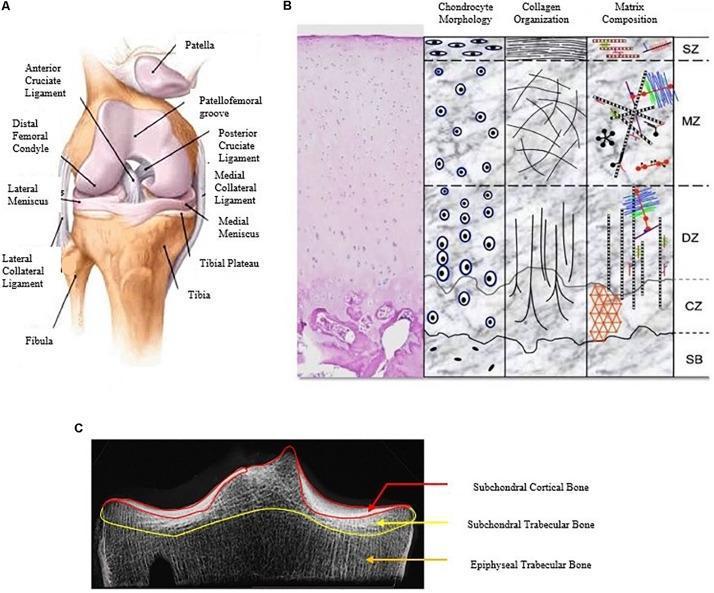
A multi-panel educational figure illustrating the macro and micro-anatomical structures of the human knee joint and articular cartilage. Panel A is an anatomical diagram of the right knee joint in an anterior view, highlighting skeletal and ligamentous structures including the femur (distal femoral condyle), tibia, fibula, and patella. Key ligaments shown include the anterior and posterior cruciate ligaments, and the medial and lateral collateral ligaments, along with the medial and lateral menisci and the patellofemoral groove. Panel B provides a histological and schematic cross-section of hyaline articular cartilage organized into four zones: Superficial (SZ), Middle (MZ), Deep (DZ), and Calcified (CZ), transitioning to Subchondral Bone (SB). It details variations in chondrocyte morphology (flattened to columnar), collagen orientation (tangential to vertical), and extracellular matrix composition across these layers. Panel C is a diagnostic radiographic image (CT/X-ray) of the tibial plateau, segmenting the subchondral bone into three regions: the subchondral cortical bone (end-plate), subchondral trabecular bone, and epiphyseal trabecular bone. This figure serves as a comprehensive reference for orthopedic anatomy and cartilage pathophysiology.
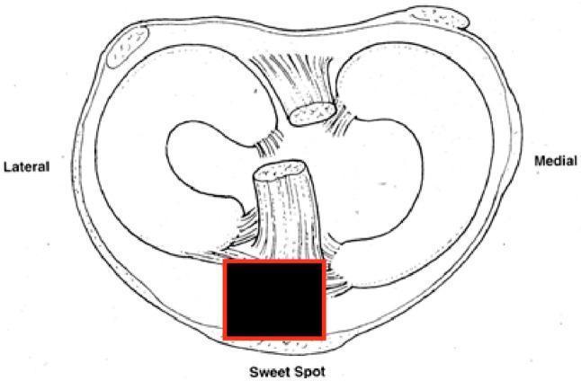
This medical anatomical diagram displays an axial cross-section of the human knee joint, focusing on the tibial plateau and its associated soft tissue structures. The illustration highlights the medial and lateral menisci as crescent-shaped fibrous structures resting on the articular surface. Centrally, the anterior and posterior cruciate ligaments are depicted, showing their relative spatial orientations. Soft tissue attachments, including the capsule and ligamentous fibers, are shown peripherally. A region labeled 'Sweet Spot' is highlighted with a red box at the anterior-inferior aspect of the joint space, corresponding to the clinical target for safe entry during suprapatellar tibial nailing (SPTN) procedures. This site is anatomically significant as the ideal starting point for intramedullary nail insertion, typically located on the medial aspect of the lateral tibial spine to minimize articular damage and optimize fracture reduction in proximal third tibial fractures. The diagram serves as an educational tool for orthopedic surgical planning and biomechanical understanding of the knee joint.
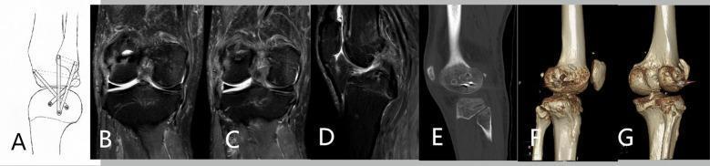
This composite figure illustrates surgical management for posterolateral corner (PLC) knee instability using a modified LaPrade technique. Panel A presents a line diagram showing the graft configuration for anatomical reconstruction of the fibular collateral ligament (FCL), popliteus tendon (PT), and popliteofibular ligament (PFL). Panels B-D are preoperative MRI scans (coronal and sagittal views) displaying the native knee anatomy and soft tissue structures including the menisci and cruciate ligaments. Panels E-G show postoperative imaging: E is a sagittal CT slice demonstrating surgical bone tunnels, while F and G are 3D CT reconstructions of the knee joint. These reconstructions highlight the precise placement of femoral, tibial, and fibular bone tunnels and the presence of interference screws used for graft fixation. The series serves as an educational resource for orthopedic surgery, specifically focusing on complex knee ligament reconstruction and the radiographic assessment of tunnel positioning and hardware integration in multi-ligamentous knee injuries.
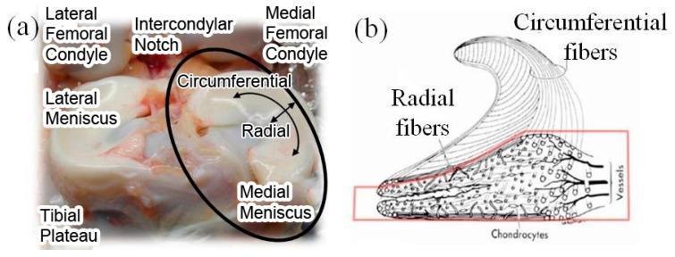
This dual-panel educational graphic illustrates the macro and micro-anatomy of the human knee meniscus. Panel (a) is an anatomical photograph of a superior view of the tibial plateau, identifying the lateral and medial femoral condyles separated by the intercondylar notch. The lateral and medial menisci are visible resting on the tibial plateau; the medial meniscus is circled to highlight its circumferential and radial axes. Panel (b) is a histological diagram of a wedge-shaped meniscal cross-section. It details the internal microstructure, emphasizing the arrangement of collagen fibers: circumferential fibers are shown running parallel to the periphery, while radial fibers act as cross-linkers to provide structural integrity. The diagram also illustrates the distribution of chondrocytes (fibrochondrocytes) throughout the tissue and the presence of peripheral vessels in the vascularized 'red zone' near the outer margin. This content is designed for orthopaedic and biomechanical education, focusing on how meniscal microstructure relates to its function in load distribution and shock absorption within the knee joint.
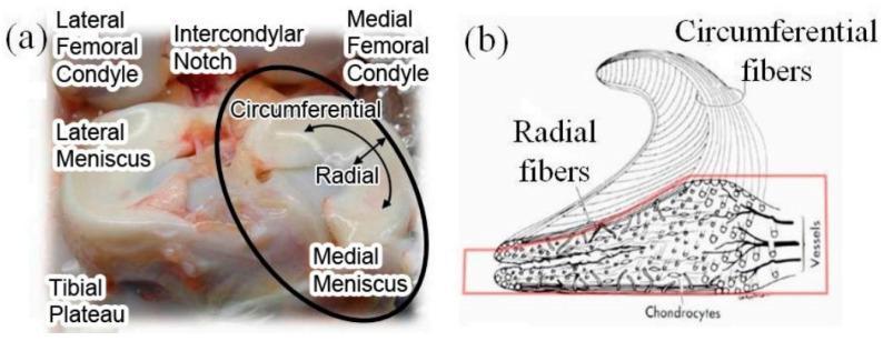
Educational figure regarding human knee anatomy and meniscal microstructure. Panel (a) is a clinical photograph of a human knee joint dissection, illustrating the anatomical relationship between the femoral condyles (lateral and medial), the intercondylar notch, and the tibial plateau. The lateral and medial menisci are shown positioned atop the tibial plateau, with labels indicating the circumferential and radial directions of the fibrocartilage. Panel (b) is a pathophysiological diagram of a meniscal cross-section, detailing the complex internal arrangement of collagen fibers. The diagram highlights the superficial circumferential fibers oriented along the periphery to resist tension and the deeper radial fibers. The lower section of the diagram illustrates the biological composition, including chondrocytes (meniscal cells) and the vascular supply (vessels) located toward the peripheral 'red zone.' This figure serves as a reference for understanding the load-bearing biomechanics and structural organization of meniscal tissue in orthopedic surgery and sports medicine.
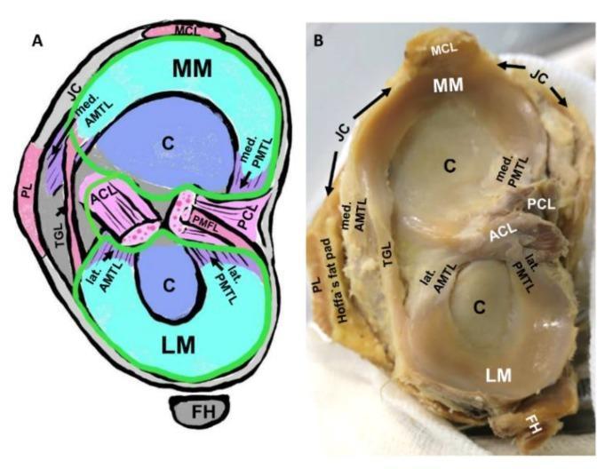
Educational visual showing the gross anatomy and schematic representation of the human tibial plateau and its intraarticular structures. The image is divided into a labeled schematic (A) and a corresponding clinical photograph of a cadaveric specimen (B), demonstrating the spatial relationships of the knee joint. Key anatomical features identified include the medial meniscus (MM) and lateral meniscus (LM) resting on the tibial articular cartilage (C). The cruciate ligaments—anterior (ACL) and posterior (PCL)—are shown centrally. Detailed focus is given to the meniscotibial ligaments, specifically the medial and lateral anterior meniscotibial ligaments (AMTL) and posterior meniscotibial ligaments (PMTL), which provide elastic fixation for the meniscal horns. Additional structures visible include the transverse genicular ligament (TGL) connecting the menisci, the medial collateral ligament (MCL), the joint capsule (JC), and the fibular head (FH). The schematic further illustrates the synovial membrane (green line) and its relation to the joint cavity. This material is designed to teach orthopedic anatomy, emphasizing the complex ligamentous network supporting knee joint stability and the entheses involved in degenerative conditions like osteoarthritis.
Knee Joint Anatomy - Postgraduate Answer
Introduction
- Bailey and Love's Short Practice of Surgery 28th Ed, p. 690
- Gray's Anatomy for Students
Type and Articulations
- Medial tibiofemoral joint - between medial femoral condyle and medial tibial plateau
- Lateral tibiofemoral joint - between lateral femoral condyle and lateral tibial plateau
- Patellofemoral joint - between the V-shaped patellar surface on the anterior femur and the posterior patella
Articular Surfaces
Femur
- Distal femur has medial and lateral condyles separated by the intercondylar notch posteriorly
- Anteriorly, a patellar groove (trochlear groove) accommodates the patella
- The medial condyle is larger than the lateral - this asymmetry is critical to the screw-home mechanism
Tibia
- The superior tibial plateau has medial and lateral facets separated by the intercondylar eminence (tibial spines)
- The medial tibial plateau is concave (congruent with medial condyle)
- The lateral tibial plateau is convex (less congruent - the lateral meniscus compensates)
Patella
- Largest sesamoid bone; lies within the quadriceps tendon
- Posterior surface has medial and lateral facets articulating with the trochlear groove
- Acts as a pulley, increasing the mechanical advantage of the quadriceps by ~30%
Joint Capsule
- Anteriorly: extensions of vastus medialis and lateralis (patellar retinacula), quadriceps tendon, and the iliotibial band (anterolaterally)
- Posteriorly: the oblique popliteal ligament (expansion of semimembranosus tendon - runs from medial to lateral) and the arcuate popliteal ligament
- The popliteus muscle tendon passes through an aperture in the posterolateral capsule
- Gray's Anatomy for Students, p. 714
- Rheumatology 2-Volume Set (2022, Elsevier), p. 737
Synovial Membrane and Bursae
Key Bursae
| Bursa | Location | Communication with Joint |
|---|---|---|
| Suprapatellar bursa | Between quadriceps tendon and femur | Yes (always) |
| Prepatellar bursa | Superficial to patella | No |
| Superficial infrapatellar | Superficial to patellar ligament | No |
| Deep infrapatellar | Between patellar ligament and tibia | No |
| Semimembranosus bursa | Between semimembranosus and medial gastrocnemius | Yes |
| Subtendinous medial gastrocnemius | Behind medial gastrocnemius head | Yes |
Menisci
Functions
- Load transmission and shock absorption - transmit ~70% of compressive load in extension and ~85-90% in flexion
- Joint congruency and stability
- Lubrication and nutrition of articular cartilage
- Proprioception
Structure
- Triangular in cross-section: thick peripherally (red zone), thin centrally (white zone)
- Composed of circumferential collagen fibers (resist hoop stress) reinforced by radial fibers
- The peripheral third (red zone) is vascularized; the inner two-thirds (white zone) is avascular and receives nutrition by diffusion

Medial Meniscus
- Larger, C-shaped, covers ~60% of the medial tibial surface
- More firmly attached: connected to the joint capsule around its periphery AND to the deep layer of the MCL (medial capsular ligament)
- Because of these firm attachments, it is less mobile and more prone to tears than the lateral meniscus
- Anterior horn attaches anterior to the ACL insertion; posterior horn attaches posterior to the tibial spines
Lateral Meniscus
- Smaller, nearly circular in shape, covers ~80% of the lateral tibial surface
- Not attached to the lateral capsule (a hiatus in the posterolateral capsule allows the popliteus tendon to pass through)
- Therefore more mobile and less frequently torn
- Anchored posteriorly by the meniscofemoral ligaments (Humphrey's - anterior to PCL; Wrisberg's - posterior to PCL) connecting its posterior horn to the medial femoral condyle
Transverse (Intermeniscal) Ligament
-
Connects the anterior horns of both menisci
-
Gray's Anatomy for Students, p. 710
-
Rheumatology 2-Volume Set (2022, Elsevier), p. 738
-
Miller's Review of Orthopaedics 9th Ed, p. 334
Ligaments
1. Patellar Ligament
- Continuation of the quadriceps femoris tendon distal to the patella
- Attaches from the apex/margins of the patella to the tibial tuberosity
- Tested clinically by the knee jerk reflex (L3-L4)
2. Tibial (Medial) Collateral Ligament (MCL)
- Superficial layer: from the medial femoral epicondyle, extends 8-12 cm below the tibial plateau (100-120 mm total length); lies deep to the pes anserinus tendons
- Femoral origin: proximal and posterior to the medial epicondyle
- Two tibial insertions: proximally onto soft tissue over the semimembranosus anterior arm; distally directly to bone 6-7 cm below the joint line
- Deep layer (mid-third medial capsular ligament): thickening of the medial capsule with meniscofemoral and meniscotibial components; intimately attaches to the medial meniscus
3. Fibular (Lateral) Collateral Ligament (LCL)
- Smaller, cord-like (unlike the flat MCL)
- From the lateral femoral epicondyle to the fibular head
- Not attached to the lateral capsule or lateral meniscus
- Function: Resists varus stress across the knee
- Part of the posterolateral corner complex
4. Anterior Cruciate Ligament (ACL)
- Tibial attachment: Broad, irregular oval area in the anterior intercondylar region, immediately medial to the anterior horn of the lateral meniscus
- Femoral attachment: Posteromedial aspect of the lateral femoral condyle (semicircular area bordered by the intercondylar ridge)
- Runs posteriorly, laterally, and upward from tibia to femur
- Average length: 33-38 mm; width: 7-12 mm
- Anteromedial (AM) bundle: originates proximal to the bifurcate ridge; tight in flexion; primarily resists anterior tibial translation
- Posterolateral (PL) bundle: tight in extension; resists rotatory instability
5. Posterior Cruciate Ligament (PCL)
- Tibial attachment: Posterior intercondylar area (most posterior point of tibia)
- Femoral attachment: Lateral surface of the medial femoral condyle
- Larger and stronger than ACL; average length 38 mm, diameter 13 mm
- Runs anteriorly, medially, and upward; perpendicular to ACL
- Anterolateral (AL) bundle: tight in flexion
- Posteromedial (PM) bundle: tight in extension
- Humphrey's ligament: anterior to PCL
- Wrisberg's ligament: posterior to PCL
- Rheumatology 2-Volume Set (2022, Elsevier), p. 737-738
- Miller's Review of Orthopaedics 9th Ed, p. 333-334
- Gray's Anatomy for Students, p. 715-716
6. Posterolateral Corner (PLC)
- Static components: LCL, popliteus tendon, popliteofibular ligament, arcuate ligament
- Dynamic components: biceps femoris, popliteus muscle, ITB, lateral head of gastrocnemius
- Augments PCL function; resists posterior translation, external rotation, and varus deformity
- Often injured with PCL (combined injury)
Movements and Kinematics
| Movement | Range | Muscle |
|---|---|---|
| Flexion | 0-135° (up to 160° passive) | Hamstrings (biceps femoris, semitendinosus, semimembranosus), gracilis, sartorius, popliteus, gastrocnemius |
| Extension | 135° to 0° (5° hyperextension possible) | Quadriceps femoris (rectus femoris, vastus medialis, lateralis, intermedius) |
| Medial rotation (unlocking) | ~10° | Popliteus |
| Lateral rotation | ~30-40° in flexion | Biceps femoris |
The "Screw-Home" Mechanism (Locking Mechanism)
- As the knee approaches full extension, the tibia externally rotates ~5° on the femur (or equivalently, the femur internally rotates on the tibia)
- This occurs because the medial femoral condyle is longer than the lateral - the lateral condyle completes its arc of rolling first, forcing the medial side to continue, causing the tibia to rotate
- The ACL tightens and winds around the PCL during this rotation, locking the joint
- Unlocking is performed by the popliteus muscle - it internally rotates the tibia to "unscrew" the knee before flexion can begin
- Gray's Anatomy for Students, p. 716
- Bailey and Love's 28th Ed, p. 690
Blood Supply
| Artery | Source |
|---|---|
| Medial superior genicular | Popliteal artery |
| Lateral superior genicular | Popliteal artery |
| Medial inferior genicular | Popliteal artery |
| Lateral inferior genicular | Popliteal artery |
| Middle genicular | Popliteal artery - pierces posterior capsule to supply cruciate ligaments |
| Descending genicular | Femoral artery |
| Anterior tibial recurrent | Anterior tibial artery |
Nerve Supply
| Nerve | Supply |
|---|---|
| Femoral nerve (L2-L4) | Anteriomedial knee; branches of saphenous nerve |
| Common peroneal nerve | Anterolateral aspect |
| Tibial nerve | Posterior aspect; lies in popliteal fossa with popliteal vessels |
| Obturator nerve | Medial aspect |
-
Motor: Quadriceps (femoral nerve L2-L4); hamstrings and popliteus (sciatic - tibial division L4-S2); biceps femoris short head (common peroneal nerve)
-
Clinical significance: The tibial nerve lies closely associated with popliteal vessels - at risk in posterior knee dislocations
-
Rheumatology 2-Volume Set (2022, Elsevier), p. 738
Relations of the Knee (Popliteal Fossa)
- Boundaries: Superolateral - biceps femoris; superomedial - semimembranosus and semitendinosus; inferolateral - lateral head of gastrocnemius; inferomedial - medial head of gastrocnemius; roof - deep fascia + skin; floor - popliteal surface of femur, posterior capsule, popliteus fascia
- Contents (superficial to deep): popliteal lymph nodes; tibial nerve; common peroneal nerve; popliteal vein; popliteal artery (deepest, most medial)
- Mnemonic: TAN from superficial to deep (Tibial nerve, Artery, then go lateral for common peroneal Nerve)
Applied Anatomy and Clinical Correlates
| Structure | Injury | Mechanism | Clinical Test |
|---|---|---|---|
| ACL | Most common sports injury | Non-contact pivot/deceleration; hyperextension | Lachman, anterior drawer, pivot shift |
| PCL | Less common | Dashboard injury; fall on flexed knee | Posterior drawer, sag sign |
| MCL | Common | Valgus force | Valgus stress test |
| LCL | Less common | Varus force | Varus stress test |
| Medial meniscus | More common than lateral | Twisting on weight-bearing knee | McMurray, Thessaly, Apley |
| Patellar ligament | Rupture | Sudden quadriceps contraction | Inability to extend knee |
| Unhappy triad (O'Donoghue) | ACL + MCL + medial meniscus | Valgus + rotatory force | Combined tests |
Baker's Cyst
- Distension of the semimembranosus-gastrocnemius bursa (which communicates with the joint)
- Common in knee joint effusion/osteoarthritis
- Located in the posteromedial popliteal fossa
The Unhappy Triad
Summary Table
| Feature | Detail |
|---|---|
| Type | Modified hinge (ginglymus) synovial joint |
| Bones | Femur, tibia, patella (fibula not part of joint) |
| Articular cartilage | Hyaline cartilage |
| Static stabilizers | Capsule, MCL, LCL, ACL, PCL, menisci |
| Dynamic stabilizers | Quadriceps, hamstrings, popliteus, gastrocnemius |
| Blood supply | Genicular anastomosis (from popliteal artery) |
| Nerve supply | Femoral, sciatic (tibial + common peroneal), obturator |
| Range of motion | 0-135° flexion; 5° hyperextension; ~30° rotation in flexion |
| Locking mechanism | Screwing - external tibial rotation in last 15° extension; unlocking by popliteus |