
Tell me step by step placenta formation
Reading File
Now let me search for placenta formation content in the medical library.
Finding Sources
Finding Sources
Good - I found relevant sources. Let me read the key sections on placenta formation from Creasy & Resnik's Maternal-Fetal Medicine and the Histology textbook.
Reading File
Reading File
Reading File
Reading File
Reading File
Reading File
I now have comprehensive information. Let me fetch the key diagram image of chorionic villi development and compile the full answer.
Loading Image
Loading Image
Excellent! I now have everything needed for a thorough, well-illustrated answer. Here is the step-by-step guide to placenta formation:
Step-by-Step Placenta Formation
Step 1 - Fertilization and Blastocyst Formation (Days 1-5)
After fertilization, the zygote undergoes cleavage divisions as it travels down the fallopian tube. By days 4-5 it becomes a blastocyst, with two distinct populations:
- Inner cell mass (ICM / embryoblast) - gives rise to the embryo proper
- Outer cell mass (trophoblast) - the precursor of the entire placenta
Fluid accumulates inside to form the blastocyst cavity; the zona pellucida dissolves, freeing the blastocyst to implant.
Step 2 - Implantation (Days 6-10)
The blastocyst attaches to the receptive endometrial epithelium via its polar trophectoderm (the TE cells overlying the ICM). This is called the implantation window - day 6 to day 10 after the LH surge.
Key molecular signals driving attachment include:
- HB-EGF (heparin-binding EGF-like growth factor) from endometrial pinopodia binding ErbB1/ErbB4 receptors on trophoblast microvilli
- Leukemia inhibitory factor (LIF), HOXA10/HOXA11, and WNT signaling in the endometrium
The trophoblast rapidly proliferates and invades the endometrium. By day 11, the blastocyst is completely embedded within the endometrium.
Step 3 - Trophoblast Differentiation: Two Layers Form
As the trophoblast invades, it differentiates into two layers:
| Layer | Features |
|---|---|
| Cytotrophoblast (CTB) | Inner layer; mitotically active; single-nucleated stem cells; produces cells that fuse upward into syncytium |
| Syncytiotrophoblast (STB) | Outer layer; multinucleate, non-dividing; actively erodes and invades endometrium; secretes hCG, progesterone, estrogen |
The STB secretes hCG, which rescues the corpus luteum and maintains progesterone production to prevent menstruation.
Step 4 - Lacunar Stage and Primitive Uteroplacental Circulation (Days 9-13)
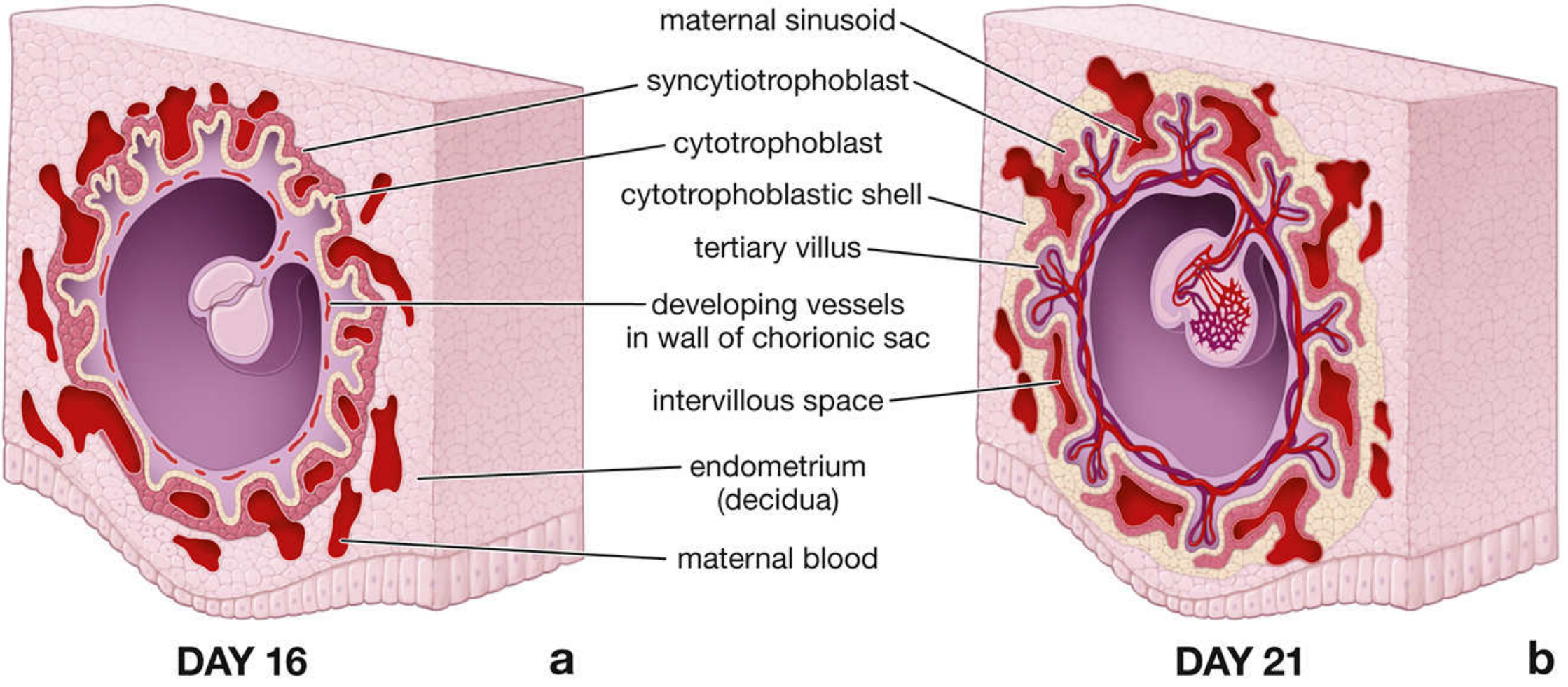
By day 9, vacuoles appear in the STB and merge to form trophoblastic lacunae (blood-filled spaces). Maternal capillaries (sinusoids) erode into these lacunae. The pressure differential between maternal arteries and veins establishes directional flow, creating a primitive uteroplacental circulation. Nutrients pass into the embryo via pinocytosis by the STB.
Step 5 - Primary Chorionic Villi (Days 11-13)
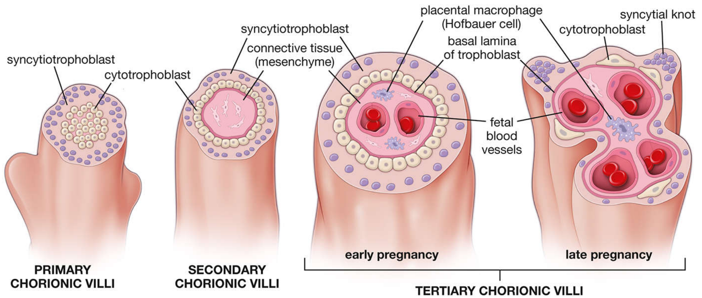
Cytotrophoblast cells proliferate rapidly and push finger-like projections into the STB and out into the lacunae - these are the primary chorionic villi. They consist of a CTB core covered by STB.
Step 6 - Secondary Chorionic Villi (~Day 16)
Extraembryonic mesoderm (ExM) - derived from the inner cell mass via the primitive streak - invades into the primary villi cores, forming the secondary chorionic villi. These have three layers:
- Outer STB
- Middle CTB
- Inner loose mesenchymal core
Step 7 - Tertiary Chorionic Villi (~Days 18-21)
Blood vessels differentiate within the mesenchymal core of the secondary villi via vasculogenesis and angiogenesis, driven by signals including VEGF from CTB. These villous capillaries:
- Coalesce and connect to the embryonic body stalk vessels
- Establish true fetal circulation by day 21
- Initial fetal red cells are yolk-sac derived; after the second month, they come from the fetal liver, later from bone marrow
Once vascularized, the villi are called tertiary chorionic villi.
Step 8 - Decidualization of the Endometrium
In parallel, the endometrium transforms into the decidua under progesterone influence. Three zones are recognized:
| Decidual Region | Location |
|---|---|
| Decidua basalis | Beneath the implanting embryo - becomes the maternal component of the placenta |
| Decidua capsularis | Covers the embryo on the uterine cavity side |
| Decidua parietalis | Lines the rest of the uterine cavity |
Step 9 - Cytotrophoblastic Shell and Villous Anchoring (~Weeks 3-4)
CTB cells at the tips of the villi (the anchoring villi) break through the STB and spread laterally to form the cytotrophoblastic shell - a continuous layer that anchors the chorionic sac to the decidua basalis. From this shell, extravillous trophoblast (EVT) cells differentiate and invade:
- Interstitial EVT - invades the decidua and myometrium
- Endovascular EVT - invades and remodels the spiral arteries, converting them from narrow, high-resistance vessels into wide, low-resistance channels to increase uteroplacental blood flow
This spiral artery remodeling peaks by 12 weeks. Failure of this step underlies conditions like preeclampsia and fetal growth restriction.
Step 10 - Villous Tree Branching and Placental Maturation (Weeks 4 onward)
The chorionic villi undergo extensive branching morphogenesis driven by FGF10 from CTB, regulated by SPRY1 from Hofbauer cells (placental macrophages). The villous tree dramatically expands its surface area.
Structural changes with gestational age:
| Early Gestation | Late Gestation |
|---|---|
| Large, edematous, loosely structured villi | Slender, branched, tightly packed villi |
| Continuous CTB layer with thick STB | CTB appears discontinuous; syncytial knots form |
| Few capillaries, nucleated fetal RBCs | Many capillaries, close to STB surface |
| Placental membrane = endothelium + CTB + STB | Placental membrane is thin (vasculosyncytial membrane) |
Step 11 - Mature Placenta Structure
By term (~38-40 weeks), the fully formed placenta consists of:
- Chorionic plate (fetal side) with umbilical vessels branching into villi
- Villous tree - branching villi bathed in maternal blood in the intervillous space
- Basal plate (maternal side) - decidua basalis with anchoring villi and remodeled spiral arteries opening into the intervillous space
- Cotyledons - 15-30 lobes, each supplied by one spiral artery
The placental membrane separating fetal and maternal blood consists of: fetal capillary endothelium → connective tissue (mesenchyme) → CTB → STB.
Summary Timeline
| Time | Key Event |
|---|---|
| Days 1-5 | Fertilization → blastocyst with trophoblast and ICM |
| Days 6-10 | Implantation window; trophoblast adheres and invades |
| Day 9 | Lacunae form; primitive uteroplacental circulation begins |
| Days 11-13 | Primary chorionic villi (CTB + STB) |
| Day ~16 | Secondary chorionic villi (mesenchyme invades core) |
| Days 18-21 | Tertiary chorionic villi (blood vessels develop; fetal circulation active) |
| Weeks 3-4 | Cytotrophoblastic shell; EVT invasion; spiral artery remodeling |
| Weeks 4+ | Branching morphogenesis; villous tree expansion |
| Term | Mature placenta with cotyledons, thin placental membrane |
Sources: Creasy & Resnik's Maternal-Fetal Medicine: Principles and Practice (Chapter 3 - Implantation and Placental Development); Histology: A Text and Atlas with Correlated Cell and Molecular Biology (Chapter 23 - Female Reproductive System)
This is a shared conversation. Sign in to Orris to start your own chat.