
Endocrine system diseases and radiological diagnostics (sonography,contrast methods,sctinigraphy,ct, mri) advantages disadvantages indications Also include anatomy and physiology in very short
Topic breakdown
- Endocrine anatomy and physiology (short summary)
- Imaging modalities: how each works, advantages, disadvantages
- Pituitary and hypothalamus (adenoma, prolactinoma, acromegaly, Cushing disease, hypopituitarism)
- Thyroid (goitre, nodules, Graves disease, thyroiditis, cancer)
- Parathyroid (hyperparathyroidism, adenoma localisation)
- Adrenal (incidentaloma, Conn, Cushing, pheochromocytoma, adrenal insufficiency)
- Pancreatic endocrine tumours (insulinoma, gastrinoma, MEN syndromes)
- Gonads, bone and others (PCOS, osteoporosis, rickets)
- Imaging selection summary table: which test for which organ
Short overview
- The hypothalamus and pituitary control the other glands. The anterior pituitary releases GH, PRL, ACTH, TSH, FSH and LH. The posterior pituitary releases ADH and oxytocin.
- The thyroid sits in front of the trachea. It makes T3 and T4, which set metabolic rate. C cells make calcitonin.
- The four parathyroids sit behind the thyroid. They make PTH, which raises serum calcium.
- The adrenals sit above the kidneys. The cortex makes aldosterone, cortisol and androgens. The medulla makes catecholamines.
- The endocrine pancreas (islets) makes insulin and glucagon. The gonads make sex steroids.
- Most axes work by negative feedback.
| Modality | Advantages | Disadvantages | Main endocrine indications |
|---|---|---|---|
| Ultrasound | No radiation, cheap, real-time, guides FNA | Operator dependent, poor for deep or retrosternal lesions, limited in obese patients | Thyroid nodules and goitre, parathyroid adenoma, thyroid FNA guidance, ovaries and testes |
| Contrast methods (CT or X-ray with contrast, angiography, venous sampling) | Show vascular detail and function. Sampling localises hormone source. | Iodinated contrast can worsen thyrotoxicosis and affects the kidneys. Radiation. Invasive. | Adrenal vein sampling (Conn), inferior petrosal sinus sampling (Cushing), selective venous sampling for parathyroid or pancreatic tumours |
| Scintigraphy (I-123, Tc-99m pertechnetate, sestamibi, MIBG, octreotide) | Shows function as well as structure, whole-body survey | Poor anatomical resolution, radiation dose, long acquisition, cost | Thyroid nodule function (hot or cold), Graves disease, ectopic thyroid, parathyroid adenoma (sestamibi), pheochromocytoma (MIBG), neuroendocrine tumours (octreotide, DOTATATE PET) |
| CT | Fast, excellent for bone and calcification, high resolution for adrenal and pancreas | Radiation, contrast risks, poor soft-tissue contrast in the sella | Adrenal masses (attenuation and washout), pancreatic NETs, retrosternal goitre, thyroid cancer staging |
| MRI | Best soft-tissue contrast, no radiation, multiplanar | Costly, long scan, contraindicated with some implants, claustrophobia | Pituitary and sella (first choice), adrenal characterisation, pancreatic NETs, hypothalamic lesions |
Which topic should I cover in detail first?
Topic breakdown
- Endocrine anatomy and physiology (short summary)
- Imaging modalities: how each works, advantages, disadvantages
- Pituitary and hypothalamus (adenoma, prolactinoma, acromegaly, Cushing disease, hypopituitarism)
- Thyroid (goitre, nodules, Graves disease, thyroiditis, cancer)
- Parathyroid (hyperparathyroidism, adenoma localisation)
- Adrenal (incidentaloma, Conn, Cushing, pheochromocytoma, adrenal insufficiency)
- Pancreatic endocrine tumours (insulinoma, gastrinoma, MEN syndromes)
- Gonads, bone and others (PCOS, osteoporosis, rickets)
- Imaging selection summary table: which test for which organ
Short overview
- The hypothalamus and pituitary control the other glands. The anterior pituitary releases GH, PRL, ACTH, TSH, FSH and LH. The posterior pituitary releases ADH and oxytocin.
- The thyroid sits in front of the trachea. It makes T3 and T4, which set metabolic rate. C cells make calcitonin.
- The four parathyroids sit behind the thyroid. They make PTH, which raises serum calcium.
- The adrenals sit above the kidneys. The cortex makes aldosterone, cortisol and androgens. The medulla makes catecholamines.
- The endocrine pancreas (islets) makes insulin and glucagon. The gonads make sex steroids.
- Most axes work by negative feedback.
| Modality | Advantages | Disadvantages | Main endocrine indications |
|---|---|---|---|
| Ultrasound | No radiation, cheap, real-time, guides FNA | Operator dependent, poor for deep or retrosternal lesions, limited in obese patients | Thyroid nodules and goitre, parathyroid adenoma, thyroid FNA guidance, ovaries and testes |
| Contrast methods (CT or X-ray with contrast, angiography, venous sampling) | Show vascular detail and function. Sampling localises hormone source. | Iodinated contrast can worsen thyrotoxicosis and affects the kidneys. Radiation. Invasive. | Adrenal vein sampling (Conn), inferior petrosal sinus sampling (Cushing), selective venous sampling for parathyroid or pancreatic tumours |
| Scintigraphy (I-123, Tc-99m pertechnetate, sestamibi, MIBG, octreotide) | Shows function as well as structure, whole-body survey | Poor anatomical resolution, radiation dose, long acquisition, cost | Thyroid nodule function (hot or cold), Graves disease, ectopic thyroid, parathyroid adenoma (sestamibi), pheochromocytoma (MIBG), neuroendocrine tumours (octreotide, DOTATATE PET) |
| CT | Fast, excellent for bone and calcification, high resolution for adrenal and pancreas | Radiation, contrast risks, poor soft-tissue contrast in the sella | Adrenal masses (attenuation and washout), pancreatic NETs, retrosternal goitre, thyroid cancer staging |
| MRI | Best soft-tissue contrast, no radiation, multiplanar | Costly, long scan, contraindicated with some implants, claustrophobia | Pituitary and sella (first choice), adrenal characterisation, pancreatic NETs, hypothalamic lesions |
"thyroid nodule"[MeSH Terms] AND "ultrasonography"[MeSH Terms]
pituitary macroadenoma MRI coronal
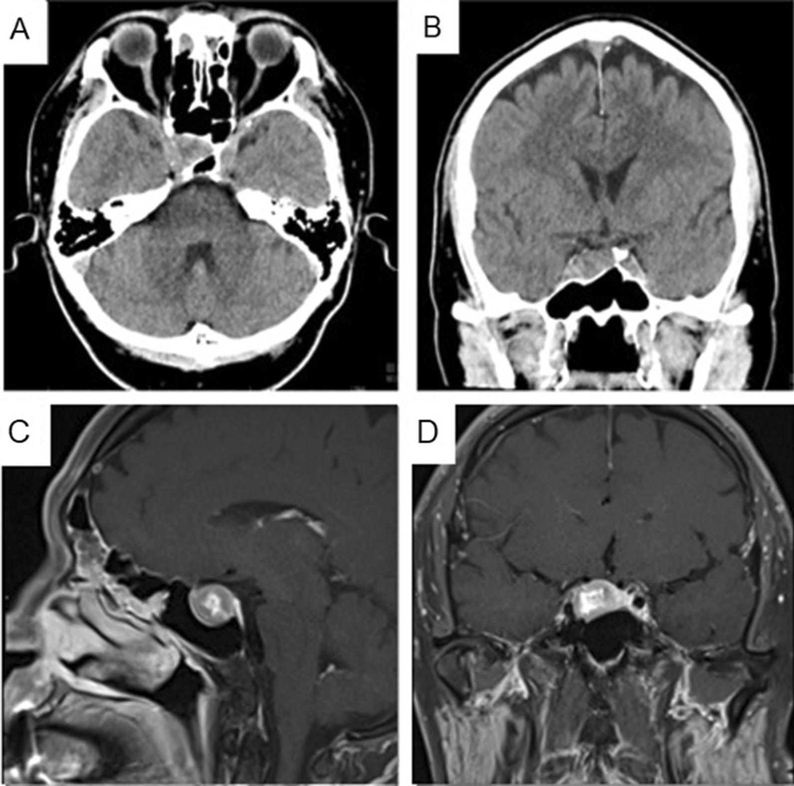
This composite diagnostic image panel displays a pituitary macroadenoma through multi-modal imaging. Panels A (axial) and B (coronal) are non-contrast brain CT scans showing a soft tissue mass within the sella turcica with associated bony remodeling of the skull base. Panels C (sagittal) and D (coronal) are T1-weighted contrast-enhanced MRI scans, providing superior soft tissue resolution. The MRI demonstrates a 1.8 x 1.6 x 1.6 cm heterogeneously enhancing lesion predominantly located in the right aspect of the pituitary gland. Key mass effects include a distinct leftward deviation of the pituitary stalk and possible invasion into the medial portion of the right cavernous sinus. The imaging illustrates the diagnostic criteria for a pituitary macroadenoma, specifically highlighting its relationship with adjacent neurovascular structures like the optic chiasm and cavernous sinuses. This is a critical educational resource for neuroradiology and endocrinology, demonstrating the utility of MRI over CT in evaluating sellar and suprasellar pathology.
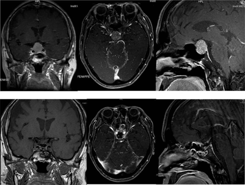
This composite of magnetic resonance imaging (MRI) of the brain and sella turcica demonstrates a large pituitary macroadenoma. The upper row features contrast-enhanced images in coronal, axial, and sagittal planes, revealing a well-circumscribed, enhancing ovoid mass measuring approximately 2.3 x 1.7 x 2.2 cm. The tumor is centered in the sella turcica with significant suprasellar extension into the suprasellar cistern and lateral invasion into the bilateral cavernous sinuses. In the coronal view, the mass is seen abutting the optic chiasm. The bottom row displays corresponding post-operative MRI views (coronal, axial, and sagittal), illustrating the surgical cavity following transsphenoidal resection, with an absence of the previously noted macroadenoma. Key anatomical landmarks visible include the sphenoid sinus, internal carotid arteries within the cavernous sinuses, and the brainstem. These images are diagnostic for a TSH-secreting pituitary macroadenoma in the context of central hyperthyroidism, highlighting the importance of pre-operative mapping of neurovascular involvement.
Graves disease thyroid scintigraphy diffuse uptake
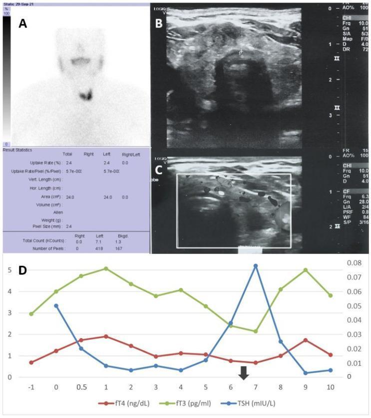
A multi-panel medical figure illustrating diagnostic findings and clinical progression of Graves' disease. Panel (A) displays a Technetium-99m (Tc99) thyroid scintigraphy scan showing a gland with diffuse increased radionuclide uptake, particularly prominent in the inferior left lobe, characteristic of hyperthyroidism. Panel (B) is a grayscale thyroid ultrasound revealing a heterogeneous echotexture of the parenchyma without discrete nodules. Panel (C) utilizes Doppler sonography to demonstrate diffuse hypervascularity (thyroid inferno), a hallmark of active Graves' disease. Panel (D) is a line graph tracking thyroid function tests over 11 months, plotting fT3 (pg/mL) and fT4 (ng/dL) against TSH (mIU/L). The timeline shows initial thyrotoxicosis, stabilization on thiamazole, and a subsequent relapse in hormone levels following a SARS-CoV-2 infection marked by an arrow. This composite serves as an educational resource for correlating nuclear medicine, ultrasonography, and longitudinal biochemical monitoring in autoimmune thyroid disorders.
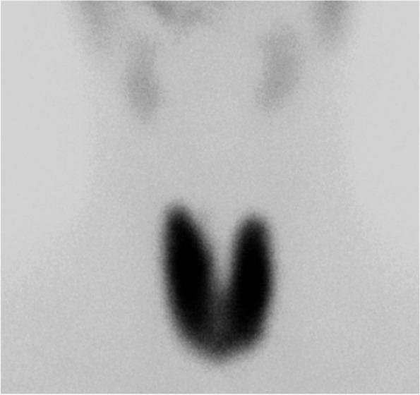
This diagnostic nuclear medicine image shows a Technetium-99m (99mTc) pertechnetate scintigraphy of the thyroid gland in the anterior view. The scan demonstrates markedly increased and diffuse radiotracer uptake throughout both the right and left lobes of the thyroid gland, which maintains its characteristic butterfly shape without evidence of focal nodules or cold zones. The intense, homogenous distribution is a classic hallmark of Graves' disease, indicating global hyperfunction. For comparison, normal physiologic uptake is visible in the salivary glands in the upper portion of the field, though its intensity is significantly lower than that of the hypermetabolic thyroid gland. This imaging modality is essential for differentiating etiologies of thyrotoxicosis, distinguishing diffuse hyperfunction from destructive thyroiditis or toxic multinodular goiter.
Endocrine diseases and imaging: anatomy, physiology, modalities, indications
1. Anatomy and physiology (very short)
| Gland | Location | Main hormones | Main role |
|---|---|---|---|
| Hypothalamus | Diencephalon | TRH, CRH, GnRH, GHRH, somatostatin, dopamine | Controls the pituitary |
| Anterior pituitary | Sella turcica | GH, PRL, ACTH, TSH, FSH, LH | Growth, lactation, and control of the thyroid, adrenals and gonads |
| Posterior pituitary | Sella turcica | ADH, oxytocin | Water balance, uterine contraction and milk ejection |
| Thyroid | Anterior neck, below the larynx | T3, T4, calcitonin | Metabolic rate, calcium (calcitonin) |
| Parathyroids (usually 4) | Behind the thyroid; ectopic glands occur | PTH | Raises serum calcium |
| Adrenal cortex | Above the kidneys | Cortisol, aldosterone, androgens | Stress response, BP and Na/K balance |
| Adrenal medulla | Above the kidneys | Adrenaline, noradrenaline | Fight or flight |
| Pancreatic islets | Pancreas | Insulin, glucagon, somatostatin | Glucose control |
| Gonads | Pelvis or scrotum | Oestrogen, progesterone, testosterone | Reproduction and secondary sex characteristics |
2. Imaging modalities: advantages, disadvantages, indications
Ultrasound (US)
- Advantages:
- No radiation, cheap and widely available.
- Real-time imaging, with Doppler for vascularity.
- Guides fine needle aspiration (FNA).
- More detailed than scintigraphy for thyroid nodules and the most cost-effective way to screen for thyroid adenoma. It also finds extra nodules in 20 to 48% of patients referred with a "solitary" nodule (K J Lee's Essential Otolaryngology, Endocrine section).
- Disadvantages:
- Operator dependent.
- Cannot see retrosternal extension or deep structures well, and is limited in obese patients.
- Gives no functional information.
- Indications:
- Thyroid nodule, goitre and thyroiditis.
- Neck lymph nodes in thyroid cancer.
- Parathyroid adenoma localisation (first-line with scintigraphy).
- Ovarian (PCOS) and testicular pathology.
- FNA guidance.
- Doppler "thyroid inferno" suggests Graves disease.
Contrast methods
- Advantages:
- Show vascular anatomy.
- Sampling localises the hormone source. Inferior petrosal sinus sampling distinguishes pituitary from ectopic ACTH. Adrenal vein sampling distinguishes unilateral from bilateral aldosterone excess. Selective venous sampling localises parathyroid and pancreatic tumours.
- Disadvantages:
- Invasive, with technical failure and complications.
- Iodinated contrast can trigger thyrotoxicosis in nodular goitre, and it delays radioiodine uptake and therapy for weeks.
- Contrast nephropathy and allergy.
- Radiation.
- Indications: Biochemically proven disease with negative or equivocal cross-sectional imaging. This applies to Cushing disease with a negative pituitary MRI (Bailey and Love, Radiological section), primary aldosteronism when surgery is considered, and occult hyperparathyroidism or insulinoma.
Scintigraphy and nuclear medicine
- Tracers:
- Tc-99m pertechnetate, or I-123/I-131 (Sabiston, Nuclear Scintigraphy).
- Tc-99m sestamibi for parathyroid.
- MIBG for pheochromocytoma and paraganglioma.
- Octreotide or Ga-68 DOTATATE PET-CT for neuroendocrine tumours (NETs).
- FDG-PET for dedifferentiated thyroid cancer.
- Advantages:
- Shows function, not just structure.
- Whole-body survey for metastases or multifocal disease.
- Single-session localisation of ectopic tissue (retrosternal goitre, ectopic parathyroid).
- Disadvantages:
- Low spatial resolution.
- Radiation dose, and long scan times.
- False positives, for example sestamibi uptake in thyroid nodules, carcinomas and lymph nodes (Fischer's Mastery of Surgery).
- Contraindicated or restricted in pregnancy and breastfeeding.
- Interference from iodine, drugs or recent contrast.
- Sestamibi is less sensitive for small (<500 mg) glands.
- Indications:
- Hyperthyroidism with a nodule. Scintigraphy establishes solitary versus multiple hyperfunctioning nodules (Plummer disease) and is paired with US (Sabiston, Ultrasound Evaluation of Thyroid Nodules).
- Graves versus thyroiditis: diffuse high uptake in Graves, low uptake in destructive thyroiditis.
- Ectopic thyroid and thyroid cancer remnant or metastases after surgery.
- Parathyroid adenoma: sestamibi SPECT/CT, sometimes with dual-isotope subtraction.
- Pheochromocytoma and paraganglioma (MIBG).
- Neuroendocrine tumours (SSTR PET).
CT
- Advantages:
- Fast, widely available, with excellent spatial resolution.
- Best for bone, calcification and the lungs.
- Measures density (Hounsfield units) and contrast washout.
- Good for adrenals, pancreas and mediastinum.
- 4D-CT localises parathyroid adenomas.
- Disadvantages:
- Ionising radiation.
- Iodinated contrast has the thyroid and renal problems described above.
- Poor soft-tissue contrast in the sella compared with MRI.
- Dental or bone artefact at the skull base.
- Indications:
- Adrenal mass characterisation. Benign adenomas are low density (<10 HU), while carcinomas tend to be hyperattenuating (>18 HU), heterogeneous and irregular (Bailey and Love, Investigation; Schwartz, Adrenal Incidentaloma).
- Pancreatic NETs.
- Retrosternal goitre and tracheal compression.
- Staging of thyroid cancer (non-iodinated contrast if radioiodine is planned).
- Ectopic ACTH search: thorax, abdomen and pelvis.
- Parathyroid 4D-CT.
- Pituitary only if MRI is contraindicated, in which case it shows bony remodelling.
MRI
- Advantages:
- Best soft-tissue contrast.
- No ionising radiation.
- Multiplanar imaging.
- Gadolinium is not nephrotoxic at standard doses and does not affect thyroid function.
- Chemical-shift sequences characterise adrenal adenomas (lipid-rich).
- Disadvantages:
- Expensive, with long scan times and motion artefact.
- Contraindicated with some devices and implants, and claustrophobia is an issue.
- Less sensitive for calcification.
- Limited lung and bone detail.
- Gadolinium caution in severe renal failure.
- Indications:
- Pituitary and hypothalamic region.
- MRI is the preferred method for pituitary anatomy. The normal gland is under 1 cm in height, and microadenomas appear hypointense on T1 (Goldman-Cecil, Diagnosis; K J Lee's, Radiograph Studies).
- Used for adenoma, craniopharyngioma, Rathke cleft cyst, hypophysitis and stalk lesions. Look for optic chiasm compression and cavernous sinus invasion.
- Adrenal masses, for example in pregnancy, children, or when CT is indeterminate.
- Pancreatic NETs and liver metastases.
- Parathyroid, as an alternative localisation tool.
- Neck, for local extension of thyroid cancer.
- Pituitary and hypothalamic region.
3. Disease-by-disease: best test
| Organ | Disease | First-line | Next / problem solver |
|---|---|---|---|
| Pituitary | Adenoma (prolactinoma, acromegaly, Cushing disease, non-functioning) | Pituitary-protocol MRI | CT if MRI is contraindicated; inferior petrosal sinus sampling for ACTH source |
| Thyroid | Nodule, goitre | US, then FNA | Scintigraphy if TSH is suppressed; CT/MRI for retrosternal extent |
| Thyroid | Graves disease | Biochemistry plus US (Doppler) | Scintigraphy: diffuse uptake |
| Thyroid | Thyroiditis | US | Scintigraphy: low uptake |
| Thyroid | Cancer | US plus FNA | CT/MRI for staging; I-131 whole-body scan; FDG-PET if radioiodine-refractory |
| Parathyroid | Primary hyperparathyroidism | US plus sestamibi SPECT/CT | 4D-CT, MRI, venous sampling |
| Adrenal | Incidentaloma | Unenhanced CT (HU) | Washout CT, chemical-shift MRI, FDG-PET |
| Adrenal | Conn syndrome | CT | Adrenal vein sampling |
| Adrenal | Cushing syndrome | By source: pituitary MRI, adrenal CT, thorax/abdomen CT | Ga-68 SSTR PET-CT for ectopic ACTH (Bailey and Love) |
| Adrenal | Pheochromocytoma | CT or MRI (T2 bright) | MIBG or DOTATATE for multifocal or metastatic disease |
| Pancreas | Insulinoma, gastrinoma, other NETs | Multiphase CT/MRI | Endoscopic US, SSTR PET-CT, venous sampling |
| Bone | Osteoporosis, rickets | DXA, X-ray | CT/MRI for complications |
4. Key principles
- Biochemistry first, imaging second. Image only after the hormonal diagnosis is confirmed, because incidentalomas are common.
- Avoid iodinated contrast before radioiodine scanning or therapy, and in thyrotoxic nodular goitre.
- Thyroid: US plus FNA for structure, scintigraphy for function.
- Pituitary: MRI.
- Adrenal: CT density, with MRI as the problem solver.
- Parathyroid: US plus sestamibi SPECT/CT.
- Neural crest and neuroendocrine tumours: MIBG or SSTR PET.