
define : Haemarthrosis
haemarthrosis joint bleeding knee swelling
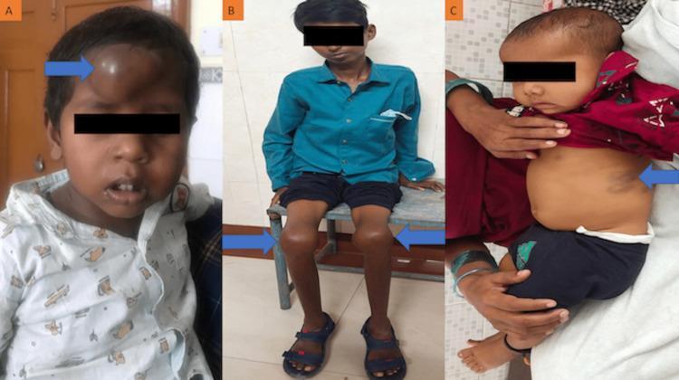
A composite of three clinical photographs (A, B, C) illustrating the varied clinical manifestations of haemophilia in pediatric patients. Image A shows a five-year-old child with a prominent, dome-shaped, tense swelling on the central forehead, identified as an extradural haematoma. The skin overlying the mass is hyperpigmented and dark reddish-brown. Image B displays a thirteen-year-old patient with significant, bilateral knee joint enlargement (haemarthrosis). The knees exhibit a bulbous, swollen contour with smooth, taut overlying skin, which is characteristic of recurrent intra-articular bleeding. Image C depicts a three-year-old child with a large, purple-to-blue ecchymosis patch on the left lateral abdominal wall. These images demonstrate the classic hemorrhagic complications associated with coagulation disorders, ranging from soft tissue hematomas and ecchymosis to chronic joint arthropathy. Blue arrows highlight the specific sites of pathology in each case.
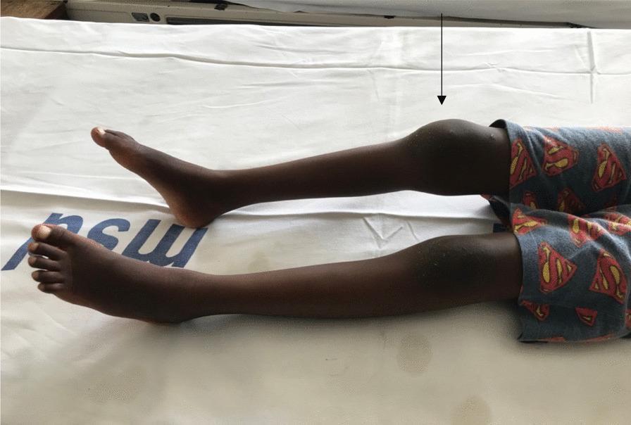
This clinical photograph displays a pediatric patient's lower extremities in a supine position, highlighting significant joint pathology. The primary finding is marked, asymmetrical swelling of the right knee joint compared to the relatively normal contralateral left knee. A black arrow indicates the site of localized joint effusion or soft tissue swelling, which manifests as a prominent, rounded protrusion that distorts the anatomical contour of the patellar and suprapatellar regions. The skin overlying the swelling appears intact without obvious erythema, though the distension is substantial. In the clinical context of a coagulopathy, such as Factor VII deficiency, this visual presentation is highly suggestive of hemarthrosis. Both legs are extended, and no deformities of the feet are visible. This image serves as a teaching aid for identifying joint swelling and understanding the physical manifestations of bleeding disorders in a musculoskeletal examination.
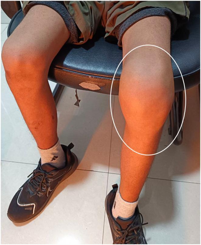
This clinical photograph displays a comparative view of the lower limbs in a seated patient, focusing on the knee joints. The primary finding is significant, diffuse swelling of the left knee (highlighted with a white circle) compared to the anatomically normal right knee. The swelling is localized around the patellar region, extending into the suprapatellar and infrapatellar areas, resulting in the loss of normal bony landmarks and joint contours. The skin overlying the affected joint appears intact without obvious erythema, bruising, or open lesions, though it appears tense due to underlying mass effect or effusion. This presentation is characteristic of hemophilic arthropathy or pseudotumor in a patient with a bleeding disorder like hemophilia A. The image serves as an educational tool for identifying joint effusion and musculoskeletal manifestations of systemic hematologic conditions. Key anatomical landmarks include the distal femur, proximal tibia, and patella, all obscured by the localized enlargement.
Haemarthrosis
- Roberts and Hedges' Clinical Procedures in Emergency Medicine, p. 4372
Most Commonly Affected Joints
Causes
- High association with ligamentous injury (e.g. ACL tear - patients often report a "pop" followed by rapid effusion) or intra-articular fracture
- Effusions can range from minor to large, painful collections that impede range of motion
- Bailey and Love's Short Practice of Surgery, 28th Ed.
-
Hereditary clotting factor deficiency - most common cause; haemophilia A and B are the classic associations. Spontaneous haemarthrosis is a hallmark of untreated haemophilia, and its severity correlates directly with the degree of factor deficiency
-
Oral anticoagulant therapy - can occur even when the prothrombin time is within the therapeutic range
-
Sickle cell anaemia
-
Pseudogout
-
Amyloidosis
-
Pigmented villonodular synovitis (PVNS) - recurrent haemarthrosis causes haemosiderin deposition in the synovium, visible as low signal on both T1 and T2 MRI sequences
-
Synovial haemangioma
-
Rheumatoid arthritis
-
Intra-articular infection
-
Roberts and Hedges' Clinical Procedures in Emergency Medicine, p. 4375; Tintinalli's Emergency Medicine, p. 135; Harrison's Principles of Internal Medicine 22E (2025), p. 617
Pathophysiology of Recurrent Haemarthrosis
- Blood accumulates in the joint cavity
- Synovial tissue cannot fully remove blood remnants
- This triggers synovial inflammation and proliferation
- The inflamed synovium develops a rich network of new, fragile blood vessels - making the joint vulnerable to further bleeding
- With enough cycles, irreversible haemophilic arthropathy develops - progressive cartilage degeneration, bone changes, chronic pain, joint stiffness, and severely limited range of motion
- Firestein & Kelley's Textbook of Rheumatology, p. 455-484
Clinical Features
| Feature | Details |
|---|---|
| Swelling | Rapid, tense joint effusion |
| Pain | Acute, often severe |
| Warmth | Present due to inflammatory response |
| Range of motion | Restricted; joint may be "locked" |
| Skin changes | Intact; taut due to distension |
Management
-
Traumatic haemarthrosis: Immobilisation, ice, and elevation; aspiration of large effusions for pain relief (no effect on long-term outcome); orthopaedic follow-up for ligamentous/articular injuries
-
Haemophilia-related: Specific clotting factor replacement first; aspiration is controversial but may be considered for large collections within the first 12 hours, only after factor replacement; haematology + orthopaedic co-management
-
Anticoagulant-related: Discontinue anticoagulant therapy (weighing risk of thromboembolism); chronic arthritis is not a typical long-term complication in this group
-
Tintinalli's Emergency Medicine, p. 130-135
Clinical Images
What are some common causes of joint effusions?
joint effusion synovial fluid classification causes
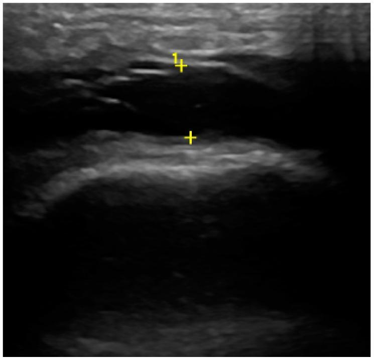
This diagnostic ultrasound image demonstrates a joint effusion, likely within the temporomandibular joint (TMJ) based on the clinical context of condylar assessment. The primary finding is an elongated, anechoic to hypoechoic fluid-filled space situated between the joint structures. This area of synovial effusion is dark and devoid of internal echoes, suggesting simple fluid without significant debris or hemorrhage. The bordering osseous surfaces are characterized by hyperechoic, bright curvilinear lines representing the bony cortex. Yellow crosshair markers (labeled '1+') indicate specific measurement points for the width of the joint space or the depth of the effusion. The bone surface appears relatively smooth and regular in the visible segment, though the field of view is localized. This imaging modality is used in rheumatology and dentistry to detect joint inflammation, synovitis, and internal derangements by visualizing soft tissue components and cortical integrity that are not visible on standard radiographs.
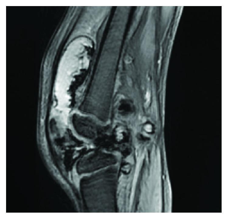
This diagnostic image is a sagittal T2-weighted magnetic resonance imaging (MRI) scan of a human knee joint. The most prominent finding is a massive, complex joint effusion characterized by a large volume of high-signal intensity fluid distending the suprapatellar bursa and joint capsule. The fluid collection demonstrates internal heterogeneity and irregular borders, which contrast with the typical homogeneous appearance of simple synovial fluid. There is visible thickening of the synovial membrane, and the posterior aspect of the joint shows signs of villonodular proliferation. The femur, tibia, and patella are visualized, with the patella displaced anteriorly due to the significant volume of the effusion. This imaging is characteristic of pigmented villonodular synovitis (PVNS), illustrating the hallmark combination of extensive hemarthrosis and synovial hyperplasia. The image serves as an educational tool for identifying pathological joint distension and distinguishing complex intra-articular fluid collections from simple effusions in the context of synovial proliferative disorders.
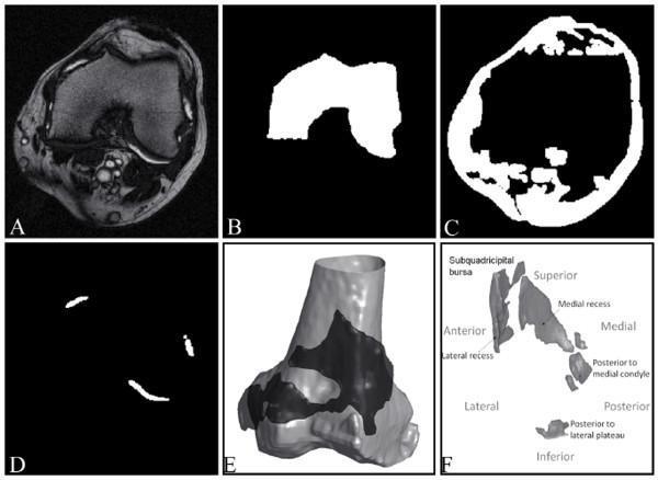
This figure illustrates the multi-stage computational segmentation process of synovial fluid in a patient with knee osteoarthritis using MRI. Panel (A) shows a source axial T2-weighted MRI slice of the knee joint, demonstrating anatomical structures including the femur, patella, and surrounding soft tissues with varying signal intensities. The joint effusion appears as high-intensity (bright) signals within the joint space. Panels (B), (C), and (D) present intermediate binary masks generated during segmentation: (B) masks the femur, (C) identifies other non-fluid structures such as patellar fat pads and cortical bone, and (D) isolates the synovial effusion as discrete white regions. Panels (E) and (F) provide 3D reconstructions of the synovial fluid volume. (E) shows the spatial relationship of the joint effusion (dark grey) as it wraps around the distal femur (light grey). (F) provides a labeled 3D map of the effusion, identifying specific anatomical compartments including the subquadricipital bursa, medial and lateral recesses, and posterior fluid pockets. This resource demonstrates the application of medical imaging processing to quantify joint inflammation and effusion volume.
Common Causes of Joint Effusions
Classification by Synovial Fluid Type
| Fluid Type | WBC Count | Key Conditions |
|---|---|---|
| Non-inflammatory | < 2,000/mm³ | Osteoarthritis, trauma, osteochondritis, sickle cell disease |
| Inflammatory | > 2,000/mm³ | RA, gout, pseudogout, psoriatic arthritis, reactive arthritis, SLE, ankylosing spondylitis |
| Septic (purulent) | > 50,000/mm³ (often > 100,000) | Bacterial septic arthritis, gonococcal arthritis |
| Haemorrhagic | Blood-stained | Trauma, haemophilia, anticoagulant therapy, haemangioma, tumours, PVNS |
- Tietz Textbook of Laboratory Medicine, 7th Ed., p. 3306
1. Trauma
2. Osteoarthritis (OA)
3. Rheumatoid Arthritis (RA)
4. Crystal Arthropathies
- Gout - Monosodium urate (MSU) crystals in synovial fluid; strongly negatively birefringent under polarised light. Typically monoarticular, affecting the first MTP joint, ankle, or knee
- Pseudogout (CPPD) - Calcium pyrophosphate dihydrate (CPPD) crystals; weakly positively birefringent. Commonly affects the knee, wrist, and other large joints
5. Septic Arthritis
6. Seronegative Spondyloarthropathies
7. Systemic Connective Tissue Diseases
- Systemic lupus erythematosus (SLE)
- Behçet disease
- Polyarteritis nodosa
8. Haemorrhagic Causes (Non-traumatic)
- Haemophilia A and B (most common non-traumatic cause of haemarthrosis)
- Oral anticoagulant therapy - can occur even within therapeutic range
- Pigmented villonodular synovitis (PVNS) - recurrent haemarthrosis + low-signal synovium on MRI
- Synovial haemangioma
- Intra-articular tumours
- Sickle cell anaemia
9. Other Notable Causes
| Cause | Notes |
|---|---|
| Lyme arthritis | Inflammatory effusion; history of tick exposure/endemic area; negative cultures |
| Viral arthritis | Often polyarticular, self-limiting |
| Acute rheumatic fever | Migratory polyarthritis following group A strep infection |
| Subacute bacterial endocarditis | Must not be missed in polyarthritis |
| Paraneoplastic syndrome | Arthritis as a cancer manifestation |
| Post-arthroplasty | Recurrent haemarthrosis after total knee replacement is a recognised complication |
Key Principle
"In acute monoarthritis, synovial fluid analysis is the most valuable laboratory test."
- Firestein & Kelley's Textbook of Rheumatology
What is a knee joint aspiration and why might you perform one?
knee joint aspiration arthrocentesis technique needle insertion
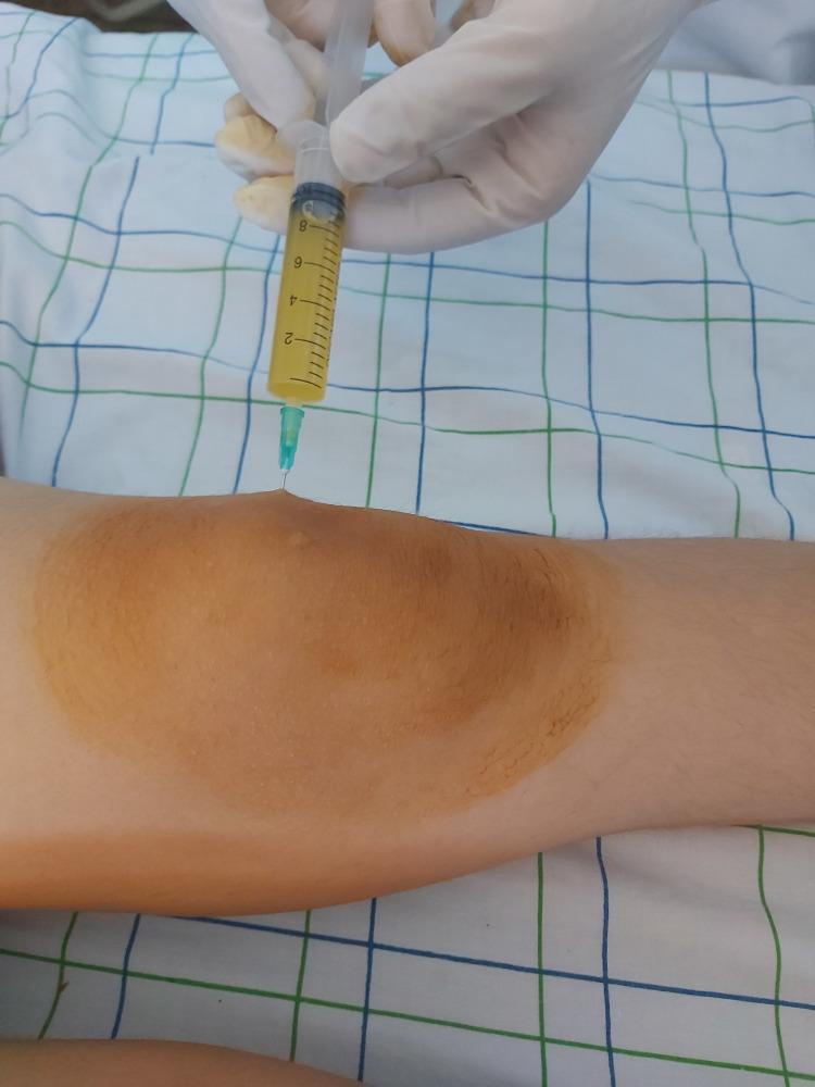
Clinical photograph demonstrating a bedside arthrocentesis (joint aspiration) of the knee. The image shows a gloved medical professional inserting a needle into the suprapatellar region of a significantly swollen knee joint. The knee exhibits a prominent joint effusion and has been prepped with a brown-tinted antiseptic solution (such as povidone-iodine), resulting in local skin discoloration. A transparent syringe attached to the needle contains approximately 3-4 mL of aspirated synovial fluid. The fluid is described as citrine yellow and appears transparent with an inflammatory quality, lacking visible turbidity, blood, or purulence. This procedure is being performed to evaluate for conditions such as septic arthritis or acute rheumatic fever in the context of polyarthritis. The visual highlights the technique for synovial fluid analysis and the physical manifestation of knee joint effusion.
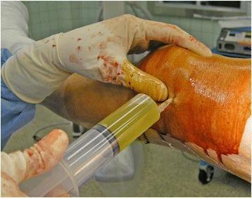
This clinical photograph captures a knee arthrocentesis (joint aspiration) being performed in an operative setting. A gloved healthcare provider is seen inserting a needle into the distended suprapatellar space of a patient's knee, which has been prepped with a povidone-iodine antiseptic solution. The procedure has successfully extracted a significant volume of synovial fluid into a large-bore syringe. The fluid exhibits a distinct yellow-amber color and a turbid (cloudy) appearance, which is a key diagnostic indicator. Such macroscopic findings typically suggest an inflammatory process, such as rheumatoid arthritis, gout, or pseudogout (calcium pyrophosphate deposition disease), rather than normal, clear, straw-colored synovial fluid. The image serves as an educational tool for demonstrating the technique of joint aspiration in the context of postoperative knee effusion and the clinical assessment of synovial fluid clarity and color for differential diagnosis in orthopedics and rheumatology.
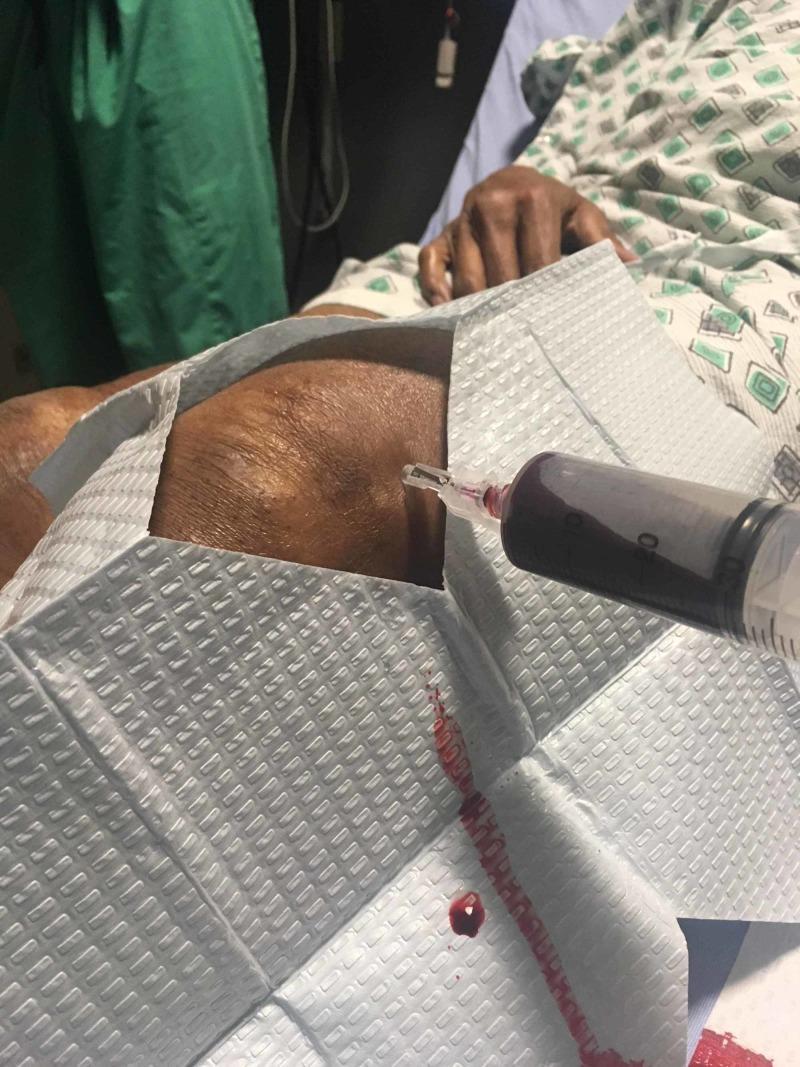
Clinical photograph depicting an arthrocentesis procedure being performed on a patient's knee. The image shows a large-bore syringe attached to a needle inserted into the suprapatellar region of the joint space. The syringe is partially filled with dark red, opaque aspirated fluid, which is visually consistent with hemarthrosis (blood within the joint). A sterile, textured gray drape surrounds the puncture site, with a small trail and droplet of sanguineous fluid visible on the drape, extending from the insertion point. The patient is positioned supine in a hospital setting, partially visible in a patterned gown. This procedural image demonstrates the diagnostic and therapeutic aspiration of a joint effusion, relevant for orthopedic and rheumatological education regarding the identification of hemarthrosis following joint trauma or coagulopathy.
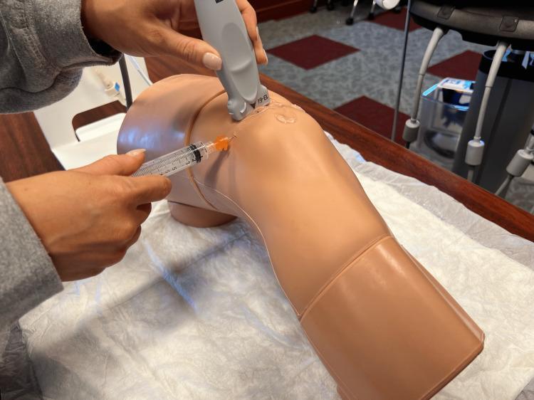
This procedural photograph illustrates a demonstration of an ultrasound-guided knee aspiration (arthrocentesis) on a medical simulator. The image shows a realistic, light-brown anatomical model of a human knee resting on a white clinical drape. A practitioner's hand is seen holding a gray linear ultrasound transducer positioned in the superolateral aspect of the patella, with clear conductive gel visible at the contact point. Simultaneously, another hand is inserting a clear plastic syringe equipped with a fine-gauge needle (indicated by the orange hub) into the joint space. The needle is oriented parallel to the transducer's long axis, demonstrating the 'in-plane' needle visualization technique common in musculoskeletal procedures. This setup teaches medical students how to utilize real-time imaging to guide needle placement into the suprapatellar bursa for diagnostic fluid aspiration or therapeutic injection. The context emphasizes procedural accuracy, landmarking, and the integration of ultrasound technology in clinical skills training for orthopedic or emergency medicine specialties.
Knee Joint Aspiration (Arthrocentesis)
Definition
- Roberts and Hedges' Clinical Procedures in Emergency Medicine, p. 4250
Why Perform It? - Indications
Diagnostic Indications
| Indication | What You Are Looking For |
|---|---|
| Suspected septic arthritis | Organisms on Gram stain, high WBC (> 50,000/mm³), positive culture |
| Crystal arthropathy | MSU crystals (gout) or CPPD crystals (pseudogout) under polarised light |
| Inflammatory arthritis | Elevated WBC 2,000-50,000/mm³ (RA, reactive arthritis, etc.) |
| Traumatic injury | Haemorrhagic fluid; fat globules indicate intra-articular fracture |
| Distinguish articular vs. periarticular disease | Confirms fluid is truly intra-articular |
| Check for communication between a laceration and the joint space | Important in open knee wounds |
"In the ED it is difficult to make an accurate assessment of an acutely painful, hot, and swollen joint without performing arthrocentesis."
- Roberts and Hedges' Clinical Procedures in Emergency Medicine, p. 4250
Therapeutic Indications
-
Relief of pain from a tense, large effusion or haemarthrosis - aspiration reduces capsular pressure and improves range of motion
-
Instillation of medications - corticosteroids (e.g. triamcinolone 20-40 mg or betamethasone 6-12 mg + lidocaine 1%) for inflammatory conditions unrespected of infection
-
Relief of haemarthrosis in haemophilia - performed after clotting factor replacement has been given
-
Textbook of Family Medicine, 9th Ed., p. 894; Roberts and Hedges', p. 4278
Contraindications
| Type | Detail |
|---|---|
| Absolute | Overlying cellulitis or soft tissue infection at the proposed puncture site |
| Relative | Bleeding diathesis (rarely an absolute bar - haemarthrosis relief in haemophilia is actually an accepted indication after factor replacement) |
| Relative | Prosthetic joint - high infection risk; avoid unless prosthetic joint infection is itself suspected |
| Relative | Known bacteraemia - theoretical risk of seeding the joint, but weighed against the value of diagnostic information |
Technique (Knee)
- Consent and position - patient supine with knee slightly flexed over a rolled towel
- Assess for effusion - a large effusion causes the patella to "float" (ballottement sign); the capsule shows an inverted U-shaped swelling
- Mark and prepare - identify the aspiration site; cleanse with chlorhexidine or povidone-iodine
- Needle insertion - a 22-gauge needle is inserted from the lateral aspect, 1 cm lateral and superior to the patella, directed 45 degrees downward and angled under the patella; aspirate during insertion
- Aspirate - when the joint space is entered, fluid enters the syringe rapidly; milking the medial and lateral recesses improves yield
- Switch syringe if injecting - the needle can be left in place and the syringe changed under sterile conditions to inject a steroid if required
- Textbook of Family Medicine, 9th Ed., p. 894
What the Fluid Tells You - Gross Examination
| Appearance | Implication |
|---|---|
| Clear, straw-coloured, highly viscous (strings on drop test) | Normal or non-inflammatory |
| Cloudy, reduced viscosity | Inflammatory (e.g. RA) |
| Floridly opaque/purulent | Septic arthritis |
| Homogeneously bloody, no clot | True haemarthrosis |
| Blood streaks in otherwise yellow fluid | Procedural/traumatic tap (not haemarthrosis) |
| Particulate debris/fat globules | Intra-articular fracture |
- Firestein & Kelley's Textbook of Rheumatology, p. 1757-1792
Synovial Fluid WBC Classification
| Category | WBC Count | Typical Causes |
|---|---|---|
| Normal | < 180 cells/mm³ | - |
| Non-inflammatory | < 2,000 cells/mm³ | OA, trauma |
| Inflammatory | 2,000-50,000 cells/mm³ | RA, gout, pseudogout, reactive arthritis |
| Septic | > 50,000 cells/mm³ (often > 100,000) | Bacterial septic arthritis |
- Firestein & Kelley's Textbook of Rheumatology, p. 1800
Complications
-
Introduction of infection (iatrogenic septic arthritis) - very rare with proper sterile technique
-
Bleeding / haemarthrosis
-
Pain at puncture site
-
Allergy to local anaesthetic
-
Roberts and Hedges', p. 4296-4302
Clinical Images
In simple terms, what is involved in aspirating a knee joint?
Aspirating a Knee Joint - In Simple Terms
What is it?
Step-by-Step: What Actually Happens
- Colour and clarity - is it clear, cloudy, or bloody?
- White blood cells - a high count suggests infection or inflammation
- Crystals - to check for gout or pseudogout
- Bacteria - to check for infection
Does it hurt?
After the procedure
- The knee may feel sore or achy for a day or two
- Most people can walk and go home immediately
- If a steroid was also injected, pain and swelling often improve within a few days
Key risks (rare)
- Introducing infection into the joint (very uncommon with proper technique)
- Minor bleeding at the puncture site
- Temporary discomfort
When should you not perform a joint aspiration/injection?
When Should You NOT Perform a Joint Aspiration or Injection?
Absolute Contraindications - Do NOT Proceed
| Contraindication | Why |
|---|---|
| Overlying cellulitis or skin infection | Inserting a needle through infected skin drives bacteria directly into the joint, risking catastrophic septic arthritis. This is the one firm rule across all textbooks. |
| Known infectious arthritis (septic joint) | An absolute bar to therapeutic injection (e.g. steroids). A septic joint must be drained diagnostically and treated with antibiotics - injecting steroids into it is dangerous. |
| Periarticular cellulitis, ulceration, or adjacent osteomyelitis | Same principle - active local infection at the site is a hard stop for any injection. |
| Hypersensitivity/allergy to the agent being injected | (Relevant to steroid injections) - allergy to the corticosteroid or its vehicle is an absolute contraindication. |
| Osteochondral fracture | Injecting a corticosteroid into a joint with a fracture risks impairing healing and causing further damage. |
| Uncontrolled bleeding disorder | If coagulation is severely deranged and uncontrolled, the risk of inducing haemarthrosis is significant. |
- Roberts and Hedges' Clinical Procedures in Emergency Medicine, p. 3016-3021
Relative Contraindications - Proceed with Caution
| Contraindication | Key Nuance |
|---|---|
| Anticoagulant therapy (warfarin, DOACs) | Studies show the risk of iatrogenic haemarthrosis is extremely low - less than 0.5% complication rate, even at INR up to 4.5. Anticoagulation alone should NOT stop you aspirating a joint when it is clinically needed. If septic arthritis is suspected, proceed regardless. |
| Prosthetic (artificial) joint | Prosthetic joints carry a high infection risk from any needle procedure. Aspiration or injection should be avoided without first discussing with the orthopaedic surgeon who placed it. However, if a prosthetic joint infection is suspected and no specialist is available, aspiration can be done. |
| Known bacteraemia (bacteria in the bloodstream) | Theoretical risk that bacteraemia seeds the joint via the needle. Weighed against the diagnostic value - if septic arthritis is suspected, this does not override the need to aspirate. |
| Poorly controlled diabetes | Relevant specifically to steroid injections - corticosteroids raise blood glucose and can cause a significant temporary spike. Must counsel the patient and arrange monitoring. |
| Bleeding diathesis (e.g. haemophilia) | A relative, not absolute, bar. Aspirating a tense haemarthrosis in haemophilia is actually an accepted therapeutic procedure - but only after the appropriate clotting factor has been replaced first. |
| Haemarthrosis | Relative caution with steroid injection into a bloody joint, as the diagnosis and cause of the bleed should be established first. |
| Joint instability or internal joint derangement | Injection may not be appropriate if structural integrity of the joint is compromised. |
| Partial tendon rupture | Corticosteroid injected near a partially torn tendon risks precipitating complete rupture. |
| Adjacent abraded skin or chronic skin breakdown (e.g. decubitus ulcer) | Local infection risk even if not frank cellulitis. |
| Inaccessible or deep joint without imaging guidance | Deep joints (hip, sacroiliac) carry greater risk when done blind - ultrasound or fluoroscopy guidance should be used. |
- Roberts and Hedges' Clinical Procedures in Emergency Medicine, p. 3024-3035; Rosen's Emergency Medicine, p. 2218
One Critical Distinction: Aspiration vs. Injection
"Suspected septic arthritis is a contraindication to therapeutic injection but an indication for diagnostic joint aspiration."
- Swanson's Family Medicine Review, p. 318
- Do NOT inject a steroid (this will make the infection dramatically worse)
- Do aspirate the joint urgently - you need the fluid for culture, and draining it relieves pressure and reduces bacterial load
Summary Table
| Situation | Aspiration | Steroid Injection |
|---|---|---|
| Overlying cellulitis | ❌ Absolute contraindication | ❌ Absolute contraindication |
| Suspected septic arthritis | ✅ Urgently indicated | ❌ Absolutely contraindicated |
| Anticoagulation therapy | ✅ Generally safe to proceed | ⚠️ Relative caution (usually safe) |
| Prosthetic joint | ⚠️ Discuss with orthopaedics first | ⚠️ Avoid unless specialist agreement |
| Haemophilia with haemarthrosis | ✅ After factor replacement | ⚠️ Relative caution |
| Poorly controlled diabetes | ✅ No specific restriction | ⚠️ Steroid will spike glucose - counsel patient |
| Osteochondral fracture | ✅ Fine | ❌ Contraindicated |
| Drug allergy to agent | N/A | ❌ Absolute contraindication |
Substances may also be injected into joint spaces to manage some medical conditions. What sorts of substances may be injected into a knee joint?
Substances Injected into the Knee Joint
1. Corticosteroids (Most Common)
- Rheumatoid arthritis (RA) - for temporary, localised flare control
- Osteoarthritis with active synovitis
- Crystal arthropathy (gout, pseudogout) flares
- General inflammatory joint disease
| Drug | Brand Name | Knee Dose |
|---|---|---|
| Hydrocortisone acetate | - | 40-100 mg |
| Prednisolone tebutate | Hydeltr-TBA | 20-30 mg |
| Methylprednisolone acetate | Depo-Medrol | 20-80 mg |
| Triamcinolone acetonide | Kenalog | 20-40 mg |
| Triamcinolone hexacetonide | Aristospan | 20-30 mg |
| Dexamethasone | Decadron | 2-4 mg |
| Betamethasone | Celestone Soluspan | 6-12 mg |
2. Local Anaesthetics
- To provide immediate pain relief while waiting for the steroid to take effect (steroids typically take 24-48 hours; lidocaine works within minutes)
- As a diagnostic tool - if injection of anaesthetic into a joint abolishes the patient's pain, it confirms the pain is originating from within that joint (rather than from surrounding structures)
- To make the procedure itself more comfortable
3. Hyaluronic Acid (Viscosupplementation)
- Moderate-to-severe knee osteoarthritis - particularly when corticosteroids have given insufficient or short-lived relief, and the patient is not yet a candidate for knee replacement
- Not used for inflammatory arthritis (RA, gout) - it is specific to degenerative joint disease
-
Must be placed strictly intra-articular (not into surrounding soft tissue) - more technically demanding than steroid injection
-
All joint effusion should be drained before injection
-
Must not be mixed with lidocaine or steroids in the same syringe
-
A 22-gauge or larger needle is required (the thick gel resists flow through smaller needles)
-
Ultrasound or fluoroscopy guidance is often used to confirm correct placement
-
Pfenninger and Fowler's Procedures for Primary Care, p. 5400-5413
4. Local Anaesthetic Alone (Diagnostic / Short-Term Therapeutic)
- Confirming the source of pain (diagnostic injection)
- Temporary pain relief when steroid injection is contraindicated (e.g. suspected infection pending results, or the steroid limit has been reached for that year)
5. Other / Emerging Agents
| Agent | Notes |
|---|---|
| Platelet-Rich Plasma (PRP) | The patient's own blood is centrifuged to concentrate growth factors; the resulting plasma is injected to promote tissue repair and reduce inflammation. Used in knee OA and sports injuries. Evidence is growing but still debated. |
| Stem cell / biological agents | Under investigation for cartilage regeneration in OA; not yet standard of care |
| Radioactive agents | Radiosynovectomy (e.g. yttrium-90) - a nuclear medicine procedure used in RA to ablate the inflamed synovial membrane when other treatments have failed; specialist use only |
| Antibiotics | Occasionally instilled directly into a joint in cases of septic arthritis (rare; usually systemic antibiotics are sufficient) |
Summary
| Substance | Primary Purpose | Main Condition |
|---|---|---|
| Corticosteroids | Reduce inflammation | RA, OA flares, crystal arthropathy |
| Local anaesthetic | Pain relief / diagnosis | Acute pain, diagnostic confirmation |
| Hyaluronic acid | Restore joint lubrication | Osteoarthritis |
| PRP | Tissue repair, anti-inflammatory | OA, sports injuries |