
Schematic drawing of micro hi test
microhemagglutination test schematic diagram MHA-TP syphilis
micro hemagglutination test schematic diagram MHA TP principle
Schematic diagram of Micro Hemagglutination Test (MHA-TP) for syphilis. Show a U-bottom microplate well layout with two columns labeled "Test" and "Control". In the Test well: red blood cells coated with Treponema pallidum antigen mixing with patient serum containing anti-treponemal antibodies, resulting in a smooth mat of agglutinated red cells covering the well bottom (positive result). In the Control well: sensitized red cells without antibodies, showing a tight compact button/pellet at the well bottom (negative result). Label each component clearly: "Antibody (patient serum)", "Antigen-coated RBCs", "Agglutination mat (Positive)", "Button pellet (Negative)". Include a small legend explaining the passive hemagglutination principle with arrows showing antibody bridging between two antigen-coated red blood cells. Clean educational medical illustration style with white background.
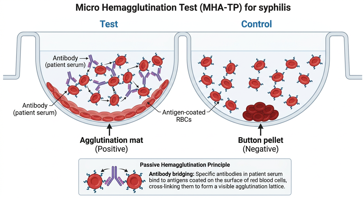
Micro Hemagglutination Test (MHA-TP) - Schematic Overview
| Component | Role |
|---|---|
| Red blood cells (avian/sheep) | Passive carrier - coated with T. pallidum (Nichols strain) antigen |
| Patient serum | Source of anti-treponemal antibodies |
| Antibody bridges | Link antigen-coated RBCs together, causing visible agglutination |
- Positive (+): Antibodies in serum bind the antigen-coated RBCs and cross-link them, forming a diffuse mat or carpet spreading across the U-bottom well
- Negative (-): No antibodies present - RBCs settle into a tight compact button/pellet at the bottom of the well
- MHA-TP is a microplate version of the TPHA (Treponema pallidum Hemagglutination Assay)
- The term "passive" means the RBCs are passive carriers - the antigen-antibody reaction does the work
- Sensitivity in primary syphilis: ~46-88% (lower than in secondary/latent stages)
- Now largely replaced by TP-PA and automated treponemal EIAs in the US, but still used internationally per CDC 2024 syphilis testing guidelines
Micro hemagglutination inhibition test
hemagglutination inhibition test schematic diagram HI test virology
micro hemagglutination inhibition test principle schematic diagram virology
Schematic diagram of the Micro Hemagglutination Inhibition (micro-HI) test in a U-bottom microplate well format. Show three side-by-side wells with clear labels: Well 1 - "Positive Control (Hemagglutination)": virus particles with hemagglutinin spikes binding to red blood cells, forming a large lattice/mat spreading across the well bottom. Label: "No antibody → RBCs agglutinate → diffuse mat (positive HA)". Well 2 - "Test Well (Inhibition - HI Positive)": patient serum antibodies (Y-shaped) bound to virus hemagglutinin spikes, blocking virus from reaching RBCs. RBCs settle as a tight compact button at bottom. Label: "Antibody blocks virus → No agglutination → button pellet = HI positive". Well 3 - "Negative Control": only RBCs, no virus, compact button at bottom. Below the wells, show a serial dilution row (8 wells left to right) with decreasing antibody concentration showing transition from button (HI positive, inhibited) to mat (HI negative, agglutination), with an arrow labeled "Increasing dilution". Mark the last inhibited well as "HI Titer endpoint". Also include a small inset box showing the molecular mechanism: a virus particle with HA spikes, one side bound by antibody (blocked), other side free and binding to RBC sialic acid receptor. Clean, color-coded educational medical microbiology illustration. White background. Blue = virus, Red = RBCs, Yellow = antibodies.
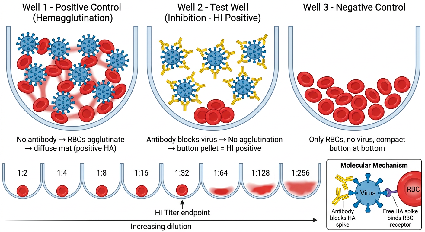
Micro Hemagglutination Inhibition (Micro-HI) Test
Core Concept
Key logic: No clumping = antibodies present = HI Positive (immunity/infection). Clumping = no antibodies = HI Negative.
Step-by-Step Procedure
| Step | What Happens |
|---|---|
| 1. Serum pretreatment | Remove non-specific inhibitors (e.g., with receptor-destroying enzyme, RDE, or kaolin) |
| 2. Serial 2-fold dilutions | Patient serum diluted across a row of U-bottom microplate wells (1:10, 1:20, 1:40...) |
| 3. Add standardized virus | Fixed dose of virus (4 HAU) added to each well; incubate 30-60 min at RT |
| 4. Add RBCs | 0.5-0.75% RBC suspension added; incubate 30-60 min at 4°C |
| 5. Read results | Observe each well for agglutination pattern |
Reading the Results
| Pattern | Meaning |
|---|---|
| Compact button pellet (RBCs settle) | Hemagglutination INHIBITED = antibodies present ✓ |
| Diffuse mat/lattice (RBCs spread out) | Hemagglutination occurred = no antibodies ✗ |
Applications
- Influenza (most classic use) - strain typing and vaccine efficacy
- Rubella - serosurveillance and antenatal immunity screening
- Arboviruses - dengue, Japanese encephalitis, West Nile
- Mumps, measles (paramyxoviruses)
- Newcastle disease virus (veterinary)
Comparison: HI Test vs. MHA-TP
| Feature | Micro-HI Test | MHA-TP |
|---|---|---|
| Disease | Viral infections (influenza, rubella) | Syphilis (T. pallidum) |
| Principle | Antibody inhibits viral HA from agglutinating RBCs | Antibody causes passive agglutination of antigen-coated RBCs |
| Positive result | Button (no agglutination) | Mat (agglutination) |
| RBC role | Natural target of virus HA | Passive carrier of treponemal antigen |
passive haemagglutination test principle applications advantages SRBC chemicals used absorbable antigens tannic acid chromic chloride
https://microbenotes.com/hemagglutination-assay-principle-ty…
Passive Haemagglutination (PHA) Test - Complete Answers
1) Principle of the Passive Haemagglutination Test
- Red blood cells (usually Sheep RBCs - SRBCs) are treated with a chemical coupling agent (e.g., tannic acid or chromic chloride) to render their surface capable of binding soluble protein antigens
- The treated RBCs are then incubated with the soluble antigen - the antigen adsorbs (sticks) to the RBC surface. These are now called sensitized RBCs
- When sensitized RBCs are mixed with patient serum containing the specific antibody, the antibody bridges the antigen-coated RBCs together, producing visible agglutination (clumping)
- If no specific antibody is present, the RBCs remain unagglutinated and settle as a compact button
Result reading:
- Agglutination (mat/lattice) = Positive - antibody present
- Button/pellet at bottom = Negative - no antibody
- Tietz Textbook of Laboratory Medicine, 7th Ed. (Agglutination Assays section)
2) Applications of the Passive Haemagglutination Test
| Disease / Use | Pathogen / Target Detected |
|---|---|
| Syphilis | Treponema pallidum antibodies (MHA-TP, TPHA) |
| Plague | Yersinia pestis antibodies (F1 antigen) - presumptive evidence |
| Coronavirus serology | Antibodies to coronavirus (strain 229E) using antigen-coated RBCs |
| Autoimmune diseases | Anti-dsDNA antibodies (SLE) - historical method |
| Blood grouping | ABO and Rh typing via direct haemagglutination variants |
| Forensic blood identification | Detecting human proteins in blood stains (tannic acid treated RBCs + anti-human globulin) |
| Thyroid disease | Anti-thyroid antibodies |
| Humoral immune response | General detection of antibodies against any infectious/pathogenic agent |
| Viral infections | Paramyxovirus, influenza (indirect detection) |
| Hepatitis B | HBsAg detection (some RDT kits) |
- P C Dikshit Textbook of Forensic Medicine and Toxicology (Immunoserological Tests)
- Goldman-Cecil Medicine (Plague diagnosis)
3) Advantages of the Passive Haemagglutination Test
| Advantage | Detail |
|---|---|
| Simple to perform | No specialized equipment needed; performed in standard microplates or test tubes |
| Inexpensive | Low-cost reagents; RBCs are readily available from sheep or other species |
| Sensitive | More sensitive than precipitation methods; can detect very small amounts of antibody |
| Versatile | Any soluble protein antigen can be coupled to RBCs, making it applicable to a wide range of diseases |
| Rapid results | Visible agglutination can be read within 30 min to 2 hours |
| Equipment readily available | Uses standard microtiter plates, pipettes, and centrifuges |
| Can detect both antigens and antibodies | Reverse passive haemagglutination (antibody-coated RBCs) detects antigens |
| Stable reagents | Sensitized cells can be prepared in advance and stored |
| Suitable for field/resource-limited settings | No requirement for ELISA readers or fluorescence equipment |
4) Chemicals Used to Make SRBCs Absorbable to Various Soluble Antigens
A. Tannic Acid (Boyden's Method, 1951)
- A polyphenolic compound that alters the RBC membrane, increasing its hydrophobicity and protein-adsorption capacity
- Mechanism: tannic acid partially denatures and cross-links surface proteins on the RBC, creating binding sites for foreign proteins
- Concentration used: 1:20,000 to 1:40,000 dilution
- Most widely used for protein antigens (e.g., thyroglobulin, bacterial extracts)
- Limitation: may be inconsistent; can cause spontaneous agglutination if used in excess
B. Chromic Chloride (CrCl₃)
- A covalent coupling agent that forms coordination bonds between the antigen and the RBC surface
- Mechanism: Cr³⁺ ions act as a bridge, covalently linking protein antigens to sialic acid residues on the RBC surface
- More stable coupling than tannic acid
- Suitable for polysaccharide as well as protein antigens
- Concentration used: 0.1 - 1 mg/mL
C. Glutaraldehyde
- A bifunctional cross-linking agent that covalently binds antigens to the RBC surface
- Produces very stable sensitized cells
- Used when long shelf-life of sensitized RBCs is required
D. Bis-Diazotized Benzidine (BDB)
- A chemical cross-linker that covalently couples proteins to the RBC surface via diazo bonds
- Used for coupling antigens that do not adsorb well by tannic acid treatment
Summary Table
| Chemical | Mechanism | Best For |
|---|---|---|
| Tannic acid | Physical adsorption (membrane alteration) | Protein antigens |
| Chromic chloride | Covalent coordination bonds | Proteins + polysaccharides |
| Glutaraldehyde | Covalent cross-linking | Stable, long-shelf-life cells |
| Bis-diazotized benzidine | Diazo covalent coupling | Proteins |
- Tietz Textbook of Laboratory Medicine, 7th Ed. (Agglutination Assays section)
- P C Dikshit Textbook of Forensic Medicine and Toxicology