
Why there is dip at 2k Hz in osteosclerosis
Reading File
The 2 kHz dip in otosclerosis (Carhart's notch) is a classic audiological finding. Let me search the medical library for a detailed explanation.
Searching sources...
Finding Sources
Finding Sources
Good. Now let me read the detailed sections from Scott-Brown's and Cummings for the full mechanism explanation.
Reading File
Reading File
Finding Sources
Now let me read more from Scott-Brown's for the detailed mechanism of why 2 kHz specifically is affected:
Reading File
I now have enough from the textbooks. Let me also fetch the audiogram image to include visually.
Loading Image
I now have everything needed for a complete, well-sourced answer.
The Carhart's Notch: Why There Is a Dip at 2 kHz in Otosclerosis
The dip in bone conduction at 2000 Hz in otosclerosis is called Carhart's notch. It is a mechanical artifact, not a true sensorineural hearing loss, and the mechanism is explained below.
How Bone Conduction Normally Works
When the skull is vibrated by a bone conduction transducer, sound reaches the cochlea via three routes (as described in Scott-Brown's):
| Route | Pathway |
|---|---|
| A | Direct vibration through skull bone to cochlea |
| B | Vibration of the ossicular chain (suspended within the skull), which adds to cochlear stimulation |
| C | Sound radiating out into the external canal and reaching the ear via air conduction |
In a normal ear, all three routes contribute to bone conduction sensitivity.
What Happens in Otosclerosis
In otosclerosis, the stapes footplate becomes fixed in the oval window due to abnormal bone growth. This fixation disrupts Route B - the ossicular chain can no longer vibrate freely and transmit energy to the cochlea during bone conduction stimulation.
The natural resonant frequency of the ossicular chain (stapes-incus-malleus system) in humans is approximately 2000 Hz. At this frequency, the ossicular chain normally contributes the maximum amount of energy to bone conduction. When the stapes is fixed:
- The contribution from the ossicular chain (Route B) is blocked at the frequency where it normally matters most - around 2 kHz
- The result is a selective reduction (dip) in apparent bone conduction sensitivity specifically at 2000 Hz
- This manifests as a notch on the audiogram
As Cummings Otolaryngology describes it, the Carhart notch occurs due to "an impedance mismatch of the cochlea from stapes fixation" - the stiffness introduced by the fixed stapes creates maximum disruption at the natural resonant frequency of the chain.
Key Characteristics of the Carhart Notch
| Feature | Detail |
|---|---|
| Frequency | 2000 Hz (the ossicular chain resonance) |
| Type | Bone conduction dip (apparent sensorineural loss) |
| Magnitude | ~15 dB at 2 kHz (Carhart, 1950); averages ~12 dB improvement after surgery |
| Nature | Mechanical artifact - NOT true cochlear/sensorineural damage |
| Reversibility | Disappears (bone conduction improves) after successful stapedectomy |
| Specificity | Not exclusive to otosclerosis - occurs in any conductive hearing loss, but is most prominent and classic in otosclerosis |
The Proof: Post-Operative Recovery
The most compelling evidence that this is a mechanical phenomenon is that after successful stapes surgery, bone conduction thresholds at 2 kHz improve and the notch is eliminated. This is called the Carhart effect - it was actually first described because stapes surgery caused "overclosure" of the air-bone gap when pre-operative bone conduction thresholds were used. When post-operative bone conduction was measured instead, it had genuinely improved, particularly at 2 kHz. - Scott-Brown's Otorhinolaryngology, Vol. 2, pp. 1113-1114
Why NOT True Sensorineural Loss?
The notch is erroneously labeled as sensorineural on the audiogram because it appears as a bone conduction dip. However:
- The cochlea is structurally normal
- The dip is proportional to the degree of stapes fixation
- It reverses with surgery
- It occurs in all conductive losses (ossicular chain disruption, middle ear effusion), not just otosclerosis
Classic Audiogram in Otosclerosis
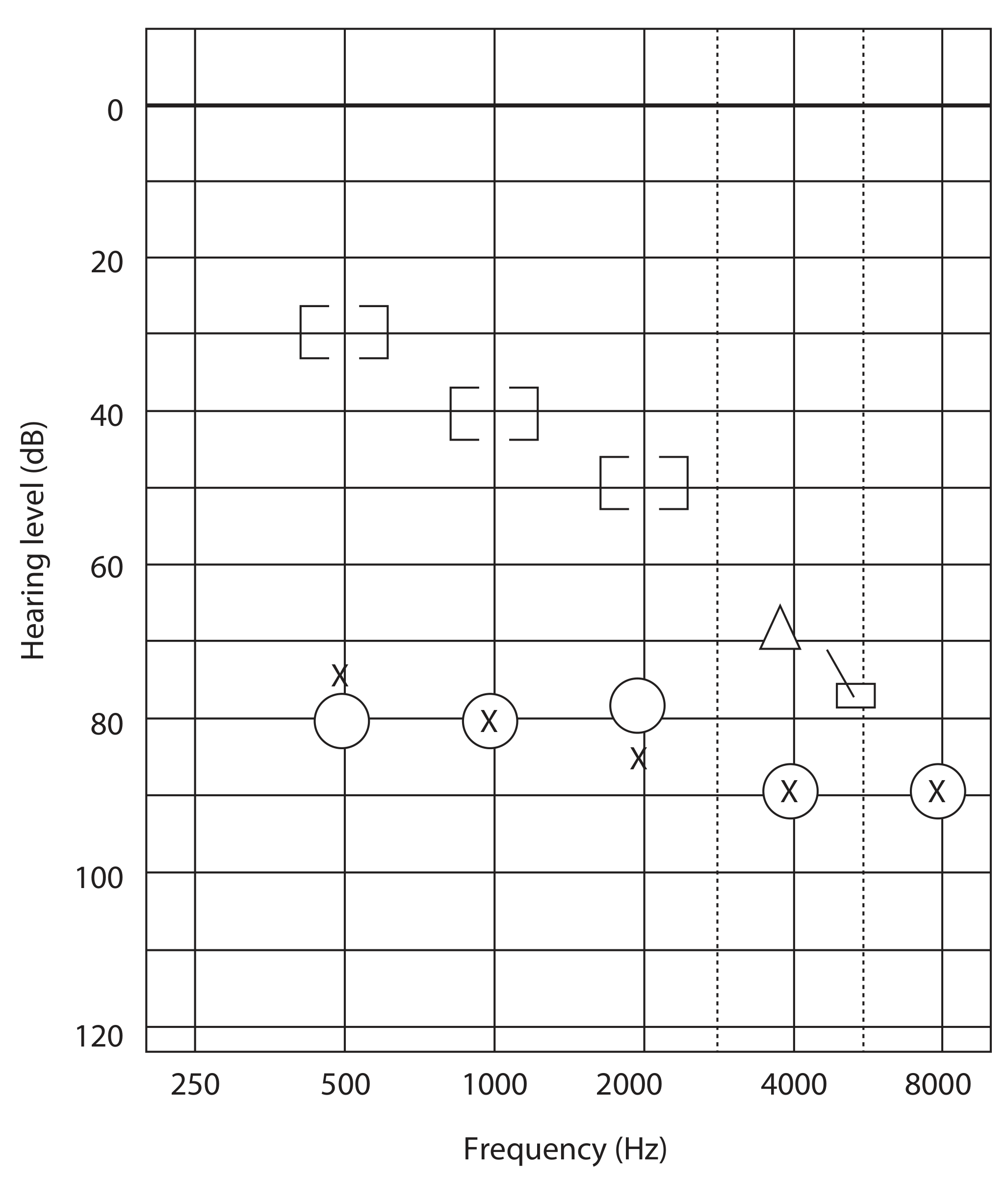
Audiogram in otosclerosis showing mixed hearing impairment. The Carhart notch at 2 kHz can be obscured when a significant sensorineural component co-exists. - Scott-Brown's Otorhinolaryngology, Vol. 2
Summary
The 2 kHz dip exists because:
- The ossicular chain (stapes) has its peak resonant contribution to bone conduction at ~2 kHz
- Stapes fixation in otosclerosis blocks this ossicular contribution
- The loss is greatest where the ossicular chain normally contributes the most - at 2000 Hz
- This is a purely mechanical, reversible artifact, not cochlear damage
Why there is rinnes positive in severe SNHL
In SNHL, both air conduction (AC) and bone conduction (BC) are reduced equally, but AC still remains better than BC, so Rinne's stays positive. Let me pull the detailed explanation from the medical library.
Searching sources...
Finding Sources
Finding Sources
Reading File
Finding Sources
Finding Sources
Reading File
Now let me also quickly search for the concept of "false negative Rinne" in profound/severe unilateral SNHL, which is the important clinical pitfall:
Finding Sources
Finding Sources
Finding Sources
I have sufficient material from the textbooks to give a complete, accurate answer. Here it is:
Why Rinne's Test is Positive in Sensorineural Hearing Loss (SNHL)
The Core Principle: What Rinne's Test Actually Measures
Rinne's test does not measure the absolute sensitivity of hearing. It only compares two pathways to the same cochlea:
- Air Conduction (AC): Sound → pinna → ear canal → tympanic membrane → ossicular chain → oval window → cochlea
- Bone Conduction (BC): Sound → mastoid → skull bone → cochlea directly
In a normal ear, AC > BC because the outer and middle ear act as a sound amplifier/impedance matcher - this gives AC roughly a 2:1 time advantage over BC.
In SNHL: Why It Stays Positive
In sensorineural hearing loss, the problem lies in the cochlea (or auditory nerve) - not in the outer or middle ear. The middle ear mechanics are completely intact.
| Normal Hearing | SNHL | Conductive HL | |
|---|---|---|---|
| Middle ear | Intact | Intact | Damaged/blocked |
| AC threshold | Normal | Raised | Raised |
| BC threshold | Normal | Raised equally | Normal |
| AC vs BC ratio | AC > BC | AC still > BC | BC > AC |
| Rinne result | Positive | Positive | Negative |
Since both AC and BC must pass through the same damaged cochlea in SNHL, both are reduced equally and proportionally. The 2:1 ratio of AC over BC is preserved. Air conduction still delivers more sound energy to the cochlea than bone conduction does - the cochlea just processes all of it poorly.
As Swanson's Family Medicine Review states: "In the presence of sensorineural hearing loss, the durations of both AC and BC are reduced; however, the 2:1 ratio remains the same (i.e., a positive Rinne test result)." - Swanson's Family Medicine Review, p. 295
The Analogy
Think of a broken loudspeaker. If you play music through a large amplifier (AC) vs a small one (BC), both will sound distorted and quiet through the broken speaker - but the large amplifier still sounds relatively louder. The defect is downstream and affects both equally.
Important Clinical Pitfall: The "False Negative Rinne" in Severe/Profound Unilateral SNHL
This is where severe SNHL creates a dangerous trap:
In a patient with a dead ear (profound unilateral SNHL), when you place the tuning fork on the mastoid of the deaf ear, the vibration travels transcranially (through the skull) and is heard by the opposite normal cochlea. The patient says "yes, I hear it via bone conduction" - but they are actually hearing it in the other ear.
When you then move the fork to the front of the deaf ear canal, no sound reaches either cochlea well - so the patient says BC > AC.
Result: The deaf ear gives a falsely negative Rinne - mimicking conductive hearing loss when none exists.
| Situation | What patient actually hears | Rinne result recorded | True pathology |
|---|---|---|---|
| Moderate SNHL | Ipsilateral cochlea (poorly) | Positive (correctly) | SNHL |
| Profound/dead unilateral SNHL | BC heard by contralateral cochlea | False Negative | SNHL misread as CHL |
This is why Weber's test and masking are essential alongside Rinne's in any severe/unilateral hearing loss, and why the Stenger test or formal audiometry must be used in suspected dead ear scenarios.
Summary
Rinne's is positive in SNHL because:
- The middle ear is structurally normal - no impedance barrier exists
- Both AC and BC are degraded equally at the level of the cochlea
- AC still delivers more acoustic energy than BC (2:1 ratio preserved)
- Therefore AC > BC remains true, giving a positive Rinne
The test is positive but at a lower absolute level - both thresholds are shifted up together. Rinne only tests the relationship between the two pathways, not their absolute values. - Shambaugh Surgery of the Ear, p. 205; Cummings Otolaryngology
This is a shared conversation. Sign in to Orris to start your own chat.