
Cerebral blood flow and inhalation agents affect on it 10 marks answer
Reading File
Now let me search the medical library for cerebral blood flow and inhalation agents.
Finding Sources
Finding Sources
Excellent results. Let me now read the most relevant sections - Miller's Anesthesia and Morgan & Mikhail's (the top anesthesia textbooks) for both CBF and inhalation agents' effects.
Reading File
Reading File
Now let me search for the specific effects of inhalation agents on CBF:
Finding Sources
Finding Sources
Let me now read the anesthesia-specific sections on inhalation agents and their effects on CBF from Miller's Anesthesia and Morgan & Mikhail:
Finding Sources
Finding Sources
Excellent. Now let me read the detailed sections from Barash's and Morgan & Mikhail's on inhalation agents and CBF:
Reading File
Reading File
Excellent! Let me also read more of the Morgan & Mikhail section and the section on nitrous oxide, ICP, and CO2 reactivity:
Reading File
Reading File
Let me also read the CO2 and O2 reactivity sections, and get the autoregulation diagram image URL:
Reading File
I now have all the material I need. Let me also fetch the key diagram showing autoregulation depression by volatile anesthetics:
Loading Image
Loading Image
I now have comprehensive, authoritative material. Let me compose a thorough 10-mark exam answer.
Cerebral Blood Flow and the Effects of Inhalation Agents
PART 1: CEREBRAL BLOOD FLOW - PHYSIOLOGY AND REGULATION
Normal Values
- Overall CBF: ~50 mL/100 g/min at PaCO₂ of 40 mmHg
- Gray matter: ~80 mL/100 g/min; White matter: ~20 mL/100 g/min
- Total CBF in adults: ~750 mL/min (15-20% of cardiac output)
Clinically critical thresholds:
| CBF (mL/100 g/min) | Clinical Consequence |
|---|---|
| 50 | Normal |
| 20-25 | EEG slowing, functional impairment |
| <20 | Isoelectric (flat) EEG |
| <10 | Irreversible brain damage / neuronal death |
The brain has no energy storage and cannot tolerate even brief interruptions in flow.
Cerebral Perfusion Pressure (CPP)
CPP = MAP - ICP (or CVP if CVP > ICP)
- Normal CPP: 80-100 mmHg
- ICP is normally <10 mmHg, so CPP is mainly MAP-dependent
- CPP <50 mmHg: EEG slowing
- CPP 25-40 mmHg: flat EEG
- CPP <25 mmHg: irreversible brain damage
Regulation of Cerebral Blood Flow
1. Cerebral Autoregulation
The cerebrovascular system maintains constant CBF over a wide range of systemic blood pressure. In normal individuals, CBF remains constant between MAPs of ~60-160 mmHg by adjusting cerebrovascular resistance - vasodilation during hypotension, vasoconstriction during hypertension. Outside these limits, CBF becomes pressure-dependent.
- Response is rapid (10-60 seconds)
- In chronically hypertensive patients, the autoregulatory curve shifts rightward
2. CO₂ Reactivity (Most Important Extrinsic Factor)
CBF is directly proportional to PaCO₂ between 20-80 mmHg.
- Flow changes ~1-2 mL/100 g/min per 1 mmHg change in PaCO₂
- Mechanism: CO₂ freely crosses the blood-brain barrier, alters CSF/brain tissue pH, and changes vascular tone
- Hypercapnia → vasodilation → ↑CBF
- Hypocapnia (hyperventilation) → vasoconstriction → ↓CBF (exploited clinically to lower ICP)
- Effect diminishes after 24-48 hrs as CSF HCO₃⁻ adjusts
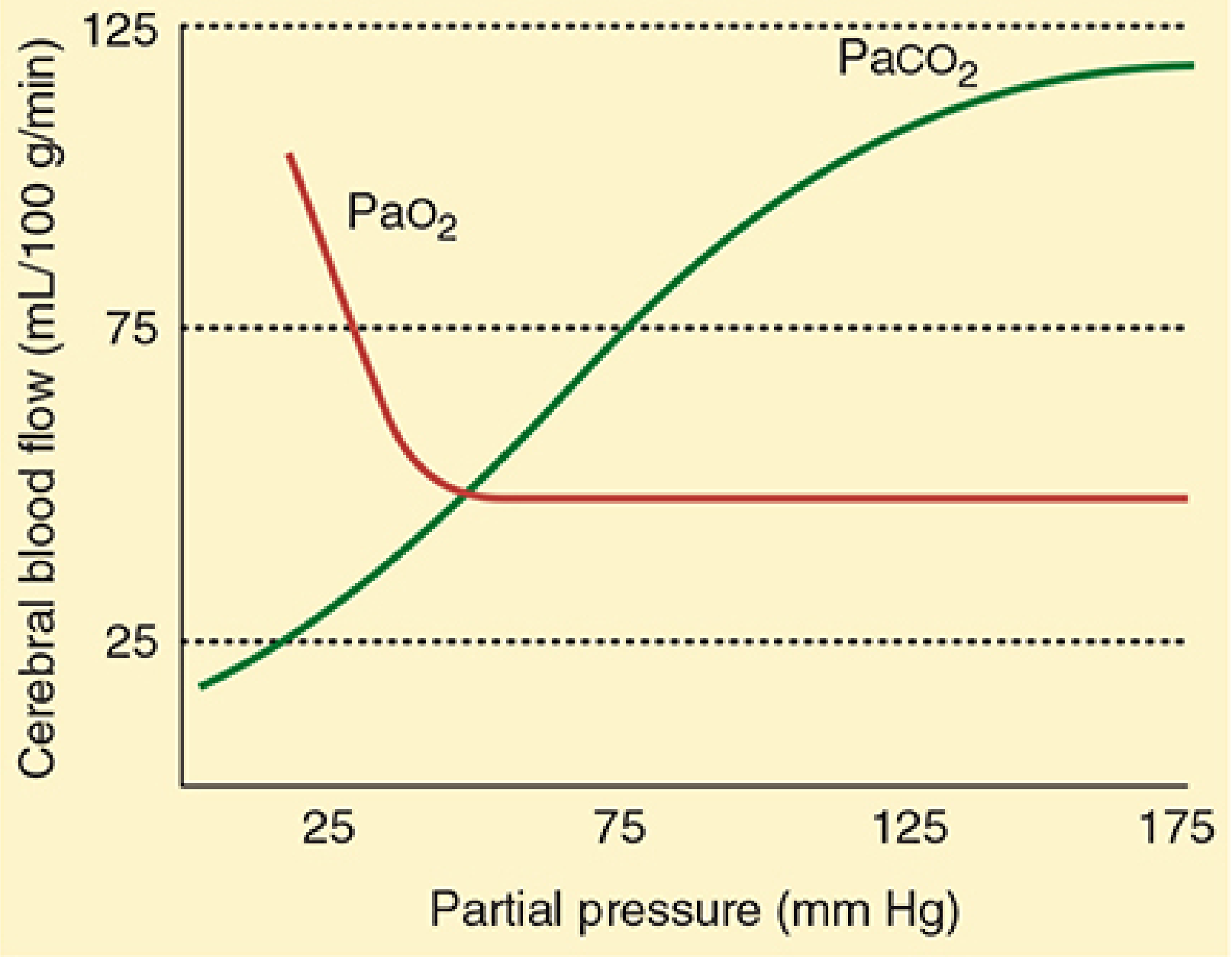
CBF vs PaCO₂ (green) and PaO₂ (red) - Morgan & Mikhail's Clinical Anesthesiology
3. Oxygen Tension
Only marked changes in PaO₂ alter CBF. Hyperoxia causes only minimal decrease (-10%) in CBF. Severe hypoxemia (PaO₂ <50 mmHg) markedly increases CBF.
4. Temperature
CBF changes 5-7% per 1°C. Hypothermia decreases both CMR and CBF. The Q10 for humans is ~2 (CMR doubles per 10°C rise). At 20°C, EEG is isoelectric but CMR continues to fall.
5. Neurovascular Coupling (Flow-Metabolism Coupling)
CBF closely tracks local cerebral metabolic rate (CMR). Regional CBF can vary 10-300 mL/100 g/min. Motor cortex CBF rises with limb movement; visual cortex CBF rises with visual stimulation.
6. Viscosity
Hematocrit is the primary determinant of viscosity. Optimal cerebral O₂ delivery occurs at hematocrit ~30%. Polycythemia increases viscosity and reduces CBF.
PART 2: EFFECTS OF INHALATION AGENTS ON CEREBRAL BLOOD FLOW
General Principle
At normocarbia, all volatile anesthetics dilate cerebral vessels and impair autoregulation in a concentration-dependent manner. This is the opposite effect of IV agents (except ketamine), which generally reduce CBF.
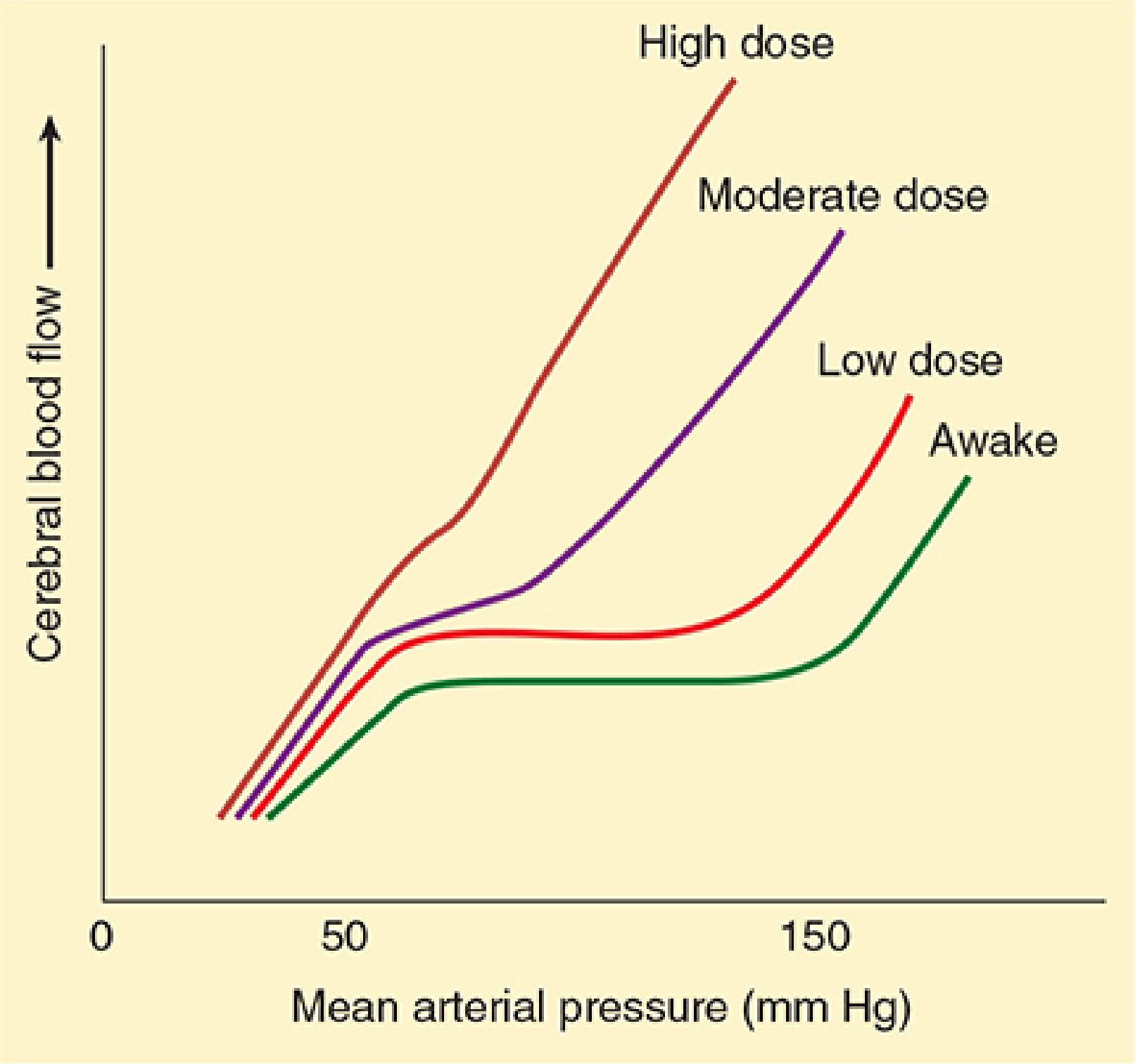
Dose-dependent depression of cerebral autoregulation by volatile anesthetics - Morgan & Mikhail's
A. Volatile Agents (Halothane, Isoflurane, Desflurane, Sevoflurane)
1. Cerebral Metabolic Rate (CMR / CMRO₂)
All volatile agents cause concentration-dependent decreases in CMR:
- Isoflurane → greatest maximal depression (up to 50% reduction), can cause isoelectric EEG at clinically tolerated doses
- Desflurane and Sevoflurane → similar depression to isoflurane (~50%)
- Halothane → least effect (≤25% reduction)
- No further CMR reduction occurs beyond the dose that renders the EEG isoelectric
Unique feature of desflurane: desflurane-induced isoelectric EEG can revert to continuous activity over time despite unchanged MAC - unlike the other agents.
2. Cerebral Blood Flow (CBF) - Comparative Effects
All volatile agents increase CBF (cerebral vasodilation), but to different degrees:
| Agent | Effect on CBF (vs. awake at equivalent MAC & BP) |
|---|---|
| Halothane | ↑↑↑ Up to 200% increase; greatest vasodilator |
| Isoflurane | ↑ ~20% increase |
| Desflurane | ↑ ~20% increase (similar to isoflurane) |
| Sevoflurane | Least cerebral vasodilation of all volatile agents |
Halothane is the most potent cerebral vasodilator:
- At >1 MAC, nearly abolishes cerebral autoregulation
- Increase in CBF is generalized throughout all brain regions
- The increase in CBF despite a falling CMR is termed "luxury perfusion"
Time dependency: With continued administration (2-5 hours), CBF begins to return toward normal levels with all volatile agents.
3. Effect on Cerebral Autoregulation
All volatile agents impair autoregulation in a dose-dependent manner:
- Low dose: partial impairment (autoregulation plateau preserved but narrowed)
- High dose: autoregulation nearly abolished; CBF becomes pressure-dependent
Order of autoregulatory impairment (greatest to least):
Halothane > Isoflurane ≈ Desflurane > Sevoflurane
4. CO₂ Reactivity
The cerebrovascular response to CO₂ is generally retained with all volatile agents. This is clinically important:
- Hyperventilation (↓PaCO₂) can abolish or blunt the initial increases in CBF caused by volatile agents
- Key difference for halothane: Hyperventilation must be initiated before halothane administration to prevent halothane-induced CBF increases. Once halothane has been given, adding hyperventilation is less effective.
- For isoflurane, desflurane, and sevoflurane: simultaneous hyperventilation can prevent increases in CBF and ICP
5. Cerebral Blood Volume and ICP
- Increases in cerebral blood volume (10-12%) generally parallel increases in CBF
- This expansion can markedly elevate ICP in patients with reduced intracranial compliance (e.g., brain tumors, head injury)
- Hypocapnia can blunt the increase in cerebral blood volume
Net effect on ICP = immediate changes in cerebral blood volume + delayed alterations in CSF dynamics + PaCO₂
6. Altered Flow-Metabolism Coupling ("Luxury Perfusion" vs. "Steal")
Volatile agents alter but do not completely uncouple the normal CBF-CMR relationship:
- CMR decreases, but CBF increases → CBF exceeds metabolic demand = "luxury perfusion"
- In global ischemia: potentially beneficial
- In focal ischemia: potentially detrimental - volatile agents increase flow in normal brain but NOT in ischemic areas (where arterioles are already maximally dilated). This can redistribute flow away from ischemic areas = "cerebrovascular steal"
7. CSF Dynamics
- Halothane: impedes CSF absorption, minimally reduces formation → unfavorable for raised ICP
- Isoflurane: facilitates CSF absorption → more favorable effect on CSF dynamics
8. Sevoflurane - Special Considerations
- Has no noteworthy adverse effects on cerebral physiology at normal CO₂ and BP
- High concentrations (1.5-2.0 MAC), sudden increase in concentration, especially in females, and/or hypocapnia can trigger EEG abnormalities with epileptiform features - though clinical significance remains debated
- The question of sevoflurane in epilepsy patients remains unsettled
B. Nitrous Oxide (N₂O)
Nitrous oxide behaves differently from the potent volatile agents:
- Alone: causes cerebral vasodilation, potentially increases ICP
- Combined with IV agents (e.g., propofol, opioids): minimal effects on CBF, CMR, and ICP
- Combined with volatile agents: can further increase CBF above the volatile agent alone
- The final effect on CBF depends heavily on the context and co-administered agents
Clinical Summary Table
| Parameter | Halothane | Isoflurane | Desflurane | Sevoflurane | N₂O |
|---|---|---|---|---|---|
| CMR | ↓ (25%) | ↓↓ (50%) | ↓↓ (50%) | ↓↓ (50%) | ↑ (mild) |
| CBF | ↑↑↑ (200%) | ↑ (20%) | ↑ (20%) | ↑ (least) | ↑ |
| Autoregulation | Severely impaired | Moderately impaired | Moderately impaired | Mildly impaired | Preserved |
| CO₂ reactivity | Retained | Retained | Retained | Retained | Retained |
| ICP | ↑↑↑ | ↑ | ↑ | ↑ (least) | ↑ |
| CSF absorption | Impaired | Facilitated | - | - | - |
Key Clinical Points for Neuroanaesthesia
- Avoid halothane in neurosurgery or patients with raised ICP - it causes the greatest CBF increase and ICP rise
- Hyperventilation before induction is the most reliable strategy to prevent inhalation agent-induced CBF increases
- Sevoflurane or isoflurane at low concentrations (<1 MAC) with normocapnia are the preferred volatile agents in neurosurgery when total IV anaesthesia is not used
- CO₂ reactivity is preserved with all volatile agents - hyperventilation can always be used to counteract CBF increases
- "Cerebrovascular steal" is a theoretical concern in focal ischemia with volatile agents; IV agents (barbiturates, propofol) produce reverse steal (preferential flow toward ischemic areas)
Sources: Morgan and Mikhail's Clinical Anesthesiology, 7e, pp. 1075-1090; Barash, Cullen, and Stoelting's Clinical Anesthesia, 9e, pp. 1410-1415; Miller's Anesthesia, 10e
Insert autoregulation dig also
The autoregulation diagram is already in the textbook library from the previous session. Let me fetch it directly.
Loading Image
Loading Image
Here is the complete answer with all three diagrams now inserted:
Cerebral Blood Flow and the Effects of Inhalation Agents
PART 1: CEREBRAL BLOOD FLOW - PHYSIOLOGY AND REGULATION
Normal Values
- Overall CBF: ~50 mL/100 g/min at PaCO₂ of 40 mmHg
- Gray matter: ~80 mL/100 g/min; White matter: ~20 mL/100 g/min
- Total CBF in adults: ~750 mL/min (15-20% of cardiac output)
Clinically critical thresholds:
| CBF (mL/100 g/min) | Clinical Consequence |
|---|---|
| 50 | Normal |
| 20-25 | EEG slowing, functional impairment |
| <20 | Isoelectric (flat) EEG |
| <10 | Irreversible brain damage / neuronal death |
Cerebral Perfusion Pressure (CPP)
CPP = MAP - ICP (or CVP if CVP > ICP)
- Normal CPP: 80-100 mmHg; Normal ICP: <10 mmHg
- CPP <50 mmHg: EEG slowing
- CPP 25-40 mmHg: flat EEG
- CPP <25 mmHg sustained: irreversible brain damage
Regulation of CBF
1. Cerebral Autoregulation
The cerebral vasculature maintains constant CBF over a wide MAP range by active vasoconstriction (at high MAP) and vasodilation (at low MAP). Response is rapid (10-60 seconds).
In normal individuals: CBF remains constant between MAPs of ~60-160 mmHg
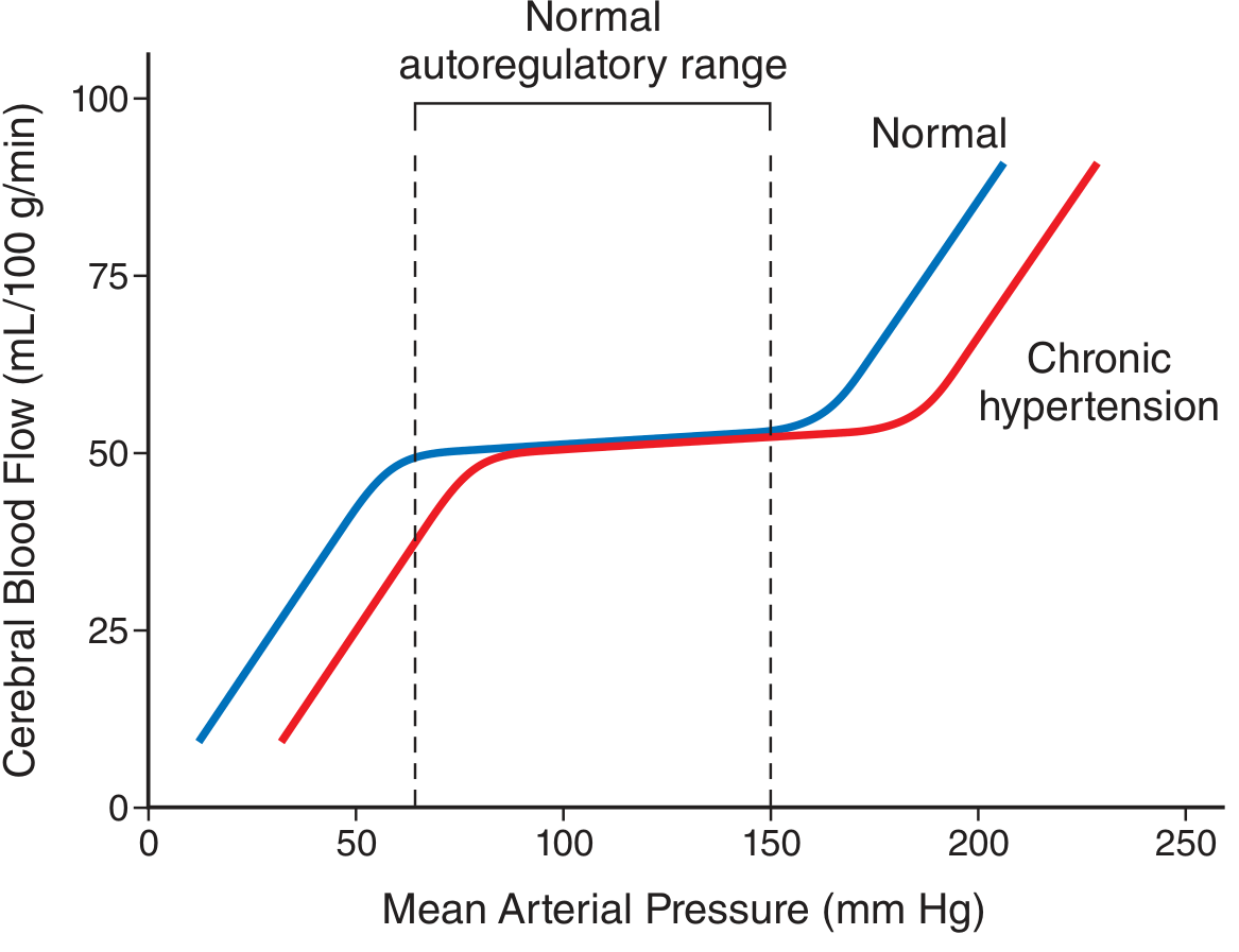
Cerebral autoregulation curve showing constant CBF (plateau) between MAP 60-150 mmHg in normals (blue). In chronic hypertension (red), the entire curve shifts rightward - both the lower and upper limits are higher, meaning these patients are vulnerable to ischemia at blood pressures that normals would tolerate well. - Guyton & Hall Medical Physiology
Key points from the diagram:
- The plateau represents the autoregulatory range where CBF stays ~50 mL/100 g/min regardless of MAP
- Below the lower limit (~60 mmHg): CBF falls passively with MAP → ischemia
- Above the upper limit (~150-160 mmHg): CBF rises with MAP → forced vasodilation, breakdown of blood-brain barrier, cerebral edema and hemorrhage
- In chronic hypertension: the plateau shifts right; the lower limit may be at MAP ~80-100 mmHg, so these patients cannot safely tolerate "normal" hypotension during anesthesia
2. CO₂ Reactivity (Most Important Extrinsic Factor)
CBF is directly proportional to PaCO₂ between 20-80 mmHg, changing ~1-2 mL/100 g/min per 1 mmHg change in PaCO₂.
- Mechanism: CO₂ freely crosses the blood-brain barrier, alters CSF pH, and changes vascular tone
- Effect diminishes after 24-48 hrs as CSF HCO₃⁻ compensates
CBF relationship with PaCO₂ (green - steep linear rise) and PaO₂ (red - only hypoxia below 50 mmHg significantly increases CBF). - Morgan & Mikhail's Clinical Anesthesiology
3. Oxygen Tension
Only marked changes in PaO₂ affect CBF. Hyperoxia causes minimal CBF decrease (-10%). Severe hypoxemia (PaO₂ <50 mmHg) markedly increases CBF.
4. Temperature
CBF changes 5-7% per 1°C. Q10 ≈ 2 (CMR doubles per 10°C rise). At 20°C the EEG is isoelectric.
5. Viscosity
Hematocrit is the main determinant. Optimal cerebral O₂ delivery occurs at hematocrit ~30%.
PART 2: EFFECTS OF INHALATION AGENTS ON CBF
General Principle
At normocarbia, all volatile anesthetics dilate cerebral vessels and impair autoregulation in a concentration-dependent manner - the opposite of most IV agents.
The autoregulatory plateau (flat region seen in the "Awake" curve) progressively disappears as volatile anesthetic dose increases. At high doses, CBF becomes entirely pressure-dependent. - Morgan & Mikhail's Clinical Anesthesiology
A. Volatile Agents (Halothane, Isoflurane, Desflurane, Sevoflurane)
1. CMR (CMRO₂)
All cause concentration-dependent CMR decreases. No further reduction occurs once EEG is isoelectric:
| Agent | Max CMR Reduction |
|---|---|
| Isoflurane | Up to 50% (greatest) |
| Desflurane | ~50% |
| Sevoflurane | ~50% |
| Halothane | ≤25% (least) |
2. CBF - Comparative Effects
All increase CBF, but to very different degrees:
| Agent | CBF Effect at Equivalent MAC |
|---|---|
| Halothane | ↑↑↑ up to 200% - greatest vasodilator |
| Isoflurane | ↑ ~20% |
| Desflurane | ↑ ~20% |
| Sevoflurane | ↑ least of all volatile agents |
Time dependency: With prolonged administration (2-5 hours), CBF returns toward baseline with all agents.
3. Autoregulation
Impaired in a dose-dependent manner (greatest → least impairment):
Halothane >> Isoflurane ≈ Desflurane > Sevoflurane
Halothane at >1 MAC nearly abolishes autoregulation entirely and increases CBF throughout all brain regions.
4. CO₂ Reactivity
Preserved with all volatile agents. This is the key clinical tool:
- Hyperventilation can blunt or abolish volatile agent-induced CBF increases
- Halothane exception: hyperventilation must be started before halothane is given to be effective. Once halothane is established, hyperventilation alone cannot fully reverse the CBF increase.
- For isoflurane, desflurane, sevoflurane: simultaneous hyperventilation effectively prevents CBF and ICP rises.
5. Cerebral Blood Volume and ICP
- Blood volume increases 10-12% parallel to CBF increases
- Can markedly elevate ICP in patients with poor intracranial compliance (trauma, tumors)
- Hypocapnia blunts this blood volume expansion
6. Luxury Perfusion vs. Cerebrovascular Steal
- CMR ↓ but CBF ↑ → supply exceeds demand = "luxury perfusion"
- In global ischemia: potentially beneficial
- In focal ischemia: volatile agents increase flow in normal tissue but not ischemic areas (maximally dilated already) → "steal" of blood away from ischemic zones
7. CSF Dynamics
- Halothane: impedes CSF absorption → unfavorable for raised ICP
- Isoflurane: facilitates CSF absorption → more favorable
8. Sevoflurane - Special Note
High concentrations (1.5-2.0 MAC), sudden dose escalation, and/or hypocapnia can trigger epileptiform EEG changes, particularly in females. The proconvulsant potential in epilepsy patients remains unsettled.
B. Nitrous Oxide (N₂O)
| Context | Effect |
|---|---|
| Given alone | Cerebral vasodilation, ↑ICP |
| Combined with IV agents | Minimal effects on CBF, CMR, ICP |
| Added to volatile agents | May further ↑CBF (additive vasodilation) |
Clinical Summary Table
| Parameter | Halothane | Isoflurane | Desflurane | Sevoflurane | N₂O |
|---|---|---|---|---|---|
| CMR | ↓ 25% | ↓↓ 50% | ↓↓ 50% | ↓↓ 50% | ↑ (mild) |
| CBF | ↑↑↑ 200% | ↑ 20% | ↑ 20% | ↑ (least) | ↑ |
| Autoregulation | Severely impaired | Moderately impaired | Moderately impaired | Mildly impaired | Preserved |
| CO₂ reactivity | Retained | Retained | Retained | Retained | Retained |
| ICP | ↑↑↑ | ↑ | ↑ | ↑ (least) | ↑ |
| CSF absorption | ↓ (impaired) | ↑ (facilitated) | - | - | - |
Key Clinical Points
- Avoid halothane in neurosurgery or raised ICP - causes maximum CBF and ICP rise
- Hyperventilate before induction with halothane to prevent CBF surge; with others, simultaneous hyperventilation suffices
- Sevoflurane or low-dose isoflurane (<1 MAC) with normocapnia are preferred volatile agents in neuroanaesthesia
- CO₂ reactivity is always preserved with volatile agents - hyperventilation is reliably effective
- Volatile agents cause steal in focal ischemia; IV barbiturates/propofol cause inverse steal (redistribute flow toward ischemic areas)
Sources: Morgan and Mikhail's Clinical Anesthesiology, 7e; Barash, Cullen, and Stoelting's Clinical Anesthesia, 9e; Guyton and Hall Textbook of Medical Physiology; Miller's Anesthesia, 10e
Cerebral autoregulation 10 mark answer
Finding Sources
Finding Sources
Reading File
Reading File
Reading File
Reading File
Finding Sources
Finding Sources
Reading File
Reading File
Now let me fetch the key diagrams:
Loading Image
Loading Image
Loading Image
Excellent - I have three outstanding diagrams plus all the textbook content needed. Here is the complete 10-mark answer:
Cerebral Autoregulation
Definition
Cerebral autoregulation is the intrinsic ability of the cerebral vasculature to maintain a relatively constant cerebral blood flow (CBF) over a wide range of systemic blood pressures, independent of passive pressure-flow relationships. It is a critical protective mechanism that ensures the brain - which has no energy stores - receives a continuous, uninterrupted blood supply.
The Autoregulation Curve
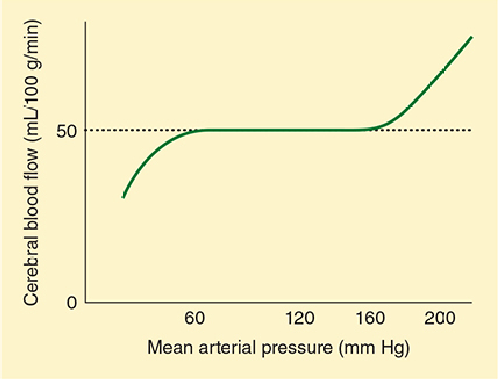
Normal cerebral autoregulation curve: CBF remains constant at ~50 mL/100 g/min (plateau) between MAP 60-160 mmHg. Below the lower limit and above the upper limit, CBF becomes pressure-passive. - Morgan & Mikhail's Clinical Anesthesiology
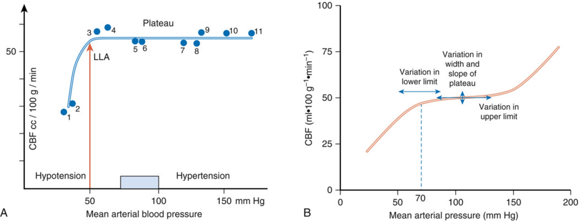
(A) Lassen's classical autoregulation curve with LLA (Lower Limit of Autoregulation) at ~50 mmHg - each dot represents a separate study group. (B) Contemporary view showing significant inter-individual variation in the lower limit, upper limit, and the width and slope of the plateau. - Miller's Anesthesia
Key Parameters of the Curve
| Component | Value |
|---|---|
| Normal CBF on plateau | ~50 mL/100 g/min |
| Lower limit of autoregulation (LLA) | MAP ~60-70 mmHg (traditional: 50 mmHg) |
| Upper limit of autoregulation (ULA) | MAP ~150-160 mmHg |
| Autoregulatory plateau width | ~60-100 mmHg range |
| Response time | 10-60 seconds |
Important contemporary view (Miller's Anesthesia): The classical autoregulatory plateau is wider and the LLA higher than originally described by Lassen. There is considerable inter-individual variability in the lower limit, upper limit, width, and slope of the plateau. The LLA in most humans is not less than 70 mmHg.
Diagram Explanation
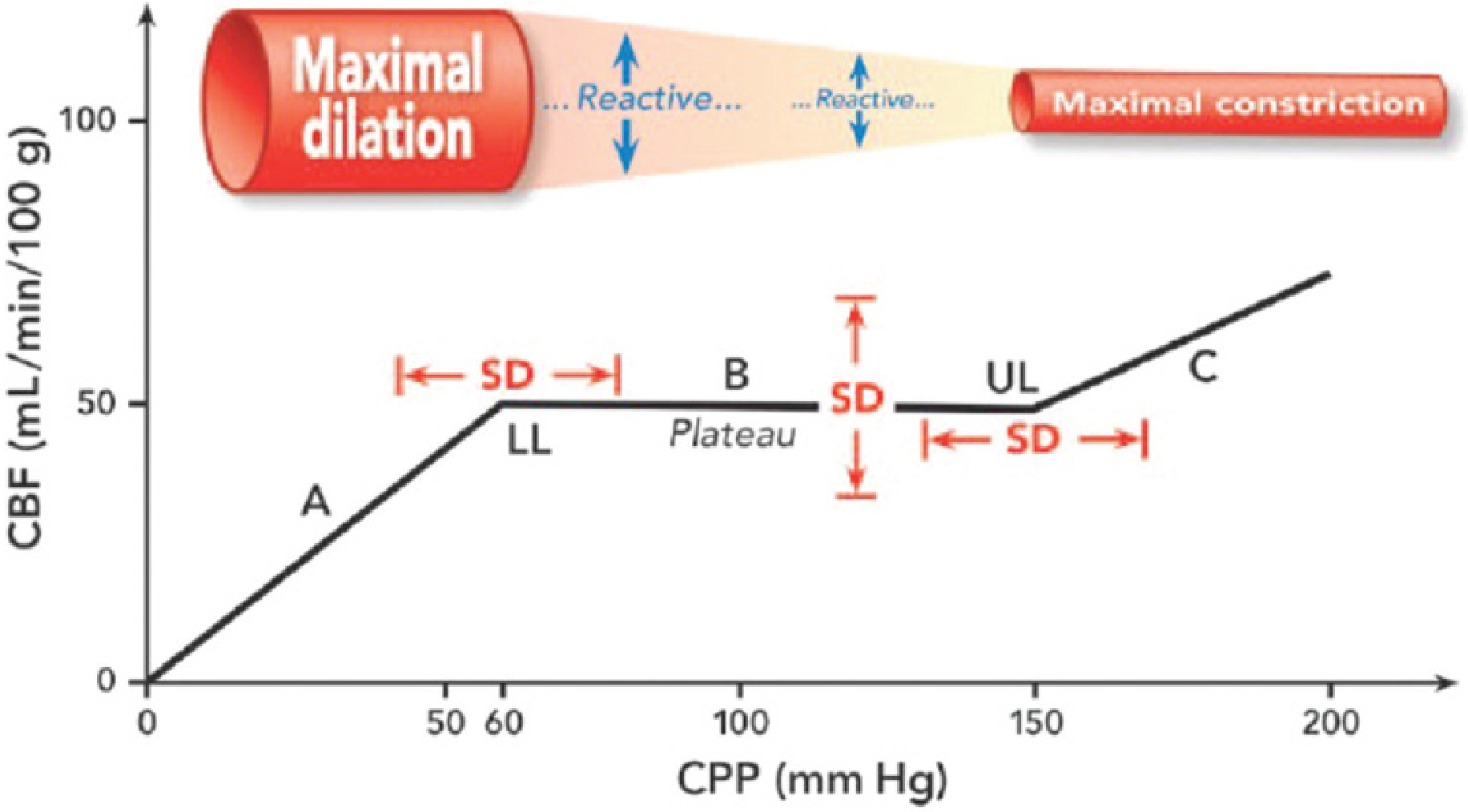
Zone A (below LL): vessels maximally dilated, CBF is pressure-passive, ischemia risk. Zone B (plateau): reactive vasomotion maintains constant CBF. Zone C (above UL): vessels maximally constricted; beyond this CBF rises passively. SD = standard deviation showing inter-individual spread. LL = lower limit, UL = upper limit. - Mulholland & Greenfield's Surgery
Three zones:
- Zone A (below lower limit, CPP <60 mmHg): Arterioles are maximally dilated. Vasomotor capacity exhausted. CBF falls passively with falling CPP → ischemia
- Zone B (plateau, CPP 60-150 mmHg): Reactive arteriolar tone adjusts. ↓CPP → vasodilation; ↑CPP → vasoconstriction. CBF stays constant
- Zone C (above upper limit, CPP >150 mmHg): Arterioles are maximally constricted. Beyond capacity → forced vasodilation → disruption of blood-brain barrier → cerebral edema, hemorrhage, hypertensive encephalopathy
Mechanisms of Autoregulation
Three mechanisms work together - no single one is sufficient alone:
1. Myogenic Mechanism
- The Bayliss effect: smooth muscle in cerebral arteriolar walls contracts in response to increased transmural pressure (stretch-induced contraction) and relaxes when pressure falls
- This is the primary rapid-response mechanism, accounting for much of the short-term autoregulation
- Acts within seconds
2. Metabolic Mechanism
- When MAP falls and CBF drops, local accumulation of metabolic byproducts (CO₂, H⁺, adenosine, K⁺, lactate) causes vasodilation, restoring flow
- When flow is excessive, washout of these metabolites leads to vasoconstriction
- This mechanism links autoregulation to neurovascular coupling (flow-metabolism matching)
3. Neurogenic Mechanism
- Intracranial vessels are innervated by:
- Sympathetic (noradrenergic): vasoconstriction - provides protection during acute hypertension by limiting flow transmission
- Parasympathetic (cholinergic): vasodilation
- Intense sympathetic stimulation shifts the autoregulatory curve rightward, protecting against hypertensive surges
- Sympathetic nerves also reduce vasodilatory capacity during hypotension
Cerebral Perfusion Pressure (CPP) and Autoregulation
CPP = MAP - ICP (or CVP if CVP > ICP)
- Autoregulation is driven by CPP, not MAP alone
- Normal CPP: 80-100 mmHg; Normal ICP: <10 mmHg
- In pathological states (raised ICP), autoregulation limits shift and the curve narrows
Static vs Dynamic Autoregulation
| Type | Assessment | Plateau Width |
|---|---|---|
| Static | Steady-state, slow BP changes over minutes | Wide (~40 mmHg or more) |
| Dynamic | Rapid BP changes (seconds) | Narrow (~10 mmHg); plateau may be absent at fastest rates |
The rate of blood pressure change critically determines autoregulatory effectiveness. Slow changes allow full compensation; rapid changes (e.g., sudden hemorrhage, bolus vasopressors) bypass autoregulation and CBF follows pressure passively.
Modification of the Autoregulation Curve
Chronic Hypertension
The entire curve shifts rightward - both lower and upper limits are at higher MAPs.
- The brain adapts to higher "normal" pressures
- The hypertensive patient is at ischemia risk at "normal" blood pressures (MAP 70-80 mmHg) that a normotensive patient tolerates easily
- Long-term antihypertensive therapy can restore the curve toward normal
Conditions That Impair Autoregulation
| Condition | Effect |
|---|---|
| Volatile anesthetic agents | Dose-dependent impairment (halothane worst) |
| Traumatic brain injury (TBI) | Disrupted in ~1/3 of severe TBI cases |
| Hypercapnia | Attenuates autoregulation |
| Hypoxia (PaO₂ <50 mmHg) | Attenuates autoregulation |
| Acute ischemic stroke | Locally lost in ischemic zone |
| Subarachnoid hemorrhage | Globally impaired |
| Second impact syndrome | Complete loss of autoregulation |
| Calcium channel blockers, nitrates, nitroprusside | Cerebral vasodilation, impair autoregulation |
| ACE inhibitors, ARBs | Shift LLA toward lower BP |
Effect of Anesthetics on Autoregulation
Volatile Agents
All volatile anesthetics impair autoregulation in a concentration-dependent manner (see previous answer). The autoregulatory plateau progressively narrows and eventually disappears at high doses, making CBF entirely pressure-passive.
Order of impairment: Halothane >> Isoflurane ≈ Desflurane > Sevoflurane
- Sevoflurane preserves autoregulation up to ~1 MAC
- At high doses of any volatile agent, CBF becomes pressure-passive (as in Zone A/C on the diagram)
IV Agents
- Barbiturates, propofol, etomidate: decrease CMR and CBF; autoregulation preserved
- Opioids, benzodiazepines: minimal impairment; autoregulation largely preserved
- Ketamine: increases CMR and CBF modestly; autoregulation effects variable
- Vasopressors (phenylephrine, norepinephrine, ephedrine): no direct effect on autoregulation; affect CBF only via MAP
Clinical Significance of Autoregulation
-
Head injury / Raised ICP: In severe TBI, loss of autoregulation means any rise in MAP directly raises CBF and ICP - target CPP of 60-70 mmHg is recommended. Any fall in MAP leads directly to ischemia.
-
Neuroanaesthesia: Choice of agent, maintaining normocarbia, and avoiding hypotension within autoregulatory limits are the cornerstones of neuroprotection.
-
Controlled hypotension: The lower limit of autoregulation defines the safe lower boundary for induced hypotension. In hypertensive patients, this limit may be at a much higher MAP.
-
Intraoperative hypotension: When cardiac output is also low (e.g., haemorrhage), both MAP and CO fall simultaneously, further compromising CBF. Ephedrine (maintains/increases CO + MAP) may be preferable to pure vasoconstrictors like phenylephrine (which can reduce CO by increasing afterload).
-
Monitoring autoregulation: The Pressure Reactivity Index (PRx) - correlation between slow-wave arterial BP and ICP oscillations - can be continuously monitored. PRx near +1 = pressure-passive (lost autoregulation); PRx near 0 or negative = intact autoregulation. Used in neurocritical care to guide individualized CPP targets.
Summary
Cerebral autoregulation is a multicomponent, dynamic process (myogenic + metabolic + neurogenic) that maintains CBF constant between MAP 60-160 mmHg. Its integrity is fundamental to normal brain function. It is impaired by volatile anesthetics, brain injury, hypercapnia, and hypoxia - and shifted rightward by chronic hypertension. Understanding autoregulation is the foundation of safe neuroanaesthesia and neurocritical care.
Sources: Morgan and Mikhail's Clinical Anesthesiology, 7e; Miller's Anesthesia, 10e; Barash, Cullen, and Stoelting's Clinical Anesthesia, 9e; Mulholland and Greenfield's Surgery, 7e
This is a shared conversation. Sign in to Orris to start your own chat.