


+11 more
I'm planning to read cleft lip and palate from mccarthy. Kindly summaries and simplify this pages in an note form. Make sure you don't miss any vital points which are important to know and study. Explain me with relevant pictures given in the book.
I can see all 10 pages from McCarthy's Chapter 46 - "Embryology of the Head and Neck." Let me analyse all the images simultaneously to build complete notes.
analyse_media
analyse_media
analyse_media
analyse_media
analyse_media
I now have sufficient content from the pages. I have the full chapter outline, all key content from pages 2451-2460, and the figure descriptions. I can see the images directly from the uploaded photos. Let me now compile the comprehensive study notes.Here are your comprehensive study notes for McCarthy Chapter 46 - Embryology of the Head and Neck (Malcolm C. Johnston):
📘 McCarthy Chapter 46 - Embryology of the Head and Neck
Study Notes (pp. 2451-2460)
CHAPTER OUTLINE (Key Topics)
- Normal Development - Overview
- Gastrulation & Organizational Plans of Ectoderm & Mesoderm
- Embryo Folding (Tubulation)
- Neural Crest
- Development of Visceral Arches & Tongue
- Vascular Development
- Origins of Voluntary Muscles
- Peripheral Nervous System Development
- Pituitary & Pharyngeal Glands
- Carotid & Aortic Bodies/Sinuses
- Eye Development
- Growth Centers in Face & Visceral Arches
- Ear Development
- Skeletal Tissues
- Pathogenesis of Craniofacial Microsomia
Key Point: Cleft lip & palate embryogenesis is covered in Chapter 48 - this chapter provides the essential embryological framework.
SECTION 1: NORMAL DEVELOPMENT - AN OVERVIEW
Why Animal Studies Matter
- Surface morphology of human and subhuman vertebrate embryos is similar through sensitive developmental stages
- This allows extrapolation of experimental animal data to human development
- Most craniofacial malformations are caused by identical teratogens or genetic defects in both animals and humans
- Molecular-level understanding now helps identify primary actions of abnormal genetic/environmental factors
Timeline of Human Embryo Development (Fig. 46-1) ⬇️
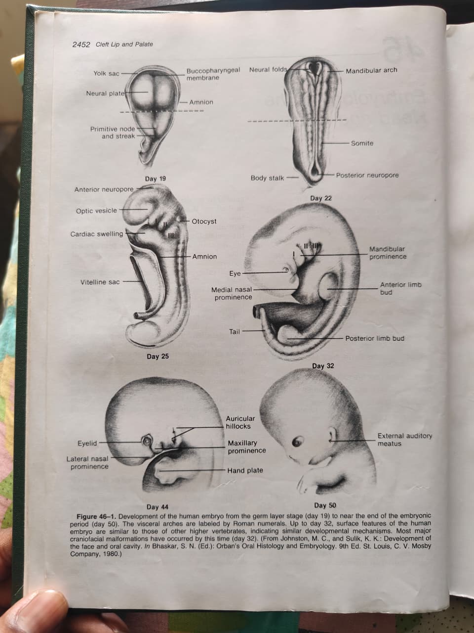
| Day | Key Structures Visible |
|---|---|
| Day 19 | Neural plate, Yolk sac, Primitive node & streak, Buccopharyngeal membrane, Amnion |
| Day 22 | Neural folds, Mandibular arch (Visceral arch I), Somites, Posterior neuropore, Body stalk |
| Day 25 | Anterior neuropore, Optic vesicle, Cardiac swelling, Otocyst, Visceral arches I-III |
| Day 32 | Eye, Medial nasal prominence, Mandibular prominence, Limb buds anterior & posterior |
| Day 44 | Eyelid, Lateral nasal prominence, Auricular hillocks, Maxillary prominence, Hand plate |
| Day 50 | External auditory meatus formed |
⚠️ Critical Point: Most major craniofacial malformations have already occurred by Day 32. Up to Day 32, human surface features are similar to other higher vertebrates.
SECTION 2: GASTRULATION & ORGANIZATIONAL PLANS
How the "Blueprint" of the Head is Set
- The structural organization of the upper face depends on the mesoderm and anterior neural plate
- Much of this blueprint is determined during or immediately after gastrulation
Gastrulation (Fig. 46-2)
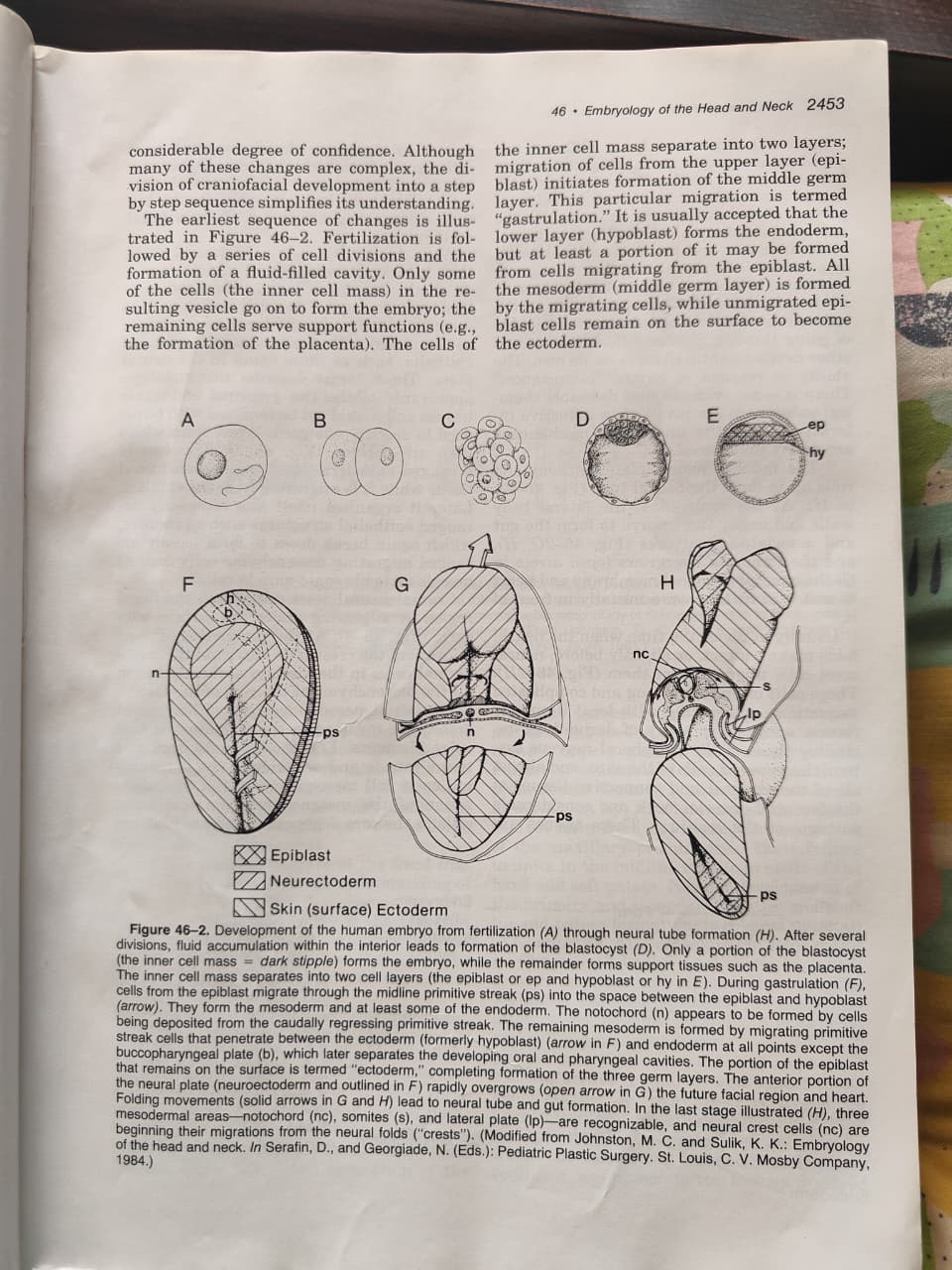
Step-by-step:
- Fertilization → cell divisions → blastocyst (fluid-filled cavity)
- Only the inner cell mass forms the embryo; remaining cells = placenta
- Inner cell mass splits into 2 layers:
- Upper layer = Epiblast
- Lower layer = Hypoblast
- Cells from the epiblast migrate through the primitive streak = GASTRULATION
- This migration forms the middle germ layer (mesoderm)
- Unmigrated epiblast cells remain on surface = Ectoderm
- Hypoblast → Endoderm (with possible contribution from epiblast cells)
Memory Tip: Gastrulation = epiblast cells migrate inward through the primitive streak to form mesoderm. Remaining epiblast = ectoderm.
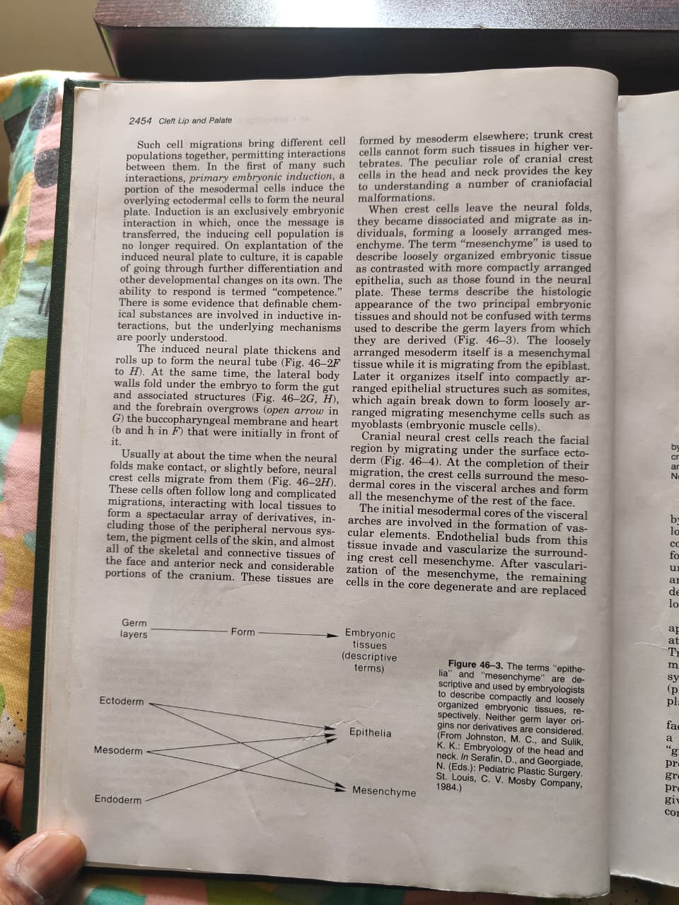
The Three Germ Layers & Their Products
| Germ Layer | Descriptive Term | Tissue Formed |
|---|---|---|
| Ectoderm | Compactly arranged epithelium | Epithelia + Mesenchyme |
| Mesoderm | Loosely arranged | Epithelia + Mesenchyme |
| Endoderm | Compactly arranged | Epithelia + Mesenchyme |
⚠️ Note: "Epithelia" and "Mesenchyme" describe tissue organization/appearance, NOT which germ layer they came from. All 3 germ layers can form both types. (Fig. 46-3)
The Organizer Mesoderm - Two Components:
- Notochord - cranial extension reaches toward the future oral plate ("prechordal plate")
- Paraxial Mesoderm - major organizing role
- Lateral Plate - mainly angiogenic (blood vessels) in the head
How Mesoderm Organizes Ectoderm (Primary Embryonic Induction):
- Mesoderm induces overlying ectoderm to differentiate into neural tissues
- This is called primary embryonic induction - exclusively an embryonic process
- Once the message is transferred, the inducing population is no longer required
- The induced neural plate has considerable ability to self-organize even with artificial inducers
Experimental Evidence:
- "Exogastrulas" - formed by high salt concentration in culture → mesoderm balloons outward instead of going inside → incomplete gastrulation → cyclopic eyes
- Removal of notochord/prechordal plate → failure of midline neural plate thinning → large cyclopic eye (2 eye fields fail to separate)
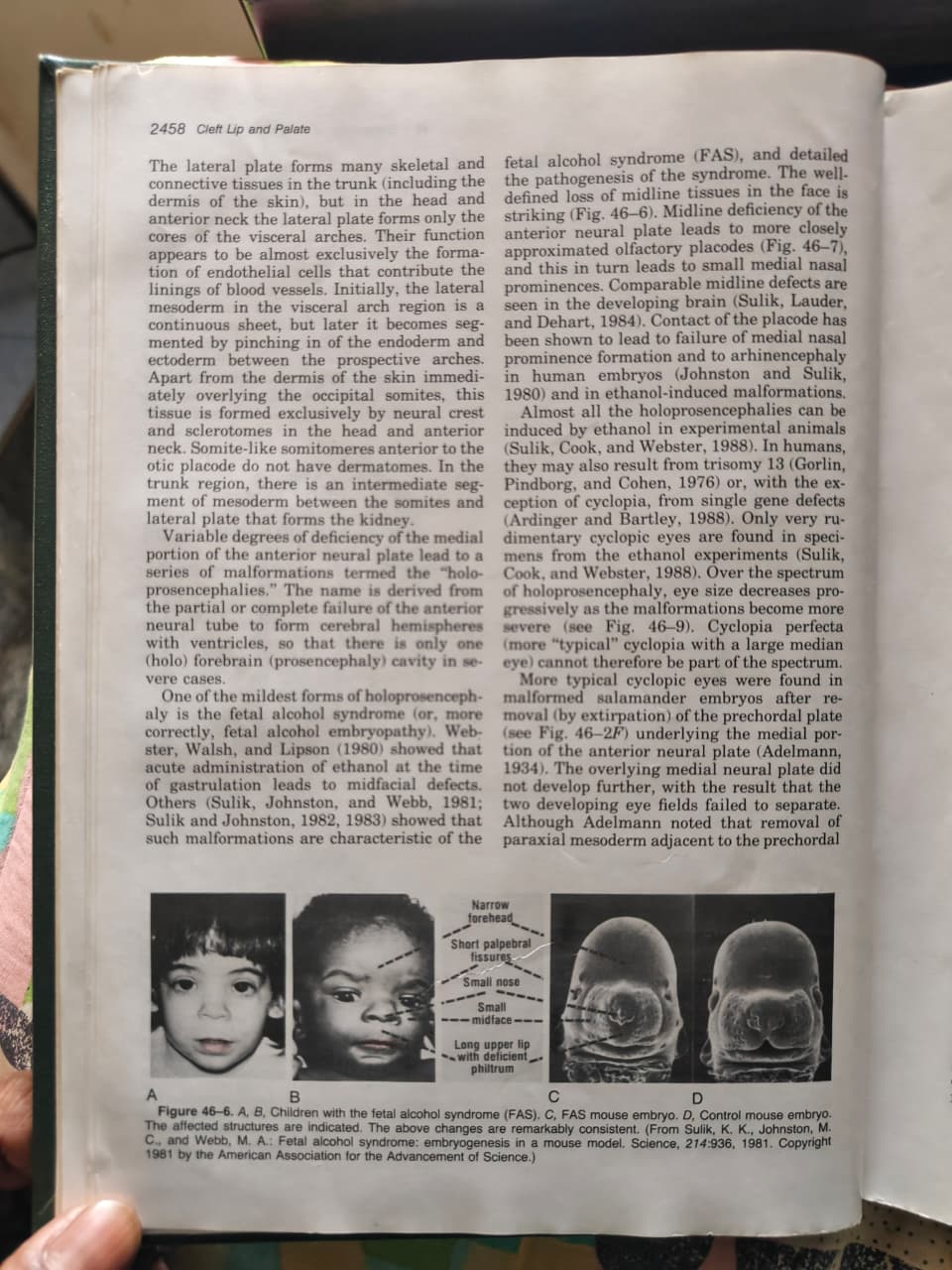
- Ethanol administration → cell death in medial portion of anterior neural plate → holoprosencephaly / FAS features
SECTION 3: THE NEURAL PLATE & ECTODERM ORGANIZATION
Ectodermal Placodes ("Thickenings")
These are ectodermal thickenings that contribute to peripheral nervous system - NOT part of the neural plate
| Placode | Develops In Relation To | Contribution |
|---|---|---|
| Olfactory | Neural folds | Neurons to olfactory system |
| Otic | Close to neural plate & tube | Inner ear structures |
| Ganglionic | Distance from neural plate/tube | Neurons to cranial sensory ganglia |
Treacher Collins Syndrome - related to defects in ectodermal thickenings (placodes) contributing to peripheral nervous system
Organization of Mesoderm (Fig. 46-5):
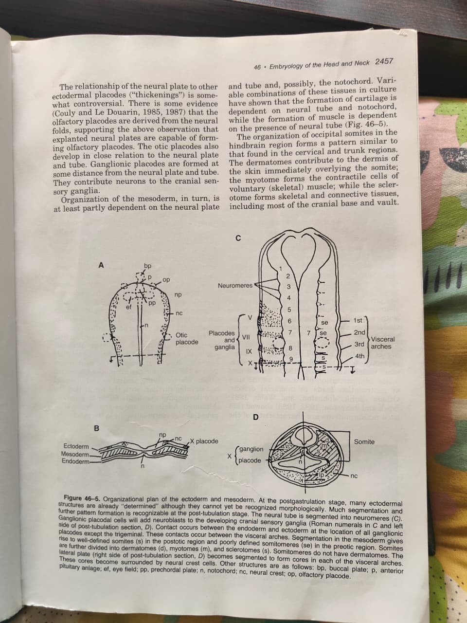
Post-gastrulation segmentation:
- Neural tube is segmented into neuromeres (C in figure)
- Mesoderm gives rise to well-defined somites in the postotic region
- Somites → divided into:
- Dermatomes (d) → dermis of skin
- Myotomes (m) → voluntary (skeletal) muscle
- Sclerotomes (s) → skeletal & connective tissue
- Somitomeres = poorly defined equivalents in the preotic (anterior) region
- Somitomeres do NOT have dermatomes
- Lateral plate (right side in figure D) → segmented into visceral arch cores
Occipital somites organization:
- Dermatomes → dermis overlying the somite
- Myotomes → contractile (voluntary/skeletal) muscle
- Sclerotomes → cranial base and vault
SECTION 4: NEURAL CREST (★★★ Most Important for Craniofacial!)
Migration & Fate (Fig. 46-4)
When: Neural crest cells migrate from neural folds at about the time folds make contact, or slightly before
What they become:
- Dissociated, loosely arranged mesenchyme
- Migrate under surface ectoderm (Fig. 46-4)
- Surround the mesodermal cores of the visceral arches
- Form ALL the mesenchyme of the rest of the face
Spectacular derivatives of neural crest cells:
- Peripheral nervous system (PNS) components
- Pigment cells of the skin
- Almost ALL skeletal and connective tissues of the face & anterior neck
- Considerable portions of the cranium
⚠️ Critical distinction: Cranial neural crest forms tissues in head/face that are formed by mesoderm elsewhere. Trunk crest cells cannot do this in higher vertebrates.
Mesenchyme vs. Epithelium (Fig. 46-3):
- Mesenchyme = loosely arranged embryonic tissue (migrating)
- Epithelium = compactly arranged embryonic tissue
- The loosely arranged mesoderm itself IS mesenchymal while migrating from the epiblast
- Later it organizes into compact somites, which again break down to form loose mesenchyme (myoblasts)
Visceral Arch Mesodermal Cores - What Happens:
- Initial mesodermal cores → involved in vascular element formation
- Endothelial buds invade and vascularize surrounding crest cell mesenchyme
- After vascularization → core cells degenerate → replaced by other mesodermal cells from near the neural tube
- New core cells = myoblasts → form contractile cells in voluntary (skeletal) muscles of face & anterior neck
- These myoblasts undergo long secondary migrations to reach final destinations
SECTION 5: PRIMARY PALATE & GROWTH CENTERS
Three Key Growth Centers for the Primary Palate:
- Medial nasal prominence
- Lateral nasal prominence
- Maxillary prominence
These three give rise to the primary palate, which upon contact forms the initial separation of the oral and nasal cavities.
Morphogenetic Movements:
- Medial and lateral nasal prominences undergo specific morphogenetic movements
- Failure of contact and fusion between these prominences = CLEFT LIP (common and rare forms)
Secondary Palate Formation:
- Palatal shelves form from the medial (inner) aspect of the maxillary prominences
- Failure to unite in the midline = clefts of the hard and soft palate
Visceral Arches - Distal Portions:
- Distal portions of 1st and 2nd arches reorganize as free-ended growth centers
- Eventually unite in the midline through merging (underlying mesenchyme becomes confluent)
- ≠ Fusions (which require breakdown of contracting epithelia)
- Other growth centers (e.g., for external ear) also involved in primary/secondary palate formation
- Skeletal tissues only begin forming at secondary palate formation time
SECTION 6: MESODERM - LATERAL PLATE & HEAD CONTRIBUTIONS
Lateral Plate Mesoderm:
- In trunk: forms skeletal AND connective tissue, dermis of skin
- In head and anterior neck: forms only the cores of visceral arches
- Function: almost exclusively angiogenic (endothelial cells → blood vessel linings)
- Initially continuous sheet in visceral arch region → later segmented
What Neural Crest Forms (Not Mesoderm):
- Dermis of skin anterior to the otic placode region
- All skeletal and connective tissues of head & anterior neck
- Exception: dermis from occipital somites; cranial base/vault from sclerotomes
SECTION 7: HOLOPROSENCEPHALY (Important Malformation Group)
Definition:
- Partial or complete failure of anterior neural tube to form cerebral hemispheres with ventricles
- Results in only ONE (holo) forebrain (prosencephaly) cavity in severe cases
Spectrum (from mild to severe):
- Mildest = Fetal Alcohol Syndrome / Fetal Alcohol Embryopathy
- With increasing severity, eye size decreases progressively
- Cyclopia perfecta (large median eye) = NOT part of the spectrum
Key Features - Fetal Alcohol Syndrome (FAS) (Fig. 46-6):
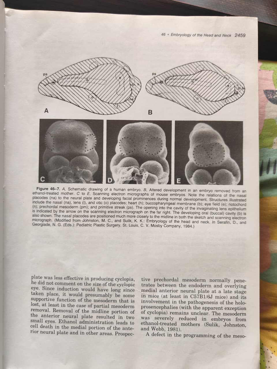
Facial features of FAS (consistent pattern):
- Narrow forehead
- Short palpebral fissures
- Small nose
- Small midface
- Long upper lip with deficient philtrum
Pathogenesis of FAS:
- Ethanol (at time of gastrulation) → midline deficiency of anterior neural plate
- This leads to more closely approximated olfactory placodes
- → Small medial nasal prominences
- → Failure of medial nasal prominence formation
- → Arhinencephaly in human embryos
- Comparable midline defects seen in developing brain
Causes of Holoprosencephaly:
- Experimental: Ethanol in animals (induces almost all forms)
- Human causes:
- Trisomy 13 (Gorlin, Pindborg, Cohen 1976)
- Single gene defects (Ardinger & Bartley, 1988) - except cyclopia
- Prechordal plate defect - deficiency of mesoderm between endoderm and overlying medial anterior neural plate → pathogenesis of holoprosencephaly
SECTION 8: EMBRYO FOLDING (TUBULATION)
Two Tubes Formed by Folding Movements:
- Neural tube (ectodermal)
- Endodermally lined gastrointestinal (GI) tube
Neural Tube Closure:
- Mechanism = coordinated contraction of a filamentous actin-myosin meshwork (terminal web) just beneath luminal lining cell surfaces (Sadler et al., 1982)
- Terminal webs also involved in many other epithelial foldings, including morphogenetic movements of the olfactory placode
- Cell proliferation & matrix formation in the mesenchymal tissue underlying the neural plate and folds are also involved
Failure of Neural Tube Closure → Neural Tube Defects (NTDs):
| NTD | Description |
|---|---|
| Anencephaly | Most severe - brain everts; brain stem portion remains; technically a misnomer as brain is not entirely absent |
| Spina bifida | Failure of closure in cervical & trunk regions |
| Meningomyelocele | Fluid-filled cavity with walls of brain tissue AND meninges |
| Meningocele | Wall contains only meninges (no brain tissue) |
Anencephaly can also be caused experimentally by postclosure rupture of the dorsal brain (associated with prior degeneration). Weakening + failure to rupture = meningomyeloceles.
SECTION 9: OTOCEPHALY (EAR HEAD) - Important Malformation
Definition:
Spectrum of malformations due to defects in the mesodermal cores of the first visceral arch
Pathogenesis:
- Mesoderm is apparently responsible for the otocephaly ("ear head") spectrum
- Breakdown of mesodermal cores in the first visceral arch (mouse mutant model - Juriloff, Sulik, and Roderick, 1980)
- Neural crest cells were normal in this study
- Failure of vascularization from the mesodermal core → complete arch breakdown
- The mesodermal core cells normally break down AFTER vascularization of the neural crest cell mesenchyme (Johnston & Listgarten, 1972)
Severity Spectrum (Fig. 46-8):
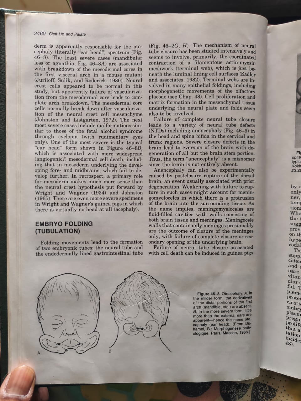
- A (Mild form) = Mandibular loss / Agnathia:
- Derivatives of the distal portions of the 1st arch (mandible, etc.) are absent
- Associated with breakdown of mesodermal cores
- B (Severe form) = True "ear head":
- Little more than external ears are apparent
- Associated with more widespread (angiogenic?) mesodermal cell death
- Including mesoderm underlying the developing fore- and midbrains → fail to develop further
- Even more severe = virtually no head at all (acephaly)
Related Malformations in Severity Spectrum:
- Next most severe: Malformations similar to fetal alcohol syndrome through cyclopia (with rudimentary eyes only)
- Shows connection between otocephaly and holoprosencephaly spectrum
SECTION 10: FIG. 46-7 - ETHANOL MODEL OF ALTERED DEVELOPMENT
Key labeled structures in the embryo diagrams:
- na = Nasal placode
- l = Lens placode
- o = Otic placode
- h = Heart
- b = Buccopharyngeal membrane (future oral/buccal cavity)
- e = Eye field
- n = Notochord
- nc = Neural crest
- pm = Prechordal mesoderm
- ps = Primitive streak
Observation: In embryo from ethanol-treated mother (Fig. B), nasal placodes are positioned much more closely together toward the midline → explains the holoprosencephaly features of FAS.
KEY CONCEPT SUMMARY TABLE
| Concept | Key Point |
|---|---|
| Gastrulation | Epiblast cells migrate through primitive streak → mesoderm |
| Primary induction | Mesoderm induces ectoderm to become neural tissue |
| Neural crest | Forms most of face, skull, PNS; unique to head region |
| Primary palate | Formed by medial & lateral nasal + maxillary prominences |
| Cleft lip | Failure of contact/fusion of nasal & maxillary prominences |
| Cleft palate | Palatal shelves fail to unite in midline |
| FAS | Ethanol → midline deficiency → holoprosencephaly spectrum |
| Otocephaly | 1st arch mesodermal core failure → ear head malformation |
| NTDs | Failure of neural tube closure → anencephaly, spina bifida |
| Visceral arches | Merge (not fuse) in midline via mesenchymal confluence |
| Somite derivatives | Dermis (dermatome) + Muscle (myotome) + Bone (sclerotome) |
MUST-KNOW ASSOCIATIONS
| Malformation | Primary Cause | Key Reference |
|---|---|---|
| Cyclopia | Prechordal plate removal; incomplete gastrulation | Adelmann, 1934; Holtfreter, 1934 |
| Holoprosencephaly | Ethanol, Trisomy 13, single gene defects | Sulik, Johnston & Webb, 1981 |
| FAS | Acute ethanol at gastrulation → midline defects | Webster, Walsh & Lipson, 1980 |
| Otocephaly | Mesodermal core failure in 1st arch | Juriloff, Sulik & Roderick, 1980 |
| Neural tube defects | Failure of actin-myosin terminal web contraction | Sadler et al., 1982 |
| Treacher Collins | Defect in peripheral nervous system placodes | - |
EXAM-READY MNEMONICS
3 Layers of Gastrulation: "Every Matron Enjoys" = Ectoderm, Mesoderm, Endoderm (outside to inside)
Somite Derivatives: "Dead Men Stand" = Dermatome, Myotome, Sclerotome
Primary Palate Prominences: "MaLM" = Medial nasal + Lateral nasal + Maxillary → Primary palate
Days of Key Craniofacial Events:
- Day 19: Germ layers form
- Day 22: Visceral arches begin
- Day 32: Most craniofacial malformations complete
- Day 50: Near end of embryonic period
Note: Chapter 48 covers cleft lip and palate embryogenesis in detail. Chapter 46 gives you the foundational embryological mechanisms - master this first, then proceed to Chapter 48 for the specific pathogenesis of clefts.
This is a shared conversation. Sign in to Orris to start your own chat.