
Location of block in Second degree AV block Mobitz Type 2
"atrioventricular block"[MeSH Terms] AND "Mobitz"
Mobitz type II second degree AV block ECG
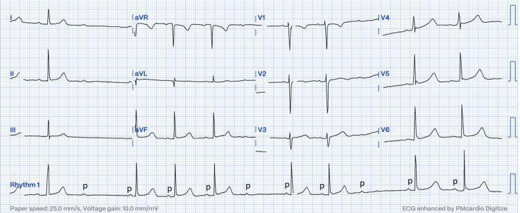
A standard 12-lead electrocardiogram (ECG) displayed on a traditional grid background with a continuous rhythm strip (Rhythm 1) along the bottom. The tracing illustrates Mobitz type II second-degree atrioventricular (AV) block. The diagnostic hallmark is visible in the rhythm strip: regular P waves are present, but there is an abrupt failure of AV conduction where a P wave (labeled with lowercase 'p') is not followed by a QRS complex. Unlike Mobitz type I (Wenckebach), the conducted beats show a constant and normal PR interval without progressive lengthening prior to the dropped beat. The ECG shows a slow heart rate (bradycardia) consistent with intermittent 2:1 or high-grade AV block. Waveform morphology includes narrow QRS complexes, suggesting the block is likely occurring at the level of the AV node or the Bundle of His. This clinical photograph serves as an educational tool for identifying advanced cardiac conduction system disease and distinguishing between subtypes of second-degree heart block in a clinical or obstetric setting.
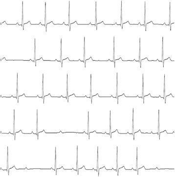
This diagnostic image consists of five continuous electrocardiogram (ECG) rhythm strips demonstrating a second-degree atrioventricular (AV) block, specifically Mobitz Type II. The strips show a regular sinus rhythm with normal P-wave morphology. The PR interval remains constant for conducted beats; however, there are intermittent, non-conducted P-waves (dropped QRS complexes) that occur without prior PR interval lengthening, which is characteristic of Mobitz Type II block. In several sections, the rhythm displays a 2:1 conduction pattern. The QRS complexes appear narrow, suggesting the site of the block is likely at the level of the Bundle of His. This finding is clinically significant as it represents a failure of conduction below the AV node and can progress to complete heart block. In this specific educational context, the arrhythmia is associated with a severe hypothyroid state and bradycardia.
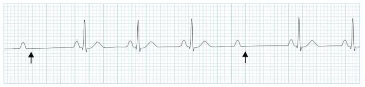
This diagnostic image is an electrocardiogram (ECG) rhythm strip illustrating Mobitz Type II second-degree atrioventricular (AV) block. The strip shows a series of P waves with a constant PR interval for conducted beats. Black arrows highlight specific P waves that are not followed by a QRS complex, indicating an intermittent failure of conduction through the AV node or His-Purkinje system. Unlike Mobitz Type I, there is no progressive lengthening of the PR interval before the dropped beat. The QRS complexes that are conducted appear narrow and maintain a consistent morphology. This rhythm strip is a classic clinical example used to teach cardiac conduction abnormalities, specifically distinguishing high-grade AV blocks that often require permanent pacemaker intervention due to the risk of progression to complete heart block.
- Usual site: The block is most often in the His-Purkinje system or the bundle branches, not the AV node. This is the textbook description in Frameworks for Internal Medicine (Mobitz II section): "The typical location of block in Mobitz II is below the AV node, within the His-Purkinje system or bundle branches."
- Narrow QRS: The block can be in the bundle of His itself (intra-Hisian).
- Wide QRS: The block is more likely in the bundle branches (infra-Hisian).
- Disease implied: Goldman-Cecil Medicine (Electrocardiography) says a sudden dropped QRS with a constant PR interval "signifies disease of the His-Purkinje system and often progresses" to complete heart block. Textbook of Family Medicine says Mobitz II and complete heart block signify marked disease in the His-Purkinje system and are always pathologic, even in athletes.
- Contrast with Mobitz I: Miller's Anesthesia (Bradyarrhythmia) says Mobitz I block is at the AV node and is often transient and asymptomatic. Mobitz II block is usually below the AV node and is often unstable.
- The conduction block is all-or-none. The PR interval stays constant and there is no Wenckebach lengthening, so the AV node's decremental conduction is not involved.
- The QRS is often wide, because bundle branch disease is common.
- Progression to complete heart block is unpredictable and can be abrupt. The escape rhythm is a slow, unreliable ventricular one.
- Atropine may worsen the block by speeding up the atria while the infranodal tissue still fails to conduct. Pacing is the definitive treatment, and a permanent pacemaker is generally indicated.
Then what's the difference between Bundle branch block and Second degree AV Block Mobitz Type 2
left bundle branch block ECG wide QRS
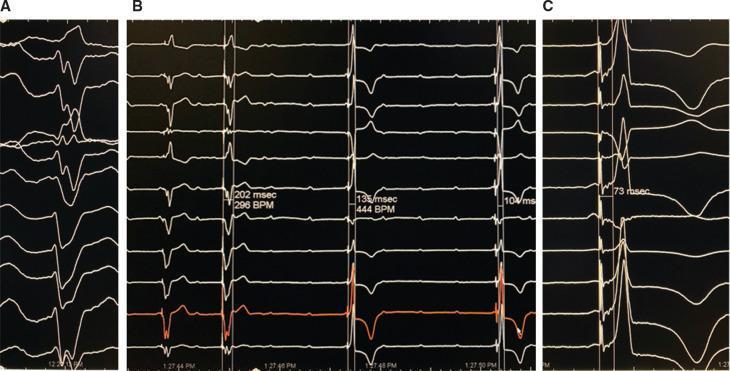
This diagnostic image consists of three panels (A, B, and C) showing multi-lead electrocardiogram (ECG) tracings recorded during a His bundle pacing (HBP) procedure in a patient with iatrogenic complete heart block. Panel A displays a Left Bundle Branch Block (LBBB) morphology with wide QRS complexes and left axis deviation. Panel B compares right ventricular (RV) pacing with non-selective His bundle pacing (NS-HBP). The first two beats show wide QRS complexes (202 msec) characteristic of RV pacing, while the subsequent beats demonstrate the transition to NS-HBP with a significantly narrower QRS duration (135 msec) and altered repolarization. Panel C illustrates a narrow QRS complex (73 msec) achieved via NS-HBP, characterized by increased voltage in lead I, normalization of the frontal QRS axis, and improved R-wave progression in the precordial leads. These findings highlight the educational concept of physiological pacing as an alternative to conventional ventricular pacing for correcting bundle branch blocks and maintaining electrical synchrony.
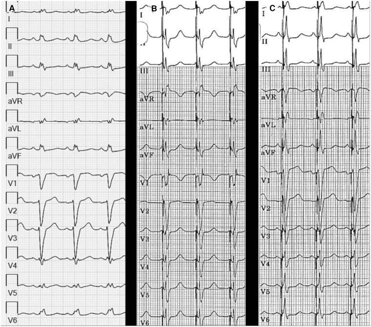
A comparison of three 12-lead electrocardiogram (ECG) strips (labeled A, B, and C) illustrating the effects of Left Bundle Branch Area Pacing (LBBAP) on ventricular activation. Panel A displays baseline sinus rhythm with Left Bundle Branch Block (LBBB), characterized by a wide QRS duration (156 ms), dominant S waves in V1-V3, and broad R waves in I, aVL, and V6. Panel B shows LBBAP with a short Atrioventricular (AV) delay of 40 ms, resulting in a reduced QRS duration of 128 ms and a Right Bundle Branch Block (RBBB) morphology in lead V1 (rSR' pattern), indicating left ventricular capture. Panel C demonstrates the normalization of the QRS complex (120 ms) by increasing the AV delay to 80 ms. This adjustment facilitates physiological fusion between the paced left ventricular activation and intrinsic anterograde conduction through the right bundle. The sequence highlights the transition from wide-complex dyssynchrony to a more narrow, synchronized ventricular activation pattern, with associated improvements in T-wave morphology and ST-segment alignment across the limb and precordial leads.
| Feature | Bundle branch block | Mobitz II (2nd-degree AV block) |
|---|---|---|
| What fails | Conduction down the right or left bundle, so ventricular depolarization is slow or abnormal | Conduction from atria to ventricles, so some P waves are not followed by a QRS |
| Main ECG sign | Wide QRS (≥120 ms) with a typical morphology | Dropped QRS after a P wave, with a constant PR interval in conducted beats |
| P-QRS relationship | Every P wave is conducted (unless there is also AV block) | Intermittent non-conducted P waves |
| QRS | Always wide (RBBB: rSR' in V1; LBBB: broad notched R in I, aVL, V6) | Wide if the block is in the bundle branches, narrow if it is intra-Hisian |
| PR interval | Normal, or prolonged if there is also first-degree AV block | Constant, often normal or prolonged |
| Heart rate | Usually normal | Often slow, because beats are dropped |
| Symptoms | Often none | Dizziness, syncope, and bradycardia are common |
| Treatment | Usually none alone. Treat the cause. CRT may be used for LBBB with heart failure. | Permanent pacemaker is generally indicated, even if asymptomatic |
- Both reflect disease of the His-Purkinje system. Mobitz II is often seen with a bundle branch block, so a wide QRS is common.
- The His-Purkinje system has a right bundle and a left bundle, and the left splits into anterior and posterior fascicles. Mobitz II happens when this system intermittently fails to conduct to the ventricles at all.
- A single bundle branch block means the other branch still conducts, so every P wave still gets through, just with delay. If the remaining branch fails intermittently, you get dropped beats, which is Mobitz II.
- Alternating BBB (RBBB on some beats, LBBB on others) is effectively the same problem. It means both bundles are diseased and is a pacing indication.
- Bifascicular block is RBBB plus left anterior or posterior fascicular block.
- Trifascicular block is bifascicular block plus first-degree AV block.
- Each is a stage of progressive infranodal disease that can progress to complete heart block.
- Goldman-Cecil Medicine (Chronic Bifascicular Block) lists these as pacing indications: advanced second-degree or intermittent third-degree AV block, type II second-degree AV block, and alternating bundle branch block.
- BBB alone is a conduction delay. Mobitz II is intermittent loss of AV conduction.
- A BBB patient who has syncope or documented Mobitz II needs a pacemaker evaluation, not just observation.
- Rosen's Emergency Medicine (Box 11.2A) lists Mobitz II second-degree block as a dysrhythmia potentially associated with syncope. Isolated BBB is not on that list.
- In acute MI, new BBB with first-degree AV block, and Mobitz II, are both listed in Pfenninger and Fowler's Procedures for Primary Care among pacing indications. This is because the infarct involves the conduction system.
Why Second degree AV Block is intermittent?
- The AV node has decremental conduction, meaning it conducts more slowly the faster it is stimulated.
- Tintinalli's Emergency Medicine explains that each depolarization lengthens the node's refractory period. The next atrial impulse arrives earlier in the relative refractory period and conducts more slowly. This is why the PR interval progressively lengthens.
- Eventually an impulse arrives while the node is still refractory and is not conducted.
- The dropped beat gives the node a pause to recover, and the cycle restarts. The intermittency is built into the mechanism.
- The His-Purkinje system does not conduct decrementally. It either conducts an impulse or it does not, so the PR interval does not lengthen before the dropped beat.
- Diseased tissue (fibrosis, ischemia, infarct, infiltration) is borderline. Its margin of safety is thin.
- Whether a given impulse conducts depends on small changes in:
- the atrial rate, because faster rates give less recovery time
- autonomic tone
- ischemia or edema at that moment
- the timing of the impulse relative to the tissue's recovery
- So the block comes and goes unpredictably, and conducted beats look normal.
- Disease that progresses leads to 2:1 or 3:1 block, then high-grade block, then complete heart block. Braunwald's Heart Disease (High-Grade AV Block) notes that high-grade block still shows some intermittent atrial-ventricular relationship, unlike complete block.
- Rosen's Emergency Medicine notes that 2:1 conduction can be normal with very fast atrial rates, such as atrial flutter. This reflects the normal refractory period of the AV node.
- Vagal tone can also cause transient block, for example during sleep or in athletes, and with drugs such as digoxin. Goodman & Gilman notes that atropine can help Wenckebach block when vagal activity is the cause.
- Because the block is intermittent, a single ECG may miss it. Ambulatory monitoring may be needed.
- A patient can be well one moment and have a syncopal pause the next.
- Mobitz II is unpredictable and can suddenly become complete block with a slow, unreliable escape rhythm. This is why it is treated with a pacemaker, even when the patient has no symptoms at the time.
Why is bundle branch block whether right or left is associated with leftward or rightward axis and left or right ventricular hypertrophy?
LBBB QRS axis left axis deviation left ventricular hypertrophy diagnosis; RBBB right axis deviation right ventricular hypertrophy ECG
1. Why BBB can shift the axis
- The LV is activated normally through the left bundle, so the early QRS looks normal.
- The right ventricle (RV) is then activated late, slowly, from cell to cell across the septum.
- Guyton and Hall (Vectorial Analysis of Right Axis Deviation in RBBB): the left ventricle depolarizes far more rapidly than the right. The late, slow forces point rightward and anteriorly. This gives a broad terminal R' in V1 and a broad S in I and V6. The mean axis can drift rightward.
- In practice the frontal axis is usually normal. StatPearls notes that determining axis in BBB is controversial. It suggests using the first 80 to 100 ms of the QRS, which reflects LV activation, to avoid the late terminal delay.
- If RBBB comes with left axis deviation, think left anterior fascicular block. If it comes with right axis deviation, think left posterior fascicular block or RV hypertrophy (RVH). Both are bifascicular patterns.
- Activation spreads from the right side. The septum is activated abnormally, then the LV is activated slowly from cell to cell.
- All the forces point leftward and posteriorly. The axis is normal or leftward. This gives broad, notched R waves in I, aVL and V6, and deep S or QS waves in V1.
- Rosen's Emergency Medicine lists LBBB and LV hypertrophy (LVH) together as causes of left axis deviation.
2. Why BBB goes with hypertrophy
- Hypertension, aortic stenosis, and dilated or ischemic cardiomyopathy enlarge and fibrose the LV. The left bundle runs through that stretched, scarred septum.
- A larger LV also takes longer to depolarize, which widens the QRS.
- LBBB itself changes the activation sequence. Long-standing LBBB can cause dyssynchrony and LV dilation.
- LITFL notes that in dilated cardiomyopathy, LBBB occurs due to cardiac dilatation, along with LVH and left axis deviation.
- Pulmonary hypertension, chronic lung disease, and congenital lesions with RV volume or pressure load stretch the RV and the right bundle.
- Creasy and Resnik notes that ASD shows incomplete RBBB. Ostium secundum defects show right axis deviation. Ostium primum defects show marked left axis deviation with incomplete RBBB and RVH.
- Braunwald's Heart Disease lists RBBB with QRS fragmentation, right axis deviation, and RVH in Eisenmenger syndrome.
- Fuster and Hurst's The Heart describes RV hypertrophy with right axis deviation in the setting of RV pressure overload.
- Incomplete RBBB in RV volume overload is partly due to delayed activation of the thickened RV outflow region (crista supraventricularis).
3. Interpretation pitfalls
- BBB distorts voltage criteria. LBBB often gives very tall QRS voltages, which can mimic LVH.
- LITFL states there are no universally accepted criteria for diagnosing RVH in the presence of RBBB. Standard voltage criteria do not apply. Right axis of about +110° or more plus a tall R in V1 and strain pattern suggests RVH.
- Ganong notes that fascicular blocks cause axis shifts without the full QRS widening of BBB. Left anterior hemiblock gives left axis deviation, and left posterior hemiblock gives right axis deviation.