
nocturnal epileptic seizures of mouth
nocturnal frontal lobe epilepsy oroalimentary automatisms
nocturnal epilepsy sleep oral automatisms treatment
nocturnal frontal lobe epilepsy EEG sleep seizure
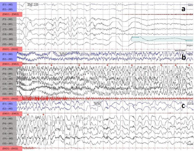
A polysomnographic (PSG) and electroencephalographic (EEG) recording displaying a sleep-hypermotor epilepsy (SHE) event, formerly known as nocturnal frontal lobe epilepsy. The content is presented in three chronological segments (a, b, and c) capturing a 30-second window. Segment (a) shows the ictal onset from N3 sleep, characterized by an abrupt transition from high-voltage slow-wave activity to rhythmic beta and theta frequencies. Segment (b) demonstrates the continuation of the event, where the EEG channels (F3-M2, F4-M1, C3-M2, C4-M1, O1-M2, O2-M1) exhibit high-amplitude, rapid, and sharp generalized epileptiform discharges, marked with red asterisks. These represent dense spikes and waves typical of frontal lobe seizures. Segment (c) captures the resolution of the event, with a gradual decrease in frequency and amplitude of discharges as the brain returns to a post-ictal state. The recording includes EOG (E1-M2, E2-M1), EMG (EMG1-EMG3), and EKG channels, providing a comprehensive view of the patient's physiological state during the seizure. This material is designed for advanced medical education in neurology and sleep medicine to illustrate EEG morphology during focal seizures.
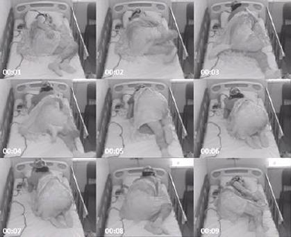
This clinical photographic sequence illustrates a focal stereotyped motor seizure captured via overnight video-EEG monitoring. The figure is presented as a 3x3 grid of grayscale frames, labeled with sequential timestamps from 00:01 to 00:09, documenting an approximately 10-second ictal event. The subject is an adult patient in a hospital bed equipped with safety rails, wearing a headwrap likely containing EEG electrodes. The sequence begins (frame 00:01) with the patient in a supine/lateral resting position. Progressive frames (00:02–00:07) demonstrate a sudden arousal followed by intense, irregular hypermotor activity involving all four limbs. The patient exhibits thrashing movements, transitioning from a recumbent position to an semi-upright, kneeling-like posture on the bed. By the final frames (00:08–00:09), the motor activity subsides as the patient returns to a resting recumbent state. This visual is representative of nocturnal frontal lobe epilepsy or similar focal seizure disorders characterized by paroxysmal motor behavior. It serves as an educational tool for identifying semiology in epileptology and clinical neurology.
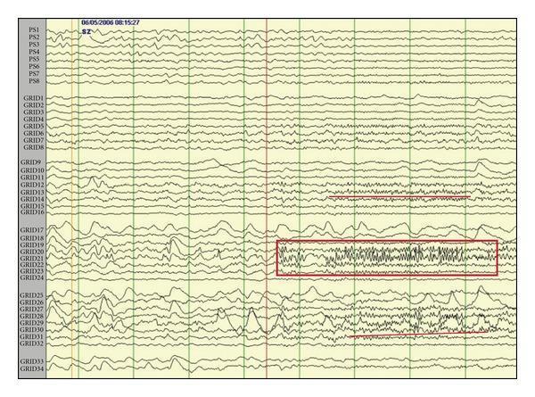
Diagnostic intracranial EEG (iEEG) trace recording a seizure onset in the frontal lobe. The image displays a referential montage from a 6 x 6 subdural grid (34 active contacts) and a 1 x 8 subdural strip (labeled PS1-PS8). A prominent red box highlights a focal gamma frequency oscillation occurring at seizure onset, localized primarily to grid contacts 21, 22, and 23. This high-frequency activity represents an ictal discharge. Early spread of this fast frequency activity is visible in adjacent contacts, including 12, 13, 28, 29, and 30. The pre-ictal phase is characterized by lower amplitude and slower background frequency activity across all electrodes. Clinically, this focal fast-frequency pattern (>13-125 Hz) on iEEG is used to identify the seizure onset zone and is associated with favorable surgical outcomes in nonlesional frontal lobe epilepsy. The recording provides critical information for neurosurgical mapping and the localization of epileptogenic zones.
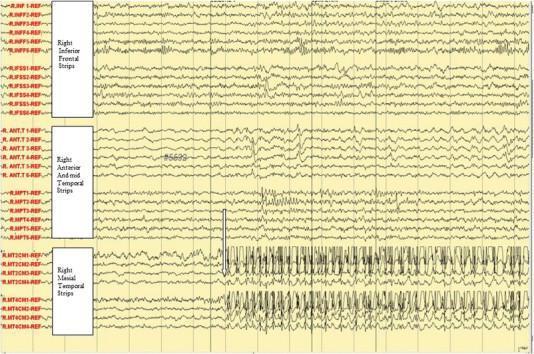
This diagnostic image displays a subdural electroencephalogram (EEG) recording, used in neurosurgery and epilepsy monitoring to localize seizure onset. The tracing is divided into three primary anatomical regions: Right Inferior Frontal Strips, Right Anterior and mid Temporal Strips, and Right Mesial Temporal Strips. The background activity across the frontal and mid-temporal leads shows relatively low-amplitude, disorganized, and irregular frequencies. A prominent ictal event is highlighted by a vertical arrow in the Right Mesial Temporal Strips, specifically localized to electrodes R-MT2 and R-MT4. At this point, the signal transitions from baseline activity to high-amplitude, rhythmic spikes and sharp waves. These paroxysmal discharges demonstrate rapid upstrokes and organized repetition, characteristic of a focal ictal onset. This visual provides clinical evidence of the electrographic correlate for a patient's aura of fear and palpitations, supporting the localization of epilepsy to the mesial temporal lobe. The image is an essential resource for understanding intracranial EEG interpretation and the electro-clinical correlation in focal epilepsy.
rolandic epilepsy centrotemporal spikes EEG childhood
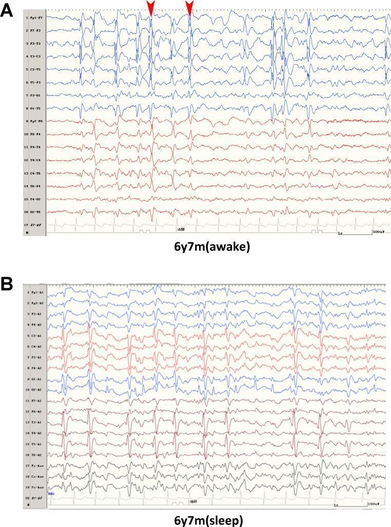
This diagnostic image displays a comparison of Electroencephalogram (EEG) recordings from a 6-year-old patient with focal epilepsy. Panel A illustrates the 'awake' state, showing irregular background activity characterized by frequent epileptiform discharges. Specifically, red arrowheads highlight spikes and spike-wave complexes concentrated in the Rolandic (centrotemporal) regions, with a more pronounced manifestation on the left hemisphere. Panel B depicts the 'sleep' state (NREM sleep), demonstrating a transition to slower, more synchronized background rhythms. In this state, the frequency of epileptiform activity increases significantly, with a discharge index of approximately 85%. The waveforms show high-amplitude, rhythmic spike-wave patterns across multiple channels, a hallmark of continuous spikes and waves during slow-wave sleep (CSWS) or related epileptic encephalopathies. The image serves as an educational tool for identifying Rolandic discharges and understanding the sleep-induced activation of epileptiform activity in pediatric focal epilepsy syndromes.
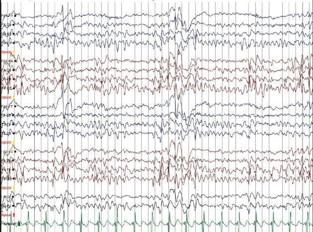
This diagnostic image shows a multi-channel electroencephalogram (EEG) tracing accompanied by a single-lead electrocardiogram (ECG) at the bottom. The EEG illustrates high-amplitude, diphasic spikes and slow-wave complexes. These epileptiform discharges are most prominent in the right centrotemporal region, appearing as sharp, transient deflections against a background of rhythmic baseline activity. The spikes exhibit a horizontal dipole morphology, characterized by distinct upward and downward deflections. A voltage scale of 70 mV/cm is utilized, highlighting the high-voltage nature of the paroxysmal activity. The distribution and morphology of these spikes are classic diagnostic features of Self-Limited Epilepsy with Centrotemporal Spikes (SLECTS). The tracing demonstrates regional predilection with maximal electronegativity in the centrotemporal channels, while the synchronous ECG lead confirms periodic cardiac rhythm alongside the neurological recording. This visual material is used to teach EEG interpretation in pediatric neurology, focusing on focal epilepsy syndromes.
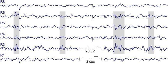
This diagnostic image displays 15 seconds of resting-state electroencephalogram (EEG) data recorded from a 7-year-old child using ear-EEG sensor technology (cEEGrid). The trace shows six active channels labeled R1 through R8, with scale indicators provided for 70 microvolts (uV) in amplitude and 2 seconds in time. The primary educational focus is the identification of interictal epileptiform discharges. Specifically, the image highlights four distinct occurrences of spike-wave activity, marked by gray shaded vertical bars. These epileptiform events are most prominent in channels R3, R4, R5, and R6, exhibiting a classic morphology consisting of sharp spikes immediately followed by slower wave components. While the subject is healthy with no seizure history, the record demonstrates subclinical epileptiform activity, which is clinically relevant in the context of a family history of Rolandic epilepsy (Benign Epilepsy with Centrotemporal Spikes). The image serves as an example of how localized ear-level EEG can capture temporal lobe epileptiform abnormalities.
Nocturnal Epileptic Seizures Involving the Mouth
1. Self-Limited Epilepsy with Centrotemporal Spikes (SeLECTS / BECTS / Benign Rolandic Epilepsy)
- Onset age 3-13 years, peak around 7-10 years
- Seizures occur predominantly during sleep (drowsiness or early sleep) or shortly after waking
- Focal clonic twitches of the face and mouth region - often one side of the face (hemifacial)
- Perioral numbness/tingling - a characteristic sensory aura
- Drooling and speech arrest - child cannot speak during the episode
- Guttural/gurgling sounds due to oropharyngeal involvement
- Secondary generalization (GTC) can occur if the seizure spreads
- Child is often conscious throughout but unable to communicate
2. Autosomal Dominant Nocturnal Frontal Lobe Epilepsy (ADNFLE) / Sleep-Related Hypermotor Epilepsy (SHE)
- Onset typically before age 20 (mean 8-11 years), but can appear at any age
- Seizures occur exclusively or predominantly during NREM sleep
- Episodes range from simple paroxysmal arousals to complex hypermotor events with thrashing, kicking, bicycling movements
- Motor and oral automatisms - including oroalimentary automatisms (lip-smacking, chewing, swallowing movements) as part of the frontal lobe ictal spread
- Usually brief (<30 seconds), stereotyped, and may cluster throughout the night
- Often misdiagnosed as parasomnias (sleepwalking, night terrors) or psychogenic events
3. Childhood Absence Epilepsy (oral automatisms during absences)
- Absence seizures can have associated mouth movements and automatisms (lip-smacking, chewing, swallowing)
- Typically occur during wakefulness but can be triggered by drowsiness
- EEG: 3-4 Hz generalized spike-and-wave, activated by hyperventilation
- Treatment: Ethosuximide (first-line), lamotrigine, or valproic acid
- Resolves by adolescence in most cases
4. Temporal Lobe Epilepsy (TLE) - Nocturnal Subtype
- Complex partial seizures arising from the mesial temporal lobe commonly feature oroalimentary automatisms - lip-smacking, chewing, swallowing, tongue movements
- A subgroup has predominantly nocturnal occurrence ("nocturnal temporal lobe epilepsy")
- Other features: staring, hand automatisms, postictal confusion
- EEG: anterior temporal sharp waves/spikes, often showing seizure onset in the hippocampal/amygdalar region
Differential Diagnosis: Parasomnias vs. Nocturnal Seizures
| Feature | Nocturnal Seizure | Parasomnia (NREM) |
|---|---|---|
| Stereotypy | High (same each time) | Variable |
| Duration | Usually <2 min | 1-30+ min |
| Frequency per night | Multiple possible | Usually 1 |
| Memory | None | Partial or none |
| Time of night | Any (NREM onset) | First third of night |
| Postictal confusion | Yes (often brief) | Yes (prolonged) |
| EEG | May show changes | Normal |
Summary of Key Syndromes by Mouth Symptom
| Symptom | Most Likely Syndrome |
|---|---|
| Perioral tingling + unilateral face twitching | BECTS/Rolandic epilepsy |
| Drooling + speech arrest during sleep | BECTS/Rolandic epilepsy |
| Lip-smacking/chewing + hypermotor movements | ADNFLE / SHE |
| Lip-smacking + staring + postictal confusion | Temporal lobe epilepsy |
| Mouth movements + brief staring spells | Childhood absence epilepsy |
- Bradley and Daroff's Neurology in Clinical Practice (Sleep and Epilepsy; ADNFLE sections)
- Adams and Victor's Principles of Neurology, 12th Ed.
- Harriet Lane Handbook, 23rd Ed. (Selected Epilepsy Syndromes table)