
Development of pharyngeal pouches 5m shortnote
Reading File
Now let me search the medical library for pharyngeal pouches development.
Excellent - I have two primary embryology textbooks. Let me read both in parallel.
Reading File
Reading File
I have rich content from both textbooks. Let me read the remaining sections (3rd, 4th pouches + clinical notes) from The Developing Human.
Reading File
Reading File
I now have comprehensive content from both textbooks. Let me fetch the key diagram image to include in my answer.
Loading Image
I have everything I need for a thorough short note. Here is the complete answer:
Development of Pharyngeal Pouches
Introduction
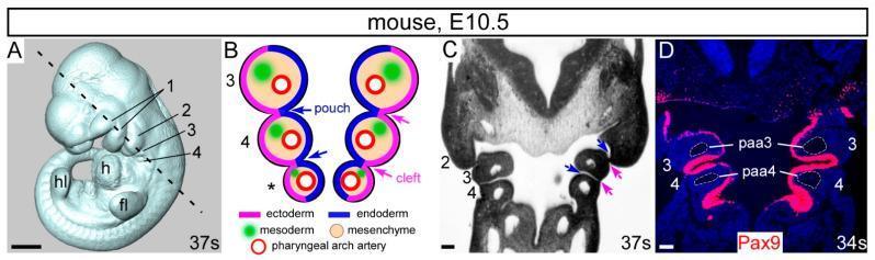
The pharyngeal (branchial) pouches are paired outpocketings of endoderm that line the lateral walls of the primordial pharynx. They arise in a craniocaudal sequence between adjacent pharyngeal arches, beginning around the 4th week of embryonic development. The human embryo has four well-defined pairs; a fifth pair, if present, is rudimentary. The endoderm of each pouch contacts the ectoderm of the corresponding pharyngeal groove (cleft), forming a thin bilateral pharyngeal membrane at each level.
Signaling molecules - retinoic acid, Wnt, and fibroblast growth factor (FGF) - are essential for pouch formation and differentiation. - The Developing Human, p. 472
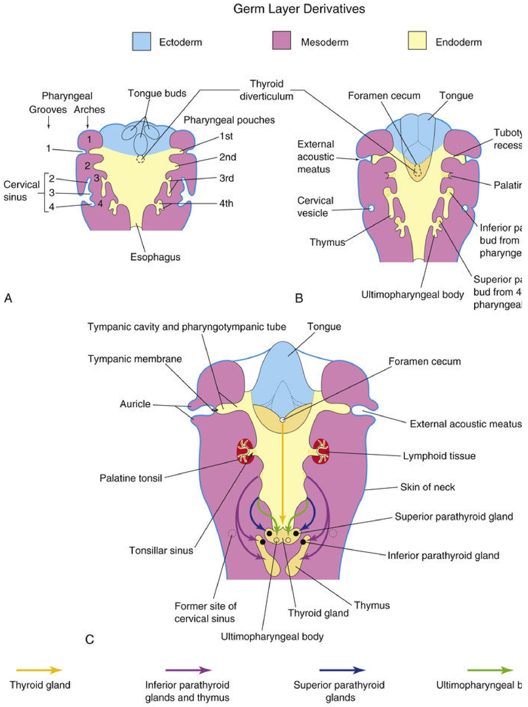
Derivatives of Each Pouch
1st Pharyngeal Pouch
The first pouch elongates into a tubular diverticulum called the tubotympanic recess (stalklike outgrowth toward the first pharyngeal groove).
- Distal expanded portion - contacts the ectoderm of the first groove and contributes to the tympanic membrane (eardrum)
- Saclike distal end - becomes the tympanic cavity (middle ear cavity) and the mastoid antrum
- Proximal narrow stalk - persists as the pharyngotympanic (auditory/Eustachian) tube, connecting the middle ear to the nasopharynx
- The Developing Human, p. 473; Langman's Medical Embryology, p. 309
2nd Pharyngeal Pouch
The second pouch is largely obliterated but makes critical contributions:
- The endoderm proliferates and forms buds that penetrate the underlying mesenchyme; these are invaded by lymphoid tissue during the 3rd-5th months to form the primordium of the palatine tonsil
- The residual cavity of the pouch persists as the tonsillar sinus (fossa) - the depression between the palatoglossal and palatopharyngeal arches in adults
- Langman's Medical Embryology, p. 309; The Developing Human, p. 473
3rd Pharyngeal Pouch
This pouch is characterized by a dorsal bulbar wing and a hollow ventral wing at its distal extremity:
- Dorsal portion - differentiates into the inferior parathyroid gland (by the 6th week)
- Ventral portion - epithelium proliferates, cavities obliterate, and both sides fuse in the midline to form the thymus (primary lymphoid organ)
The thymus migrates caudally and medially into the anterior thorax, dragging the inferior parathyroid with it initially. The inferior parathyroid ultimately separates from the thymus and settles on the dorsal surface of the thyroid gland as the inferior parathyroid. Because the inferior parathyroid travels so far (from 3rd pouch) before landing below the superior parathyroid (from 4th pouch), their positions are considered paradoxical but are explained by their embryological origins.
Thymic histogenesis: Epithelial cords sprout side branches (forming lobule cores), and some cells organize into Hassall (thymic) corpuscles. Lymphocytes populate from haematopoietic stem cells. Neural crest cells also contribute. The thymus grows until puberty, then undergoes involution with fat infiltration. FGF signaling via FRS2 drives thymus and parathyroid development.
- The Developing Human, pp. 475-477; Langman's Medical Embryology, p. 310
4th Pharyngeal Pouch
Also has dorsal and ventral wings:
- Dorsal portion - differentiates into the superior parathyroid gland, which attaches to the dorsal surface of the migrating thyroid as it descends - ending up above the inferior parathyroid in the adult
- Ventral portion - forms the ultimobranchial body (ultimopharyngeal body), which fuses with and is incorporated into the thyroid gland. Its cells become the parafollicular (C) cells of the thyroid, which secrete calcitonin (regulates serum calcium)
- Langman's Medical Embryology, p. 310; The Developing Human, p. 476
5th Pharyngeal Pouch
Rudimentary and inconsistently present. If it forms, it is usually incorporated with the 4th pouch and contributes to the ultimobranchial body.
Summary Table
| Pouch | Dorsal derivative | Ventral derivative |
|---|---|---|
| 1st | Tympanic cavity + mastoid antrum | Pharyngotympanic (Eustachian) tube |
| 2nd | Palatine tonsil / tonsillar fossa | (largely obliterated) |
| 3rd | Inferior parathyroid gland | Thymus |
| 4th | Superior parathyroid gland | Ultimobranchial body → C cells of thyroid |
| 5th | Rudimentary; joins 4th | Rudimentary |
Clinical Correlates
- DiGeorge syndrome (22q11.2 deletion): Failure of development of the 3rd and 4th pouches → absent/hypoplastic thymus (T-cell immunodeficiency) and absent parathyroid glands (hypocalcaemia, tetany). Caused by failure of neural crest cell migration.
- Cervical sinus: The 2nd arch overgrowing and burying the 2nd, 3rd, and 4th clefts forms the cervical sinus, which normally obliterates. Failure of obliteration results in a branchial (cervical) cyst, sinus, or fistula - a lateral neck swelling.
- Ectopic parathyroid/thymic tissue: Due to the long migratory path, parathyroid or thymic tissue can be found anywhere along the migration route (e.g., thymic nests in the thyroid, ectopic parathyroid in the mediastinum).
- Pharyngeal (Zenker's) diverticulum: A posterior herniation through Killian's dehiscence at the inferior constrictor of the pharynx - clinically important but a structural (not developmental pouch) anomaly.
Sources: The Developing Human: Clinically Oriented Embryology, 11th ed., pp. 472-477 | Langman's Medical Embryology, 14th ed., pp. 308-311
This is a shared conversation. Sign in to Orris to start your own chat.