Primary skin lesions
Reading File
Finding Sources
Finding Sources
Reading File
Reading File
Reading File
Reading File
Loading Image
Loading Image
Loading Image
Loading Image
Loading Image
Loading Image
Reading File
Reading File
Loading Image
I have all the content and images needed. Here is the complete, well-illustrated guide to primary skin lesions.
Primary Skin Lesions
Primary skin lesions are the original, unmodified lesions arising directly from the disease process - before any trauma, scratching, or secondary change alters them. Identifying them correctly is the most important step in the dermatologic examination. They can be modified by regression, trauma, or external factors to produce secondary lesions.
"The original lesions are known as the primary lesions, and identification of such lesions is the most important aspect of the dermatologic physical examination."
- Andrews' Diseases of the Skin, p. 410
Classification Overview
Primary lesions fall into three main groups based on whether they are palpable:
| Group | Lesion Types |
|---|---|
| Flat (non-palpable) | Macule, Patch |
| Raised (palpable/solid) | Papule, Plaque, Nodule, Tumor |
| Fluid-filled | Vesicle, Bulla, Pustule, Wheal |
Flat (Non-Palpable) Lesions
1. Macule
A flat, circumscribed area of color change - not raised or depressed, perceptible only as a color difference from surrounding skin. Diameter < 1 cm. Does not alter skin texture. Examples: freckle (ephelid), petechiae, early vitiligo.
"A macule is flat, even with the surface level of surrounding skin or mucous membranes, and perceptible only as an area of color different from the surrounding skin." - Fitzpatrick's Dermatology
2. Patch
Essentially a large macule - ≥ 1 cm in diameter. Same characteristics as a macule (flat, color change only). Examples: nevus flammeus (port-wine stain), large café-au-lait spot, vitiligo.
Note: The only distinction between a macule and a patch is size. - Harrison's Principles of Internal Medicine 22E
Raised (Solid) Lesions
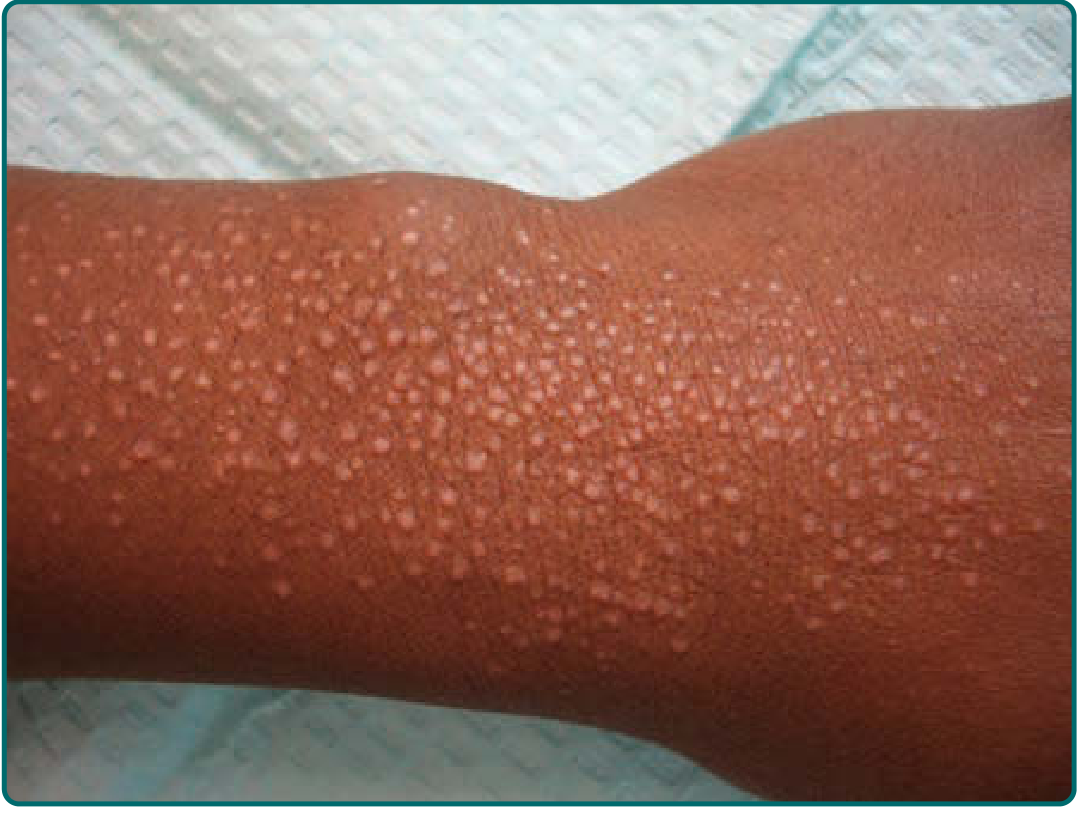
3. Papule
A small (< 1 cm), solid, circumscribed elevation above the skin surface - palpable. May be sessile, pedunculated, dome-shaped, flat-topped, filiform, or umbilicated. Can be white (milium), red (eczema), yellow (xanthoma), or pigmented (melanoma). Examples: closed comedone, warts, lichen nitidus.
Important: The term "maculopapular" should be avoided - there is no such thing as a "maculopapule." Such eruptions are better described as morbilliform. - Andrews' Diseases of the Skin
4. Plaque
A broad, flat-topped, raised lesion ≥ 1 cm - essentially a large papule or confluence of papules. Edges may be distinct (psoriasis) or blend gradually into surrounding skin (eczematous dermatitis). May sometimes be centrally depressed.
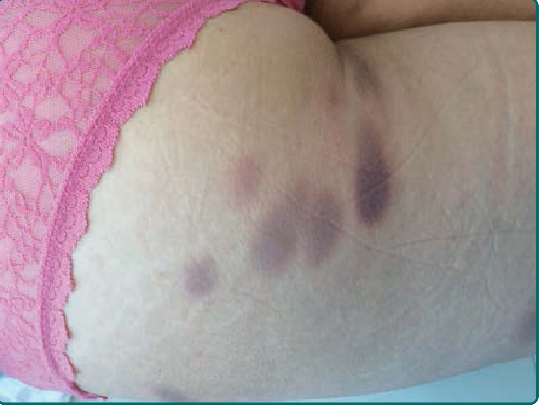
5. Nodule
A palpable lesion > 1 cm with a domed, spherical, or ovoid shape - either solid or cystic. Deeper than a papule, often centered in the dermis or subcutaneous fat. Described by 5 anatomic types: (1) epidermal, (2) epidermal-dermal, (3) dermal, (4) dermal-subdermal, (5) subcutaneous. Texture (firm, soft, fluctuant) and surface (smooth, keratotic, ulcerated) help direct diagnosis. Examples: lipoma, dermal nevus, lymphoma cutis.
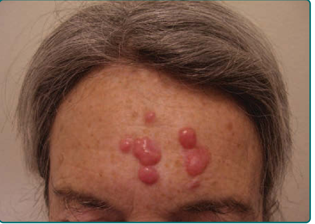
6. Tumor
A solid, raised growth or mass > 2 cm (some sources say > 5 cm per Harrison's), freely movable or fixed. May be elevated or deep-seated, sometimes pedunculated (e.g., neurofibromas). Generally rounded; consistency depends on constituents. May be benign or malignant.
Fluid-Filled Lesions
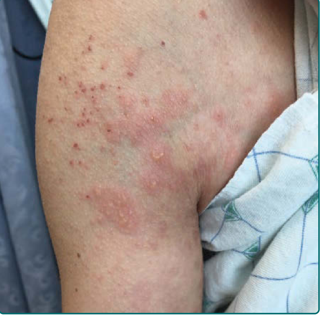
7. Vesicle
A small (< 0.5-1 cm), fluid-filled, raised lesion - the wall is thin and translucent enough to visualize the contents (clear, serous, or hemorrhagic). Arises from cleavage at various epidermal levels (intraepidermal) or at the dermal-epidermal interface (subepidermal). Tenseness vs. flaccidity helps indicate depth. Examples: contact dermatitis (poison ivy), herpes simplex/zoster, varicella.
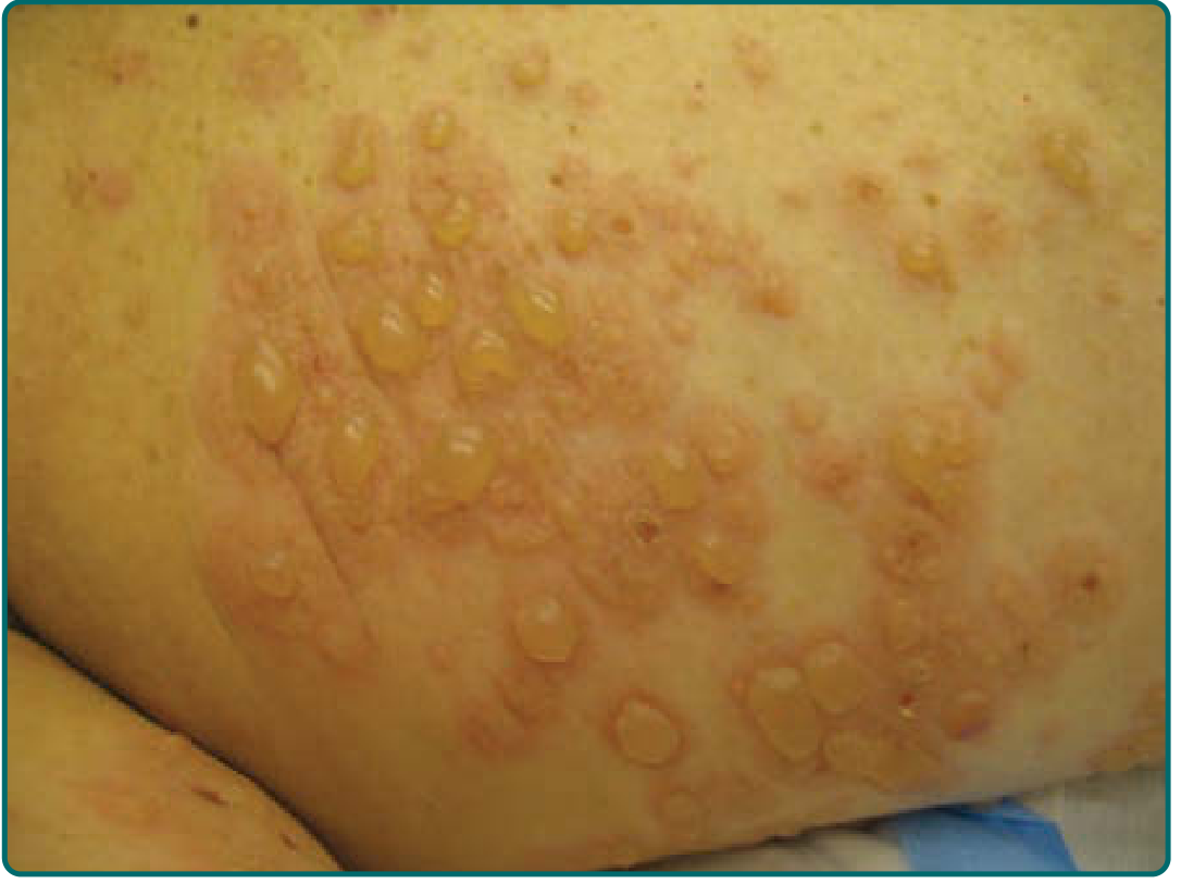
8. Bulla (Blister)
A fluid-filled lesion ≥ 0.5-1 cm - same characteristics as a vesicle, but larger. Contents may be clear, serous, or hemorrhagic. Subepidermal bullae tend to be tenser than intraepidermal ones. Examples: pemphigus vulgaris, bullous pemphigoid, thermal burns, linear IgA disease.
9. Pustule
A circumscribed, raised lesion containing visible pus (leukocytes ± cellular debris). The purulent exudate may be white, yellow, or greenish-yellow. May be sterile (pustular psoriasis) or infectious. When follicle-associated, appears conical with a central hair. Size < 1 cm. Examples: acne vulgaris, folliculitis, pustular psoriasis.
Key point: The presence of pustules does not necessarily signify infection. - Harrison's Principles of Internal Medicine 22E
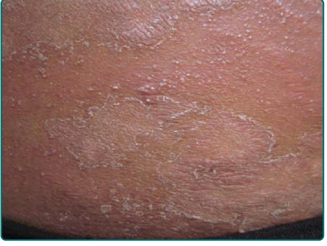
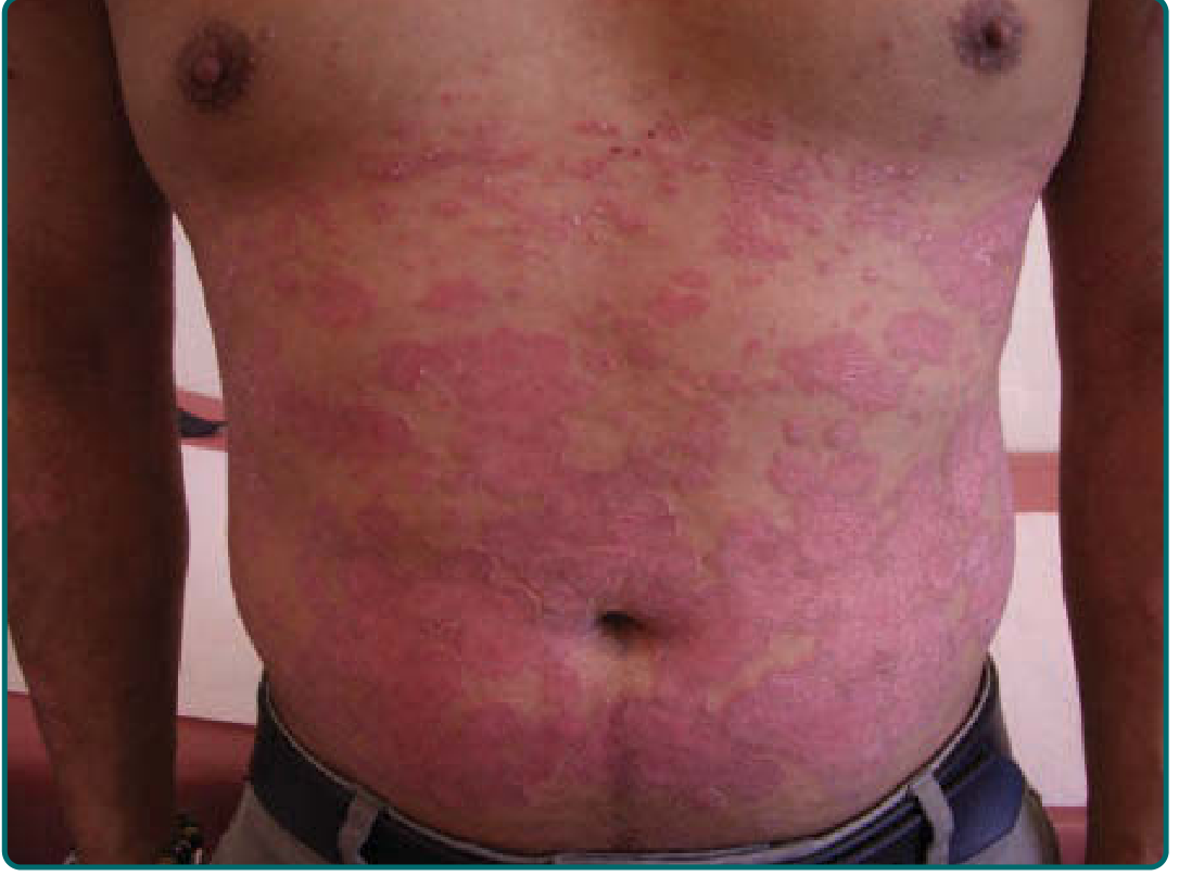
10. Wheal (Urtica)
A transient, raised, erythematous or pale, edematous papule or plaque with central pallor and erythematous periphery (or vice versa). Represents short-lived vasodilation and increased vascular permeability in the superficial dermis. Characteristically pruritic and lasts minutes to hours. Examples: urticaria (hives), dermographism, angioedema (deeper dermis/subcutis).
Size Thresholds at a Glance
| Lesion Pair | Small | Large |
|---|---|---|
| Macule / Patch | < 1 cm | ≥ 1 cm |
| Papule / Plaque | < 1 cm | ≥ 1 cm |
| Vesicle / Bulla | < 0.5-1 cm | ≥ 0.5-1 cm |
| Papule / Nodule | < 1 cm | ≥ 1 cm |
| Nodule / Tumor | 1-2 cm | > 2-5 cm |
Note: Exact size cut-offs vary slightly between sources (Harriet Lane, Harrison's, Fitzpatrick's) but the relative hierarchy is consistent.
Additional Descriptors for Papules
Fitzpatrick's describes papule topography in detail - useful for narrowing differentials:
| Topography | Description | Example |
|---|---|---|
| Sessile | Broad base | Seborrheic keratosis |
| Pedunculated | On a stalk | Skin tag (acrochordon) |
| Dome-shaped | Rounded, hemispherical | Intradermal nevus |
| Flat-topped | Flat upper surface | Lichen planus |
| Filiform | Thread-like, elongated | Verruca filiformis |
| Umbilicated | Central depression | Molluscum contagiosum |
| Acuminate | Conical, pointed | Verruca vulgaris |
| Mammillated | Nipple-like projections | - |
Clinical Approach
Correctly identifying primary lesions is the foundation of dermatologic diagnosis. After identifying the primary lesion type, further characterize by:
- Color (red, brown, white, yellow, violaceous, blue)
- Surface (smooth, scaly, hyperkeratotic, ulcerated)
- Distribution (localized, generalized, flexural, extensor, photo-distributed)
- Arrangement (discrete, grouped/herpetiform, linear, annular, confluent)
- Border (well-defined vs. indistinct)
Secondary changes (scale, crust, excoriation, lichenification, erosion, ulcer, fissure, atrophy) arise when primary lesions are modified.
Sources:
- Fitzpatrick's Dermatology, 9th ed., Ch. 1 (pp. 33-36)
- Harrison's Principles of Internal Medicine, 22E (2025), Table 39-1
- Andrews' Diseases of the Skin: Clinical Dermatology, pp. 410-415
- Harriet Lane Handbook, 23rd ed., Section 39 (Dermatology)
This is a shared conversation. Sign in to Orris to start your own chat.