
Mechanism of action of insulin
I'll start by reading the medical library skill, then search for insulin mechanism of action content in parallel.
Reading File
Now let me search the library and PubMed in parallel:
Finding Sources
Finding Sources
Searching PubMed
insulin mechanism action receptor signaling
Excellent results. Now let me read the key sections from Costanzo Physiology, Goodman & Gilman's, and Tietz in parallel, and also fetch the insulin signaling diagram:
Reading File
Reading File
Reading File
Loading Image
I now have comprehensive, authoritative content from three major textbooks and the signaling diagram. Here is the full answer:
Mechanism of Action of Insulin
(Sources: Costanzo Physiology 7e, Tietz Textbook of Laboratory Medicine 7e, Goodman & Gilman's Pharmacological Basis of Therapeutics)
1. The Insulin Receptor
The insulin receptor is a heterotetrameric transmembrane glycoprotein consisting of:
- Two α-subunits (MW ~135,000 Da each) - located entirely extracellularly; contain the insulin-binding site
- Two β-subunits (MW ~95,000 Da each) - span the plasma membrane and extend intracellularly; contain intrinsic tyrosine kinase activity
The two α-subunits are linked to each other by disulfide bonds, and each α-subunit is connected to a β-subunit by a disulfide bond. - Costanzo Physiology 7e, p. 447
2. Step-by-Step Signaling Cascade
Step 1 - Ligand Binding & Receptor Activation
Insulin binds to the α-subunits of the receptor, inducing a conformational change that activates the tyrosine kinase domains of the β-subunits.
Step 2 - Autophosphorylation
The β-subunits phosphorylate themselves (autophosphorylation) on multiple tyrosine residues in the presence of ATP. This amplifies kinase activity further.
Step 3 - Phosphorylation of Intracellular Substrates
The activated receptor tyrosine kinase phosphorylates several key docking proteins:
- IRS proteins (IRS-1, IRS-2, IRS-3, IRS-4) - Insulin Receptor Substrate family; the most important downstream substrates
- Shc (Src homology 2 domain-containing protein)
- Grb2 (Growth factor receptor-bound protein 2)
The phosphorylated tyrosines on these proteins act as docking sites for intracellular signal transducers that contain SH2 (Src Homology 2) domains - ~100 amino acid sequences that specifically recognize phosphotyrosine. - Tietz 7e, p. 1721
3. Two Main Downstream Signaling Arms
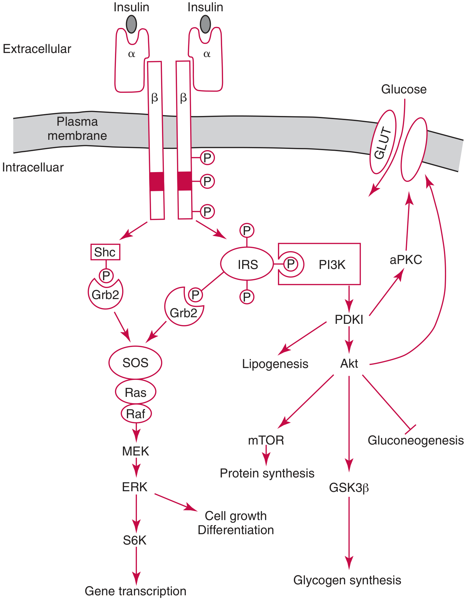
FIGURE: Mechanism of insulin action (Tietz Textbook of Laboratory Medicine 7e)
A. Metabolic Arm - PI3K/Akt Pathway
This is the primary pathway for insulin's metabolic effects:
- Phosphorylated IRS proteins recruit and activate PI3K (Phosphatidylinositol 3-kinase)
- PI3K generates PIP3 (phosphatidylinositol 3,4,5-trisphosphate) at the membrane
- PIP3 recruits and activates PDK1 (3-phosphoinositide-dependent protein kinase-1)
- PDK1 phosphorylates and activates Akt (also known as PKB)
- Akt then mediates the key metabolic effects:
| Effect | Mechanism |
|---|---|
| GLUT4 translocation (muscle & adipose) | Akt promotes translocation of GLUT4 vesicles to plasma membrane → glucose uptake |
| Glycogen synthesis (liver) | Akt phosphorylates and inactivates GSK-3β → relieves inhibition of glycogen synthase |
| Suppression of gluconeogenesis (liver) | Akt inhibits FOXO transcription factors → reduces expression of gluconeogenic enzymes |
| Lipogenesis (liver) | Akt activates lipogenic pathways |
| Protein synthesis | Akt activates mTOR → ribosomal S6 kinase (S6K) → increased translation |
B. Mitogenic Arm - Ras/MAPK Pathway
This pathway mediates insulin's growth-promoting effects:
- Shc and Grb2 (bound to phosphorylated IRS or directly to the receptor) activate Ras
- Ras activates Raf → MEK → ERK (MAP kinase cascade)
- ERK activates S6K and nuclear transcription factors
- End effects: cell growth, differentiation, and gene transcription
C. Caveolar Pathway (Alternative)
A separate pool of insulin receptors in caveolae (lipid raft microdomains) phosphorylates caveolin (CAV), APS, and Cbl. This pathway also drives GLUT4 translocation via the CrkII-C3G-TC10 signaling axis. - Goodman & Gilman's, Figure 51-4
4. Receptor Internalization and Down-regulation
After insulin-receptor binding, the complex is internalized by receptor-mediated endocytosis. The receptor is then either:
- Degraded by intracellular proteases
- Stored intracellularly
- Recycled back to the cell membrane
Insulin down-regulates its own receptor by decreasing receptor synthesis and increasing degradation. This phenomenon is a key mechanism underlying the decreased insulin sensitivity seen in obesity and Type 2 diabetes mellitus. - Costanzo Physiology 7e, p. 447
5. Nuclear and Organelle Effects
Beyond the plasma membrane cascade, insulin also binds to elements in the nucleus, Golgi apparatus, and endoplasmic reticulum, where it directly stimulates gene transcription - an action similar to that of IGF-1 and IGF-2. - Costanzo Physiology 7e
Summary Table
| Level | Key Players | Effect |
|---|---|---|
| Receptor | α/β tetramer, tyrosine kinase | Binding + autophosphorylation |
| Docking proteins | IRS-1 to 4, Shc, Grb2 | Signal branching |
| Metabolic branch | PI3K → PDK1 → Akt | GLUT4, glycogen synthesis, anti-gluconeogenesis |
| Mitogenic branch | Ras → Raf → MEK → ERK | Cell growth, gene transcription |
| Termination | Receptor internalization, phosphatases (PTP1B) | Signal attenuation |
Clinical relevance: Defects at any step in this cascade (particularly at IRS-1/2, PI3K, or Akt levels) contribute to insulin resistance in Type 2 diabetes and metabolic syndrome. PTP1B (protein tyrosine phosphatase 1B) is a key phosphatase that terminates insulin signaling and is an active drug target for insulin-sensitizing therapies.
Refer from guyton
Finding Sources
Good, Guyton is available. Now let me read all the key sections in parallel:
Reading File
Reading File
Reading File
I found the key section. Let me now read the full receptor activation section and the downstream effects sections in parallel:
Reading File
Reading File
Reading File
Now let me fetch Guyton's own insulin receptor diagram:
Loading Image
All the content from Guyton is now in hand. Here is the complete answer directly from Guyton and Hall Textbook of Medical Physiology:
Mechanism of Action of Insulin
Guyton and Hall Textbook of Medical Physiology - Chapter 79
Insulin Chemistry
Human insulin (MW 5808) consists of two amino acid chains (A and B) connected by disulfide linkages. It is synthesized as preproinsulin (MW ~11,500) in the beta cells, cleaved to proinsulin (MW ~9000) in the ER, then further cleaved in the Golgi apparatus into:
- Insulin (A + B chains linked by disulfide bonds)
- C peptide (connecting peptide, secreted in equimolar amounts)
Insulin circulates in unbound form with a plasma half-life of ~6 minutes and is degraded primarily by insulinase in the liver. - Guyton, Ch. 79
The Insulin Receptor - Structure
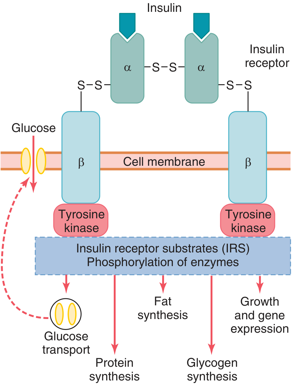
Fig. 79.3 - Guyton and Hall: Insulin binds to the α subunit, causing autophosphorylation of the β-subunit, which induces tyrosine kinase activity leading to a cascade of phosphorylation events.
To initiate its effects, insulin must first bind to and activate a membrane receptor protein (MW ~300,000). The receptor is a combination of four subunits held together by disulfide linkages:
| Subunit | Location | Function |
|---|---|---|
| 2 α-subunits | Entirely extracellular | Insulin-binding site |
| 2 β-subunits | Span the membrane, protrude into cytoplasm | Contain intrinsic tyrosine kinase activity |
"Insulin binds with the alpha subunits on the outside of the cell, but because of the linkages with the beta subunits, portions of the beta subunits protruding into the cell become autophosphorylated. Thus, the insulin receptor is an example of an enzyme-linked receptor." - Guyton, p. 963
Receptor Activation Cascade
Step 1: Insulin binds to α-subunits → conformational change
Step 2: β-subunits autophosphorylate → local tyrosine kinase is activated
Step 3: Tyrosine kinase phosphorylates multiple intracellular enzymes, especially the Insulin Receptor Substrate (IRS) proteins (IRS-1, IRS-2, IRS-3), which are expressed differently across tissues
Step 4: IRS phosphorylation activates some enzymes and inactivates others, directing intracellular metabolic machinery for carbohydrate, fat, and protein metabolism
Time-Course of Cellular Effects
Guyton uniquely organizes the downstream effects by the time of onset:
| Time | Effect |
|---|---|
| Within seconds | Membranes of ~80% of body's cells markedly increase glucose uptake via GLUT4 translocation to cell membrane (mainly muscle and adipose - NOT most brain neurons). When insulin is withdrawn, GLUT4 vesicles return to cell interior within 3-5 minutes. |
| Seconds | Cell membrane becomes more permeable to amino acids, K⁺, and phosphate ions |
| 10-15 minutes | Changed activity levels of intracellular metabolic enzymes via altered phosphorylation states |
| Hours to days | Changed rates of mRNA translation and DNA transcription in the nucleus → new protein synthesis, remodeling of cellular enzymatic machinery |
"Insulin can increase the rate of transport of glucose into resting muscle cells by at least 15-fold." - Guyton, p. 964
Metabolic Effects - Three Major Systems
A. Carbohydrate Metabolism
- Stimulates glucose uptake into muscle and adipose tissue
- Promotes glycogen storage in muscle (up to 2-3% concentration) and liver (up to 5-6%)
- Inhibits glycogenolysis (breakdown of glycogen)
- Inhibits gluconeogenesis in the liver (by suppressing gluconeogenic enzymes)
- During the fed state, muscles use glucose preferentially over fatty acids
B. Fat Metabolism
- Acts as a fat sparer - by increasing glucose utilization, it automatically decreases fat utilization
- Promotes fatty acid synthesis in the liver (excess glucose → pyruvate → acetyl-CoA → fatty acids via malonyl-CoA)
- Activates lipoprotein lipase in capillary walls of adipose tissue to allow fatty acid uptake
- Inhibits hormone-sensitive lipase in fat cells → prevents triglyceride hydrolysis and fatty acid release
- Promotes glucose transport into fat cells → forms α-glycerol phosphate → combines with fatty acids to form and store triglycerides
"When insulin is not available, even storage of the large amounts of fatty acids transported from the liver in lipoproteins is almost blocked." - Guyton, p. 966
C. Protein Metabolism
- Stimulates transport of amino acids into cells (especially valine, leucine, isoleucine, tyrosine, phenylalanine)
- Increases translation of mRNA - "turns on" ribosomal machinery (acts like an on/off switch)
- Increases rate of DNA transcription → more RNA → more protein synthesis
- Inhibits protein catabolism - reduces lysosomal degradation of proteins
- Suppresses gluconeogenesis from amino acids → conserves protein stores
"In summary, insulin promotes formation of protein and prevents degradation of proteins." - Guyton, p. 967
Insulin and Growth
Because insulin is required for protein synthesis, Guyton states it is "as essential as growth hormone for the growth of an animal." Insulin and growth hormone act synergistically to promote growth - neither alone produces normal growth in a depancreatized, hypophysectomized rat.
Key Clinical Point - Down-Regulation
Chronic hyperinsulinemia (as in obesity) leads to down-regulation of insulin receptors, reducing insulin sensitivity. This is a key mechanism in the development of Type 2 diabetes mellitus - target tissues become less responsive despite normal or elevated insulin levels.
This is a shared conversation. Sign in to Orris to start your own chat.