
Poplital fossa with diagram 5 marks
Reading File
Now let me search the medical library for popliteal fossa content and images simultaneously.
Finding Sources
Searching the Web
popliteal fossa anatomy diagram boundaries contents
Excellent! Now let me read the Gray's Anatomy for Students content on popliteal fossa and the THIEME Atlas content simultaneously.
Reading File
Reading File
I have the image URLs. Let me fetch the key diagrams and read more content simultaneously.
Loading Image
Loading Image
Loading Image
Reading File
I now have comprehensive textbook content and excellent diagrams. Let me compile a thorough 5-mark answer.
Popliteal Fossa
Introduction
The popliteal fossa is a diamond-shaped space located on the posterior aspect of the knee joint. It serves as the major route through which structures pass between the thigh and leg.
Boundaries
The fossa has the shape of a diamond with four borders and a roof and floor:
Superomedial border: Semimembranosus and semitendinosus muscles (supplemented by gracilis and sartorius)
Superolateral border: Biceps femoris muscle
Inferomedial border: Medial head of gastrocnemius muscle
Inferolateral border: Lateral head of gastrocnemius muscle + plantaris muscle
Roof (posterior wall): Popliteal fascia (deep fascia continuous with fascia lata above and deep fascia of leg below), superficial fascia, and skin
Floor (anterior wall):
- Upper part - popliteal surface of femur
- Middle part - capsule of knee joint (with oblique popliteal ligament)
- Lower part - popliteus muscle covered by its fascia
Diagram
(A) Boundaries, (B) Nerves and vessels, (C) Superficial structures - from Gray's Anatomy for Students:
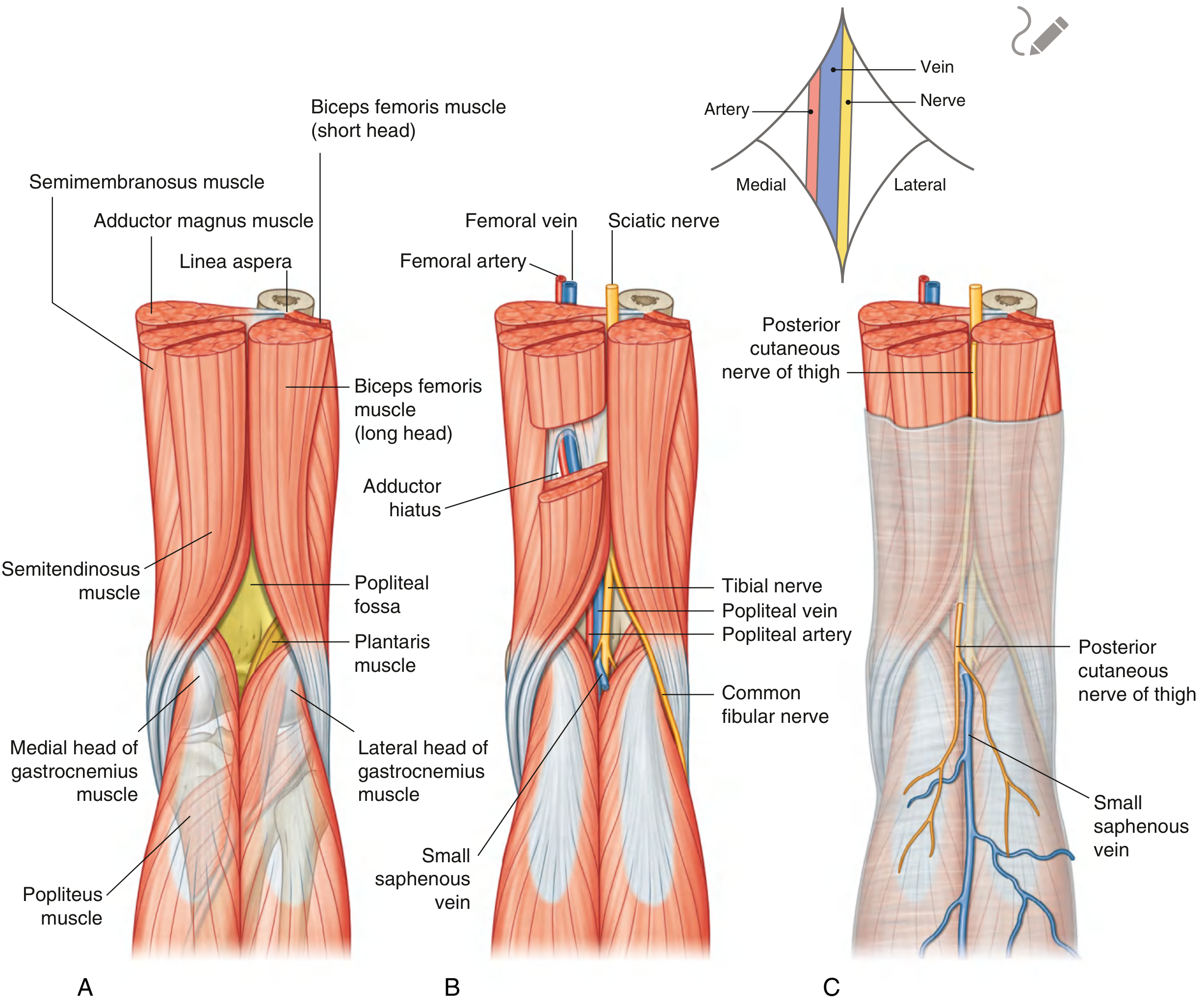
Fig. 6.85 - Popliteal Fossa. Gray's Anatomy for Students, p. 720-721
Contents (from superficial to deep)
The neurovascular order from superficial to deep is "Nerve - Vein - Artery" (NVA):
1. Nerves (most superficial)
Tibial nerve:
- Larger terminal branch of the sciatic nerve
- Enters the fossa from above under the biceps femoris
- Descends vertically through the center of the fossa
- Exits deep to the plantaris margin to enter the posterior compartment of the leg
- In the fossa it gives branches to gastrocnemius, soleus, plantaris, and popliteus
Common fibular (peroneal) nerve:
- Smaller terminal branch of sciatic nerve
- Follows the biceps femoris tendon along the lateral margin of the fossa
- Exits by winding around the neck of the fibula into the lateral compartment
2. Popliteal vein (intermediate)
- Formed by union of the anterior and posterior tibial veins at the lower border of the popliteus
- Lies superficial to the popliteal artery
- Receives the small saphenous vein (which pierces the roof)
- Exits superiorly through the adductor hiatus to become the femoral vein
3. Popliteal artery (deepest)
- Continuation of the femoral artery after it passes through the adductor hiatus
- Deepest structure in the fossa - difficult to palpate directly
- Ends at the lower border of popliteus by dividing into anterior and posterior tibial arteries
- Branches in the fossa:
- Medial and lateral superior genicular arteries
- Middle genicular artery (pierces the joint capsule to supply cruciate ligaments)
- Medial and lateral inferior genicular arteries
- Sural arteries (to the heads of gastrocnemius)
4. Other contents
- Popliteal lymph nodes (4-5 nodes receiving lymphatics from the knee region and from the small saphenous vein territory)
- Fat and areolar tissue
- Small saphenous vein (in the roof/superficial fascia - drains into popliteal vein)
- Posterior cutaneous nerve of the thigh (passes through the roof)
Detailed Neurovascular Diagram (THIEME Atlas)
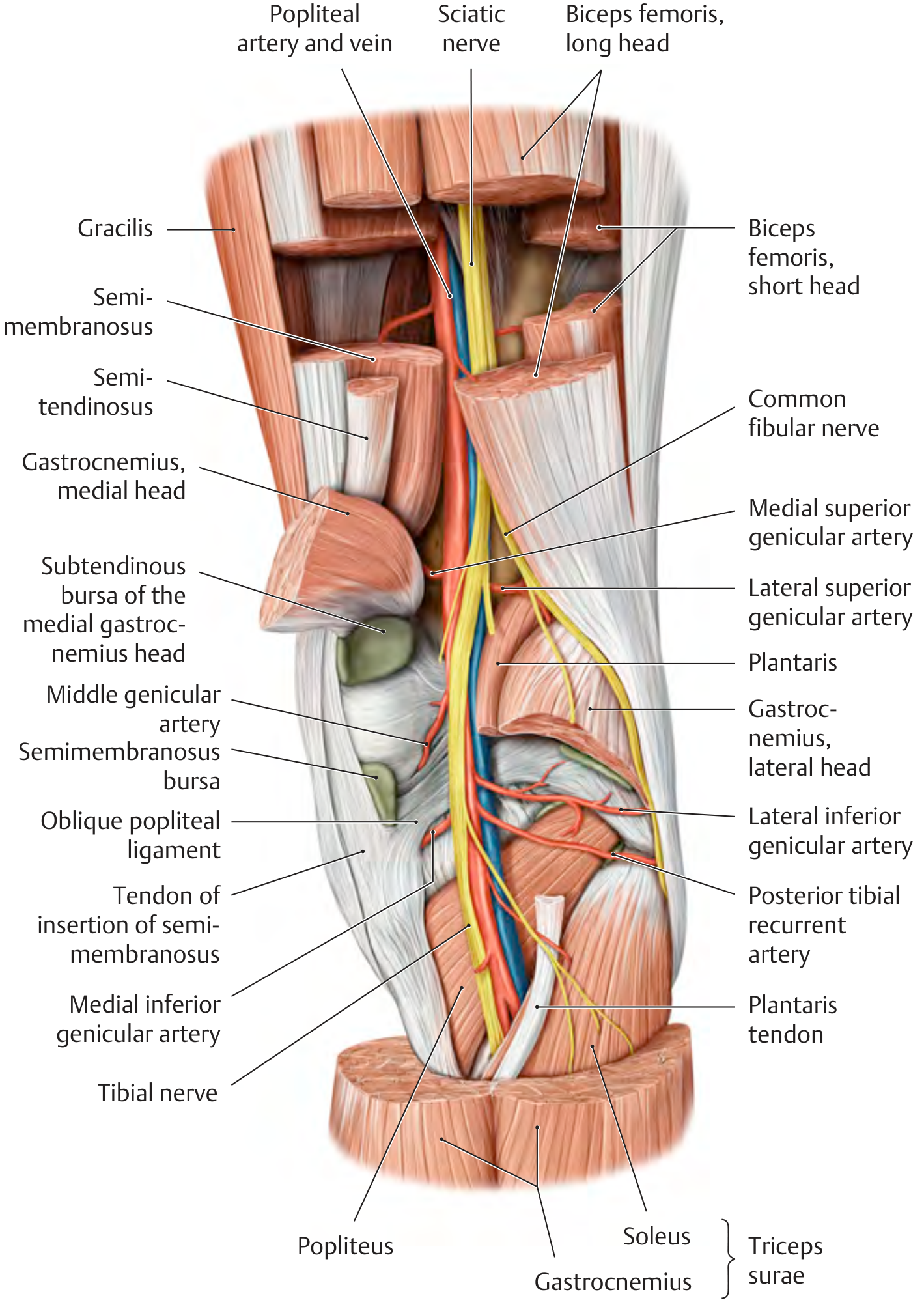
THIEME Atlas of Anatomy - Deep neurovascular structures of the right popliteal fossa, posterior view
Popliteal Artery Branches Diagram
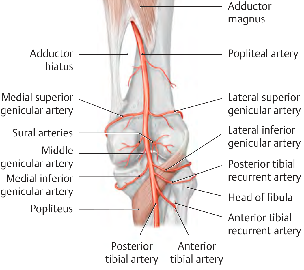
THIEME Atlas of Anatomy - Branches of the popliteal artery, right knee, posterior view
Clinical Significance
-
Baker's cyst (Popliteal cyst): A synovial cyst that bulges posteriorly into the popliteal fossa through the capsule. The subtendinous bursa of the medial head of gastrocnemius communicates with the knee joint and can form a Baker's cyst in inflammatory arthritis. Presents as a swelling behind the knee.
-
Popliteal artery aneurysm: Most common peripheral artery aneurysm. The artery is considered aneurysmal at >7 mm diameter. Often associated with aneurysms elsewhere (femoral artery, aorta). Complications are mainly from distal thromboembolism rather than rupture, potentially causing lower limb ischemia. Diagnosed with duplex Doppler ultrasound.
-
Common fibular nerve injury: The nerve is vulnerable as it winds around the fibular neck just at the exit of the fossa. Fracture of the fibular neck or pressure from a plaster cast can cause foot drop (loss of dorsiflexion and eversion).
-
Popliteal artery compression: The rigid popliteal fascia (roof) means that any expanding mass (aneurysm, Baker's cyst, popliteal abscess) compresses the neurovascular contents, which can compromise circulation distal to the knee.
-
Popliteal pulse: The popliteal artery is the deepest structure; it requires firm, deep palpation with the patient prone and the knee slightly flexed. Absence of popliteal pulse is significant in peripheral vascular disease.
Memory Aid for order of contents (lateral to medial at apex):
- Common fibular nerve (lateral)
- Tibial nerve (medial)
- Popliteal vein (between nerves, superficial to artery)
- Popliteal artery (deepest, medial)
Sources: Gray's Anatomy for Students, pp. 720-722; THIEME Atlas of Anatomy - General Anatomy and Musculoskeletal System
This is a shared conversation. Sign in to Orris to start your own chat.