
Do you have access to the content of Thomas m delvin seventh edition biochemistry textbook?
Let me check the library database to see what textbooks are available.
Finding Sources
Finding Sources
Unfortunately, Thomas M. Devlin's "Textbook of Biochemistry with Clinical Correlations" (7th edition) is not in the library. That specific title does not appear in the database.
However, the library does have three biochemistry textbooks you can draw on:
| Title | Edition |
|---|---|
| Basic Medical Biochemistry - A Clinical Approach (Marks et al.) | 6th edition |
| Biochemistry - Lippincott Illustrated Reviews | 8th edition |
| Harper's Illustrated Biochemistry | 32nd edition |
All three are authoritative, widely used biochemistry references. If you have a specific biochemistry topic or question in mind, I can search across these textbooks to find the relevant content for you.
Explain Tricarboxylic acid cycle
Reading File
Searching sources...
Finding Sources
Finding Sources
Good - found detailed TCA sections in Basic Medical Biochemistry. Let me read the key blocks now.
Reading File
Reading File
Found the TCA cycle diagram image. Let me fetch it and read more content from the textbook.
Loading Image
Reading File
Reading File
Reading File
Reading File
I now have comprehensive content. Let me compile the full explanation.
Tricarboxylic Acid (TCA) Cycle
Source: Basic Medical Biochemistry - A Clinical Approach, 6th Edition
Overview
The TCA cycle goes by three names:
- Krebs cycle - named after Sir Hans Krebs who first formulated it
- Citric acid cycle - because citrate was one of the first known participants
- Tricarboxylic acid (TCA) cycle - named for the tricarboxylates citrate and isocitrate
The cycle takes place in the mitochondrial matrix and serves as the central hub of energy metabolism. Its core function is to accept the 2-carbon acetyl group (from acetyl CoA) and oxidize it completely to CO2, capturing the released energy as NADH, FAD(2H), and GTP.
The TCA Cycle Diagram
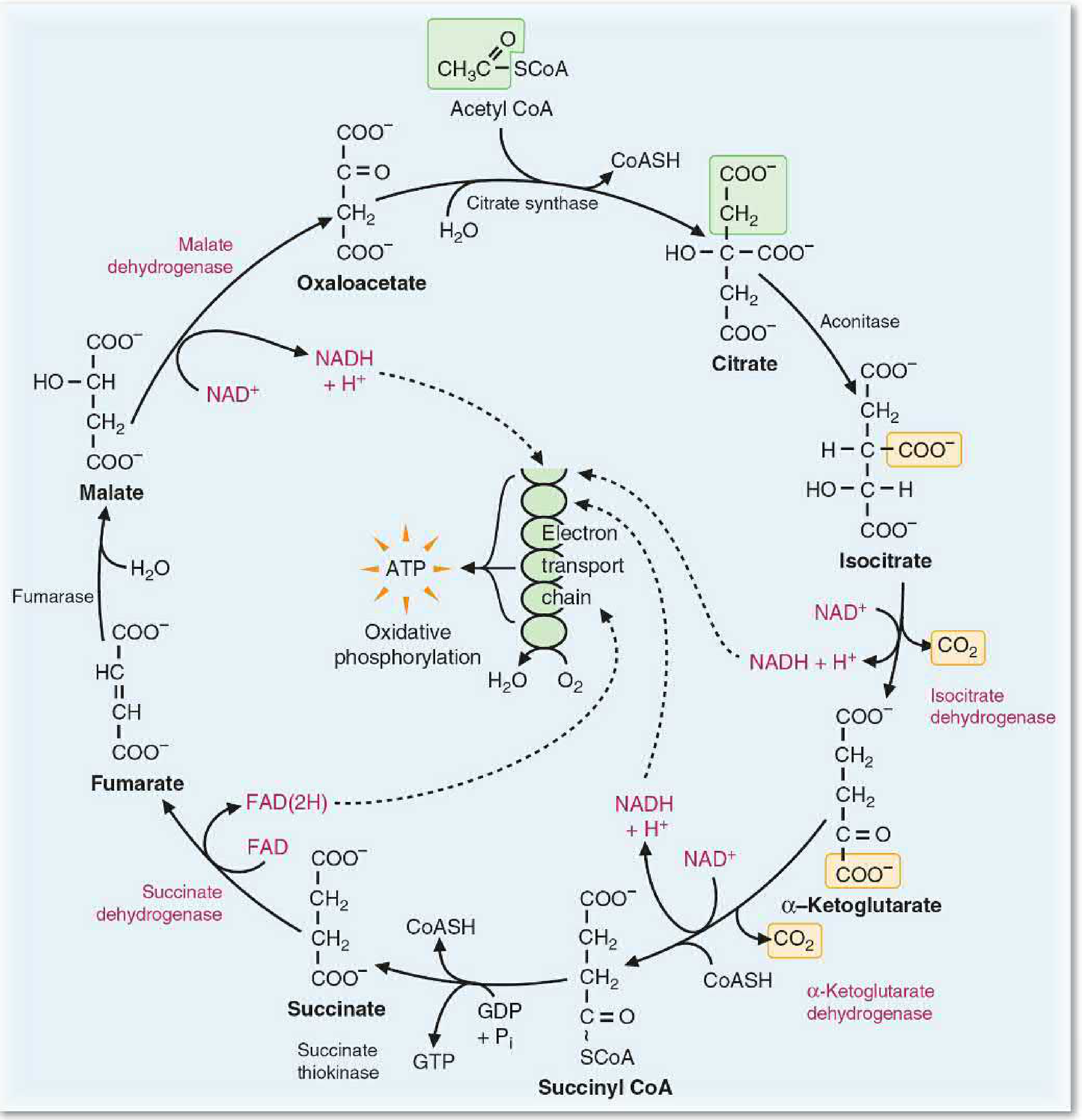
Figure 23.3 from Basic Medical Biochemistry, 6e - The complete TCA cycle. Oxidation-reduction enzymes and coenzymes are shown in red. Entry of acetyl CoA carbons is shown in green. Carbons released as CO2 are shown in yellow.
Entry Substrate: Acetyl CoA
The major pathways of fuel oxidation (glycolysis, beta-oxidation of fatty acids, amino acid catabolism) all funnel their products into acetyl CoA, which is the substrate for the TCA cycle. The acetyl group donates 8 electrons (four per carbon) to the cycle.
The 8 Reactions Step by Step
Step 1 - Citrate formation
Acetyl CoA + Oxaloacetate (4C) → Citrate (6C)
- Enzyme: Citrate synthase
- Water is added; CoASH is released
- This is a condensation reaction - no high-energy phosphate is required
Step 2 - Isomerization
Citrate → Isocitrate
- Enzyme: Aconitase (an isomerase, requires Fe2+ cofactor)
- The hydroxyl group is moved to an adjacent carbon so it can be oxidized in the next step
Step 3 - First oxidative decarboxylation
Isocitrate → α-Ketoglutarate (5C) + CO2
- Enzyme: Isocitrate dehydrogenase
- NAD+ is reduced to NADH (1st NADH produced)
- First CO2 is released
Step 4 - Second oxidative decarboxylation
α-Ketoglutarate (5C) → Succinyl CoA (4C) + CO2
- Enzyme: α-Ketoglutarate dehydrogenase complex (requires TPP, lipoic acid, FAD, NAD+, CoA)
- NAD+ is reduced to NADH (2nd NADH produced)
- Second CO2 is released
- Energy is conserved in the high-energy thioester bond of succinyl CoA
Step 5 - Substrate-level phosphorylation
Succinyl CoA → Succinate + GTP
- Enzyme: Succinate thiokinase (succinyl CoA synthetase)
- Energy from the thioester bond drives formation of GTP from GDP + Pi
- This is the only step of substrate-level phosphorylation in the TCA cycle
- GTP is energetically equivalent to ATP
Step 6 - Oxidation of succinate
Succinate → Fumarate
- Enzyme: Succinate dehydrogenase (embedded in inner mitochondrial membrane)
- FAD is reduced to FAD(2H) (the only step using FAD)
- Electrons pass directly into the electron transport chain
Step 7 - Hydration
Fumarate + H2O → Malate
- Enzyme: Fumarase
- Water is added across the double bond
Step 8 - Final oxidation
Malate → Oxaloacetate
- Enzyme: Malate dehydrogenase
- NAD+ is reduced to NADH (3rd NADH produced)
- Oxaloacetate is regenerated to accept another acetyl group
Net Equation Per Turn of the Cycle
Acetyl CoA + 3 NAD⁺ + FAD + GDP + Pᵢ + 2H₂O → 2 CO₂ + 3 NADH + FAD(2H) + GTP + CoASH + 3H⁺
Energy Yield
| Product | Quantity | Energy (kcal/mol) |
|---|---|---|
| NADH | 3 | 3 × 53 = 159 |
| FAD(2H) | 1 | 41 |
| GTP | 1 | 7 |
| Total | ~207 kcal/mol |
The total energy available from the acetyl group is ~228 kcal/mol, meaning the TCA cycle conserves approximately 90% of that energy - an extraordinary efficiency.
The net ΔG° for the full cycle is -13 kcal/mol, a small value that ensures the process runs to completion. The oxidation of NADH and FAD(2H) in the electron transport chain pulls the cycle forward.
Coenzymes Required
| Coenzyme | Vitamin Precursor | Enzyme(s) |
|---|---|---|
| NAD+ | Niacin (B3) | Isocitrate DH, α-KG DH, Malate DH |
| FAD | Riboflavin (B2) | Succinate DH, α-KG DH |
| CoA | Pantothenic acid (B5) | α-KG DH, Succinyl CoA synthetase |
| TPP | Thiamine (B1) | α-KG DH complex |
| Lipoic acid | - | α-KG DH complex |
Minerals required: Mg2+, Ca2+, Fe2+, phosphate
Regulation
The TCA cycle is regulated to match the cell's ATP demands. Three key enzymes are regulated:
| Enzyme | Inhibitors | Activators |
|---|---|---|
| Citrate synthase | Citrate (product inhibition), NADH | ADP |
| Isocitrate dehydrogenase | NADH, ATP | ADP, Ca2+ |
| α-Ketoglutarate dehydrogenase | Succinyl CoA, NADH | Ca2+, ADP |
Two major regulatory signals:
- ATP/ADP ratio: When ATP is consumed (ADP rises), the cycle accelerates
- NADH/NAD+ ratio: Accumulating NADH inhibits multiple enzymes, slowing the cycle when the electron transport chain is backed up
During exercise or muscular contraction, rising Ca2+ concentrations independently activate isocitrate dehydrogenase and α-ketoglutarate dehydrogenase, increasing ATP production.
Clinical Significance
- Thiamine (B1) deficiency - impairs the α-ketoglutarate dehydrogenase complex, causing Wernicke's encephalopathy and beriberi
- Riboflavin (B2) deficiency - impairs FAD-dependent reactions (succinate dehydrogenase)
- Aconitase inhibition - fluorocitrate (a fluoroacetate metabolite, found in some poisons) blocks aconitase, halting the cycle
- The TCA cycle intermediates also serve as biosynthetic precursors: α-ketoglutarate for amino acids, oxaloacetate for gluconeogenesis, succinyl CoA for heme synthesis, citrate for fatty acid synthesis
Basic Medical Biochemistry - A Clinical Approach, 6e, Chapter 23 (pp. 827-840)
This is a shared conversation. Sign in to Orris to start your own chat.