LONG ESSAY (1×10= 10 Marks) 1. A 56-year-old man presented with altered bowel habits, bleeding per rectum and progressive weight loss. Colonoscopy revealed an ulcer proliferative growth in the sigmoid colon. a) What is the most likely diagnosis? (1) b) Describe the etiopathogenesis. (3) c) Describe the gross and microscopic morphology. (4) d) Add a note on Familial Adenomatous Polyposis (FAP). (2) - 15 Morks)

Reading File
Finding Sources
Finding Sources
Searching Images

colorectal carcinoma sigmoid colon gross morphology ulcero-proliferative
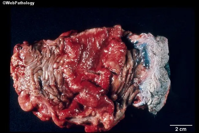
Gross pathology image of a resected sigmoid colon segment showing a bulky colorectal carcinoma. The tumor mass is exophytic, lobulated, and red-purple in color, with irregular, friable surfaces and clear demarcation from adjacent normal mucosa. The anal mucosa is visible to the right of the lesion, illustrating the tumor’s distal extent toward the rectosigmoid junction. A 2 cm scale bar is provided for size reference. The lesion distorts the colonic wall with thickening and luminal narrowing, consistent with invasive growth through the mucosa and into the submucosa and muscularis propria in a gross examination. Gross variants described in literature include exophytic polypoid masses, flat/depressed ulcerating infiltrative lesions, napkin-ring constrictions in distal colon, mucinous gelatinous appearances, and signet-ring cell related linitis plastica in advanced disease; these features are not all simultaneously present here, but the image exemplifies exophytic-type colorectal carcinoma. This macroscopic morphology correlates with potential lymphovascular invasion and nodal metastasis risk, and it supports clinical staging and surgical decision-making. The specimen’s appearance provides valuable educational context for understanding colorectal cancer pathology, morphology, gross differential diagnoses (polypoid adenocarcinoma, ulcerative cicatrix), and correlation with imaging and endoscopic findings. This image supports educational reference for residents, pathologists, and gastroenterology trainees in clinic and research settings.
Gross pathology image of a resected sigmoid colon segment showing a bulky colorectal carcinoma. The tumor mass is exophytic, lobulated, and red-purple in color, with irregular, friable surfaces and clear demarcation from adjacent normal mucosa. The anal mucosa is visible to the right of the lesion, illustrating the tumor’s distal extent toward the rectosigmoid junction. A 2 cm scale bar is provided for size reference. The lesion distorts the colonic wall with thickening and luminal narrowing, consistent with invasive growth through the mucosa and into the submucosa and muscularis propria in a gross examination. Gross variants described in literature include exophytic polypoid masses, flat/depressed ulcerating infiltrative lesions, napkin-ring constrictions in distal colon, mucinous gelatinous appearances, and signet-ring cell related linitis plastica in advanced disease; these features are not all simultaneously present here, but the image exemplifies exophytic-type colorectal carcinoma. This macroscopic morphology correlates with potential lymphovascular invasion and nodal metastasis risk, and it supports clinical staging and surgical decision-making. The specimen’s appearance provides valuable educational context for understanding colorectal cancer pathology, morphology, gross differential diagnoses (polypoid adenocarcinoma, ulcerative cicatrix), and correlation with imaging and endoscopic findings. This image supports educational reference for residents, pathologists, and gastroenterology trainees in clinic and research settings.

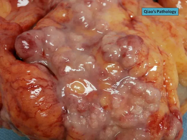
Gross pathology photograph of a resected sigmoid colon specimen showing a serosal surface densely studded with grayish-white, firm to soft nodules. The nodules are subserosal in location, compressing underlying adipose tissue and focal serosal irregularity is evident. The appearance is characteristic of serosal implants seen with advanced colorectal neuroendocrine carcinoma. The primary tumor is in the sigmoid colon; the nodules on the outer serosal surface suggest peritoneal dissemination or subserosal metastatic spread. The color is pale gray to pink-turquoise with a glistening, lobulated contour in places. The surrounding colonic wall shows irregular thickening and mucosal involvement consistent with an aggressive neuroendocrine tumor; vascular congestion is mild. In the clinical setting, such gross features raise concern for metastatic disease and peritoneal carcinomatosis; histopathology would reveal sheets of neuroendocrine cells with immunohistochemical positivity for chromogranin A and synaptophysin, high mitotic activity, and a high Ki-67 index. This image is valuable for education on gross morphology of colorectal neuroendocrine carcinomas and their serosal metastases, contributing to differential diagnosis with carcinoid tumors, adenocarcinoma metastases, and granulomatous processes. It supports imaging-pathology correlation, surgical pathology review, and teaching scenarios in gastrointestinal oncology. These features underscore the importance of correlation with clinical staging.
Gross pathology photograph of a resected sigmoid colon specimen showing a serosal surface densely studded with grayish-white, firm to soft nodules. The nodules are subserosal in location, compressing underlying adipose tissue and focal serosal irregularity is evident. The appearance is characteristic of serosal implants seen with advanced colorectal neuroendocrine carcinoma. The primary tumor is in the sigmoid colon; the nodules on the outer serosal surface suggest peritoneal dissemination or subserosal metastatic spread. The color is pale gray to pink-turquoise with a glistening, lobulated contour in places. The surrounding colonic wall shows irregular thickening and mucosal involvement consistent with an aggressive neuroendocrine tumor; vascular congestion is mild. In the clinical setting, such gross features raise concern for metastatic disease and peritoneal carcinomatosis; histopathology would reveal sheets of neuroendocrine cells with immunohistochemical positivity for chromogranin A and synaptophysin, high mitotic activity, and a high Ki-67 index. This image is valuable for education on gross morphology of colorectal neuroendocrine carcinomas and their serosal metastases, contributing to differential diagnosis with carcinoid tumors, adenocarcinoma metastases, and granulomatous processes. It supports imaging-pathology correlation, surgical pathology review, and teaching scenarios in gastrointestinal oncology. These features underscore the importance of correlation with clinical staging.

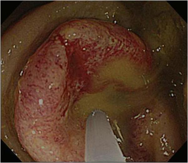
This endoscopic clinical image displays a colonoscopy view of the sigmoid colon, revealing an advanced, obstructive colorectal carcinoma. The pathology presents as a bulky, circumferential mass that causes severe luminal stenosis, nearly completely occluding the intestinal passage. The tumor surface is markedly irregular and nodular, exhibiting a predominantly pinkish-red color with dark red hyperemic patches suggestive of friability and potential hemorrhage. Areas of yellowish-tan discoloration are visible on the mass, which may indicate surface necrosis or mucinous components. Surrounding the lesion, the lumen contains yellowish-brown, translucent fluid with small air bubbles and suspended particulate matter. The image illustrates a critical case of obstructive colitis, highlighting the characteristic morphology of a moderately differentiated adenocarcinoma in a geriatric clinical context.
This endoscopic clinical image displays a colonoscopy view of the sigmoid colon, revealing an advanced, obstructive colorectal carcinoma. The pathology presents as a bulky, circumferential mass that causes severe luminal stenosis, nearly completely occluding the intestinal passage. The tumor surface is markedly irregular and nodular, exhibiting a predominantly pinkish-red color with dark red hyperemic patches suggestive of friability and potential hemorrhage. Areas of yellowish-tan discoloration are visible on the mass, which may indicate surface necrosis or mucinous components. Surrounding the lesion, the lumen contains yellowish-brown, translucent fluid with small air bubbles and suspended particulate matter. The image illustrates a critical case of obstructive colitis, highlighting the characteristic morphology of a moderately differentiated adenocarcinoma in a geriatric clinical context.

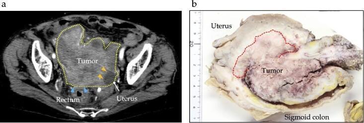
Educational medical composite featuring preoperative imaging and gross pathology of a sigmoid colon carcinoma. Panel (a) is an axial contrast-enhanced CT scan of the pelvis showing a large, heterogeneous, hypodense mass approximately 12 cm in diameter (delineated by a yellow dotted line). The imaging demonstrates suspicion of direct invasion into the adjacent rectum (blue arrows) and uterus (yellow arrows). Panel (b) shows a macroscopic photograph of the resected surgical specimen alongside a measurement scale. The specimen contains the sigmoid colon and uterus, revealing a bulky, irregularly shaped Type 1 tumor (outlined with a red dotted line) measuring 130 x 40 x 25 mm. The gross findings confirm direct local invasion into the uterine corpus, ovary, and small intestine. This case illustrates clinical Stage IIC (T4bN0M0) colorectal adenocarcinoma, emphasizing the visual characteristics of locally advanced disease and the importance of multidisciplinary surgical planning for multi-organ resection.

Educational medical composite featuring preoperative imaging and gross pathology of a sigmoid colon carcinoma. Panel (a) is an axial contrast-enhanced CT scan of the pelvis showing a large, heterogeneous, hypodense mass approximately 12 cm in diameter (delineated by a yellow dotted line). The imaging demonstrates suspicion of direct invasion into the adjacent rectum (blue arrows) and uterus (yellow arrows). Panel (b) shows a macroscopic photograph of the resected surgical specimen alongside a measurement scale. The specimen contains the sigmoid colon and uterus, revealing a bulky, irregularly shaped Type 1 tumor (outlined with a red dotted line) measuring 130 x 40 x 25 mm. The gross findings confirm direct local invasion into the uterine corpus, ovary, and small intestine. This case illustrates clinical Stage IIC (T4bN0M0) colorectal adenocarcinoma, emphasizing the visual characteristics of locally advanced disease and the importance of multidisciplinary surgical planning for multi-organ resection.

Finding Sources
Finding Sources
Searching Images

colorectal adenocarcinoma histology microscopy glands mucin
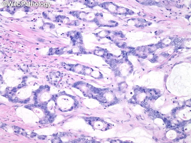
Histopathology slide of colorectal tissue examined under light microscopy after Hematoxylin and Eosin staining. The specimen architecture shows a mucinous variant of colorectal adenocarcinoma characterized by moderately differentiated neoplastic glands embedded in abundant extracellular mucin pools within a conspicuous desmoplastic stroma. Neoplastic glands display epithelial atypia with nuclear pleomorphism, hyperchromasia, and increased mitotic activity, arranged in back-to-back glands with cribriform or clustered patterns interspersed within mucin lakes. The tumor produces copious mucin which displaces adjacent stroma and forms lakes that are partially lined by neoplastic epithelium. The surrounding stroma is fibrous and dense (desmoplastic), with inflammatory infiltrates and occasional hyalinization. No evident invasion of vessels or nerves is clearly visible at this low magnification, but the desmoplastic reaction supports invasive carcinoma. The mucinous phenotype has clinical implications: often associated with higher stage at presentation, potential for peritoneal dissemination, and variable response to chemoradiation. This image is indicative of colorectal cancer of mucinous subtype and underscores the importance of recognizing mucin-rich histology as a distinct clinicopathologic entity with prognostic and therapeutic relevance. Correlation with immunohistochemical profiles (CK20, CK7, MUC2, MUC5AC, CEA) can aid differential diagnosis, confirm lineage, and guide targeted therapy decisions in multidisciplinary tumor boards for optimal patient management today.
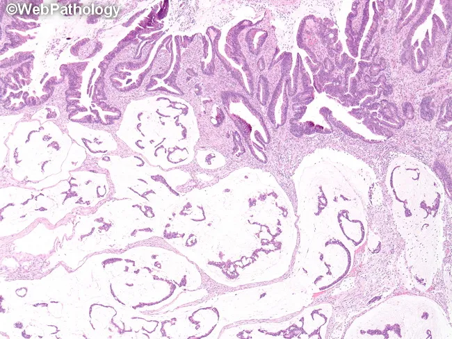
Histopathology slide of colorectal tissue examined under light microscopy after Hematoxylin and Eosin staining. The specimen architecture shows a mucinous variant of colorectal adenocarcinoma characterized by moderately differentiated neoplastic glands embedded in abundant extracellular mucin pools within a conspicuous desmoplastic stroma. Neoplastic glands display epithelial atypia with nuclear pleomorphism, hyperchromasia, and increased mitotic activity, arranged in back-to-back glands with cribriform or clustered patterns interspersed within mucin lakes. The tumor produces copious mucin which displaces adjacent stroma and forms lakes that are partially lined by neoplastic epithelium. The surrounding stroma is fibrous and dense (desmoplastic), with inflammatory infiltrates and occasional hyalinization. No evident invasion of vessels or nerves is clearly visible at this low magnification, but the desmoplastic reaction supports invasive carcinoma. The mucinous phenotype has clinical implications: often associated with higher stage at presentation, potential for peritoneal dissemination, and variable response to chemoradiation. This image is indicative of colorectal cancer of mucinous subtype and underscores the importance of recognizing mucin-rich histology as a distinct clinicopathologic entity with prognostic and therapeutic relevance. Correlation with immunohistochemical profiles (CK20, CK7, MUC2, MUC5AC, CEA) can aid differential diagnosis, confirm lineage, and guide targeted therapy decisions in multidisciplinary tumor boards for optimal patient management today.

Imaging modality and technique: light microscopy of a formalin-fixed colorectal tissue section stained with Hematoxylin and Eosin (H&E). In this mucinous variant of colorectal adenocarcinoma, the tumor demonstrates abundant extracellular mucin pools separating irregular, branching or anastomosing neoplastic glands. The glands are lined by tall, columnar epithelium with goblet-cell–like mucin cytoplasm; cytologic atypia is generally minimal and mitotic activity is relatively low compared with high-grade conventional adenocarcinoma. The neoplastic cells project into mucin lakes, producing a characteristic sign of mucinous histology. The surrounding stroma may be looser or inflammatory, and residual non-neoplastic mucosa is variably present at the periphery. The histologic pattern may be accompanied by mucin-positive secretions within glands and throughout the extracellular matrix. This morphological subtype frequently correlates with distinctive molecular features, including mucin expression patterns such as MUC2 predominance, and possibly MUC1/MUC3 in other variants; serrated variants may show MUC2/MUC5AC/MUC6 upregulation. Clinically, mucinous colorectal carcinomas tend to present at a different stage distribution and may have different prognostic implications compared with conventional colorectal adenocarcinoma. Immunohistochemical profiling for mucin core proteins can aid classification and guide targeted therapeutic considerations, with potential implications for surgical planning and adjuvant therapy choices.
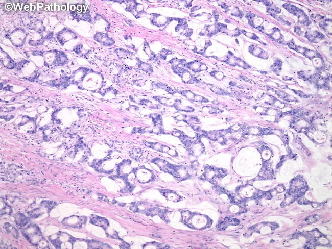
Imaging modality and technique: light microscopy of a formalin-fixed colorectal tissue section stained with Hematoxylin and Eosin (H&E). In this mucinous variant of colorectal adenocarcinoma, the tumor demonstrates abundant extracellular mucin pools separating irregular, branching or anastomosing neoplastic glands. The glands are lined by tall, columnar epithelium with goblet-cell–like mucin cytoplasm; cytologic atypia is generally minimal and mitotic activity is relatively low compared with high-grade conventional adenocarcinoma. The neoplastic cells project into mucin lakes, producing a characteristic sign of mucinous histology. The surrounding stroma may be looser or inflammatory, and residual non-neoplastic mucosa is variably present at the periphery. The histologic pattern may be accompanied by mucin-positive secretions within glands and throughout the extracellular matrix. This morphological subtype frequently correlates with distinctive molecular features, including mucin expression patterns such as MUC2 predominance, and possibly MUC1/MUC3 in other variants; serrated variants may show MUC2/MUC5AC/MUC6 upregulation. Clinically, mucinous colorectal carcinomas tend to present at a different stage distribution and may have different prognostic implications compared with conventional colorectal adenocarcinoma. Immunohistochemical profiling for mucin core proteins can aid classification and guide targeted therapeutic considerations, with potential implications for surgical planning and adjuvant therapy choices.

Imaging modality: Light microscopy of a hematoxylin and eosin (H&E) stained colorectal tissue section. Primary subject: Mucinous variant of colorectal adenocarcinoma with moderately differentiated neoplastic glands surrounded by abundant extracellular mucin in a desmoplastic stroma. The histology shows clusters of malignant glandular units lined by columnar epithelial cells with moderate cytologic atypia, nuclear pleomorphism, and irregular contours. These glands are interspersed with large lakes of mucin, which displace and pull apart the surrounding fibrous stroma. The desmoplastic reaction appears as dense, fibrous connective tissue with activated myofibroblasts and sparse inflammatory cells. Glandular architecture is irregular and commonly displays cribriform or back-to-back patterns, with variable mitotic activity. The tumor demonstrates notable mucin production both within gland lumina and as extracellular mucin pools; this mucinous pattern typically lacks cohesive invasion in some areas but can invade through the muscularis propria. Diagnostic significance: histologic confirmation of a mucin-producing colorectal carcinoma, with implications for prognosis and therapeutic planning; mucin-rich tumors can have distinct molecular features (e.g., mismatch repair deficiency) and may influence response to chemotherapy. The image is relevant for diagnostic pathology review, tumor classification, educational illustrate mucinous differentiation, and oncologic decision-making. Correlation with clinical presentation and imaging aids comprehensive assessment and management planning.

Imaging modality: Light microscopy of a hematoxylin and eosin (H&E) stained colorectal tissue section. Primary subject: Mucinous variant of colorectal adenocarcinoma with moderately differentiated neoplastic glands surrounded by abundant extracellular mucin in a desmoplastic stroma. The histology shows clusters of malignant glandular units lined by columnar epithelial cells with moderate cytologic atypia, nuclear pleomorphism, and irregular contours. These glands are interspersed with large lakes of mucin, which displace and pull apart the surrounding fibrous stroma. The desmoplastic reaction appears as dense, fibrous connective tissue with activated myofibroblasts and sparse inflammatory cells. Glandular architecture is irregular and commonly displays cribriform or back-to-back patterns, with variable mitotic activity. The tumor demonstrates notable mucin production both within gland lumina and as extracellular mucin pools; this mucinous pattern typically lacks cohesive invasion in some areas but can invade through the muscularis propria. Diagnostic significance: histologic confirmation of a mucin-producing colorectal carcinoma, with implications for prognosis and therapeutic planning; mucin-rich tumors can have distinct molecular features (e.g., mismatch repair deficiency) and may influence response to chemotherapy. The image is relevant for diagnostic pathology review, tumor classification, educational illustrate mucinous differentiation, and oncologic decision-making. Correlation with clinical presentation and imaging aids comprehensive assessment and management planning.

Reading File
Reading File
Reading File
Reading File
Reading File
Reading File
Reading File
Reading File
Searching Images

familial adenomatous polyposis colon hundreds polyps gross specimen
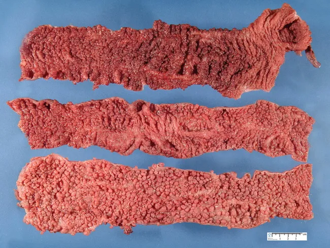
Imaging modality: Gross pathology photograph of a colectomy specimen with extensive mucosal polyposis. Primary subject: Familial Adenomatous Polyposis (FAP) characterized by hundreds to thousands of colonic polyps. Specimen type: colectomy (colon) showing a broad, polyp-dense mucosal surface. Imaging perspective: exterior view of longitudinally oriented colon segments, highlighting the mucosal polyp burden. Detailed description: The colon shows a remarkable burden of tiny polypoid lesions distributed across the mucosa. Most polyps are diminutive, under 1 cm in greatest diameter, giving a rough, nodular surface with a cobblestone appearance. The tissue coloration is a uniform reddish-pink. The polyps arise from the colonic mucosa with variable pedunculation; adenomatous-type appearance is suggested by their regular rounded contours and granular surface. This gross phenotype aligns with classic FAP, often associated with APC gene mutations, particularly around codons 1250-1464 (notably near codon 1300), producing profuse polyp formation. Mutations toward the extreme 5' and 3' ends yield attenuated polyposis. In this case, the description notes that approximately 25% of patients harbor colorectal cancer at colectomy. Clinically, such specimens illustrate the rationale for prophylactic colectomy to mitigate near-certain cancer risk. Credit: image courtesy of Dr. Jean-Christophe Fournet; humpath.com; Used with permission.
Imaging modality: Gross pathology photograph of a colectomy specimen with extensive mucosal polyposis. Primary subject: Familial Adenomatous Polyposis (FAP) characterized by hundreds to thousands of colonic polyps. Specimen type: colectomy (colon) showing a broad, polyp-dense mucosal surface. Imaging perspective: exterior view of longitudinally oriented colon segments, highlighting the mucosal polyp burden. Detailed description: The colon shows a remarkable burden of tiny polypoid lesions distributed across the mucosa. Most polyps are diminutive, under 1 cm in greatest diameter, giving a rough, nodular surface with a cobblestone appearance. The tissue coloration is a uniform reddish-pink. The polyps arise from the colonic mucosa with variable pedunculation; adenomatous-type appearance is suggested by their regular rounded contours and granular surface. This gross phenotype aligns with classic FAP, often associated with APC gene mutations, particularly around codons 1250-1464 (notably near codon 1300), producing profuse polyp formation. Mutations toward the extreme 5' and 3' ends yield attenuated polyposis. In this case, the description notes that approximately 25% of patients harbor colorectal cancer at colectomy. Clinically, such specimens illustrate the rationale for prophylactic colectomy to mitigate near-certain cancer risk. Credit: image courtesy of Dr. Jean-Christophe Fournet; humpath.com; Used with permission.

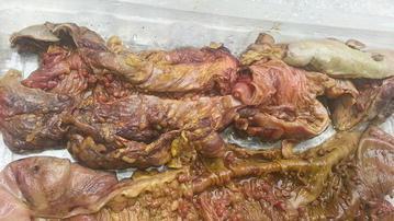
This clinical photograph shows a gross pathological total colectomy specimen from a patient with Familial Adenomatous Polyposis (FAP). The resected colon is opened longitudinally to reveal the mucosal surface. The image displays the caecum and ascending colon in the lower section, with the descending colon positioned above. The most prominent feature is the extensive, dense distribution of hundreds of adenomatous polyps across the mucosal lining, creating a characteristic 'carpeted' appearance. Centrally located in the upper portion of the specimen is a visible surgical site corresponding to a previous anastomosis. The tissue appears erythematous and fleshy, typical of a fresh surgical specimen. This image serves as a classic educational example of the phenotypic manifestation of FAP, illustrating the necessity of radical surgical intervention due to the high density of precancerous lesions throughout the colorectal tract.

This clinical photograph shows a gross pathological total colectomy specimen from a patient with Familial Adenomatous Polyposis (FAP). The resected colon is opened longitudinally to reveal the mucosal surface. The image displays the caecum and ascending colon in the lower section, with the descending colon positioned above. The most prominent feature is the extensive, dense distribution of hundreds of adenomatous polyps across the mucosal lining, creating a characteristic 'carpeted' appearance. Centrally located in the upper portion of the specimen is a visible surgical site corresponding to a previous anastomosis. The tissue appears erythematous and fleshy, typical of a fresh surgical specimen. This image serves as a classic educational example of the phenotypic manifestation of FAP, illustrating the necessity of radical surgical intervention due to the high density of precancerous lesions throughout the colorectal tract.

I now have all the material I need for a comprehensive answer. Let me compose the full long-essay answer.

Long Essay Answer: Carcinoma of the Sigmoid Colon (10 Marks)


a) Most Likely Diagnosis (1 Mark)

Carcinoma of the Sigmoid Colon - Adenocarcinoma
The clinical triad of altered bowel habits, bleeding per rectum, and progressive weight loss in a 56-year-old man, with colonoscopic finding of an ulcero-proliferative growth in the sigmoid colon, is the classic presentation of colorectal adenocarcinoma. The sigmoid colon accounts for approximately 20% of all colorectal cancers, making it the second most common site after the rectum.

b) Etiopathogenesis (3 Marks)

Risk Factors

Dietary factors are most closely linked to sporadic colorectal cancer: low intake of unabsorbable vegetable fiber, high intake of refined carbohydrates and fat. Reduced fiber leads to decreased stool bulk, altered intestinal microbiota, increased synthesis of toxic oxidative byproducts, and prolonged mucosal contact time. High fat intake enhances hepatic synthesis of cholesterol and bile acids, which are converted by intestinal bacteria into carcinogens. Other risk factors include:
  • Age >50 years (peak incidence 60-70 years)
  • Inflammatory bowel disease (Crohn's disease, ulcerative colitis)
  • Hereditary syndromes (FAP, HNPCC/Lynch syndrome)
  • Physical inactivity, obesity, smoking, alcohol

Molecular Pathways of Carcinogenesis

Three major pathways drive colorectal adenocarcinoma (see Table 17.10, Robbins Pathologic Basis of Disease):

1. APC/β-Catenin (Wnt) Pathway - the Classic Adenoma-Carcinoma Sequence (~80% of sporadic cancers)

This is the predominant pathway for left-sided and sigmoid colon cancers. It proceeds through stepwise accumulation of mutations:
  • Step 1 - APC loss: Both copies of the tumor suppressor gene APC (chromosome 5q21) are inactivated. Normally, APC protein binds β-catenin and promotes its degradation. Loss of APC allows β-catenin to accumulate and translocate to the nucleus, where it forms a complex with TCF and activates transcription of proliferative genes including MYC and cyclin D1 - resulting in formation of early adenomas.
  • Step 2 - KRAS mutation: Activating mutations in KRAS (found in 50% of adenomas >1 cm and 50% of invasive carcinomas) promote growth and prevent apoptosis. This is a late event in small adenomas (<1 cm, <10% KRAS mutation) but increases with progression.
  • Step 3 - SMAD2/SMAD4 loss: Loss of these TGF-β signaling effectors (chromosome 18q21) removes the cell cycle brake and allows unrestrained growth.
  • Step 4 - TP53 mutation: Inactivation of p53 (mutated in ~70% of invasive carcinomas) removes the DNA damage checkpoint, enabling malignant transformation.
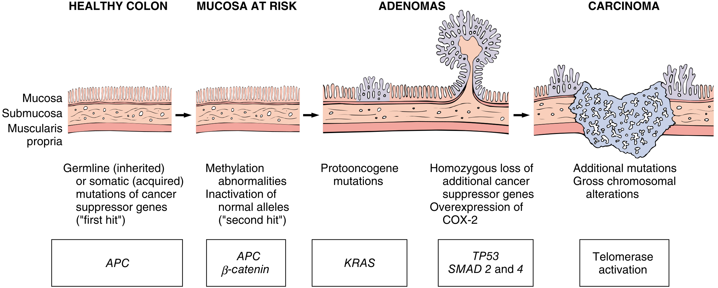
This sequence is illustrated in Fig. 17.52 of Robbins Pathologic Basis of Disease - progression from normal mucosa → early adenoma → late adenoma → adenocarcinoma, each step driven by accumulating genetic hits.
Adenoma-carcinoma sequence molecular diagram
Fig. 17.52 - Robbins Pathologic Basis of Disease: Morphologic and molecular changes in the adenoma-carcinoma sequence

2. Mismatch Repair (MMR) Deficiency / Microsatellite Instability (MSI) Pathway (~15%)

Defects in MMR genes (most commonly MSH2 or MLH1) cause failure to correct DNA replication errors. Mutations accumulate at rates up to 1000x normal, particularly in microsatellite repeat regions (50,000-100,000 microsatellites in the human genome). This leads to microsatellite instability (MSI). MSI tumors tend to be right-sided, show mucinous differentiation, prominent peritumoral lymphocytic infiltrates, and are associated with better response to checkpoint inhibitor therapy. This pathway underlies Lynch syndrome (HNPCC) germline cases.

3. CpG Island Methylation Phenotype (CIMP) (~5-10%)

Epigenetic silencing of genes (including MLH1) via hypermethylation of CpG islands. Often involves BRAF mutations. Also right-sided and associated with serrated polyp pathway.

c) Gross and Microscopic Morphology (4 Marks)

Gross Morphology

The gross appearance depends on anatomical location:
  • Left colon / sigmoid colon (as in this case): Tumors tend to grow as annular, circumferential lesions producing "napkin-ring" constrictions and progressive luminal narrowing, sometimes leading to obstruction. The ulcero-proliferative pattern (as described colonoscopically in this patient) is characteristic - with a raised, irregular proliferative edge surrounding a central ulcerated crater.
  • Right colon / proximal colon: Tumors grow as polypoid, exophytic masses extending along one wall; these rarely cause obstruction due to the large lumen.
  • Both forms invade the bowel wall over time. Cut surface shows firm, white-gray tumor tissue due to desmoplastic response; areas of chalky necrosis may be present within the wall.
  • Mucinous (colloid) variant shows gelatinous, glistening cut surface.
Gross sigmoid colon carcinoma - exophytic type
Gross pathology of resected sigmoid colon adenocarcinoma showing bulky exophytic tumor with luminal narrowing
Endoscopic view of obstructive sigmoid carcinoma
Colonoscopic appearance of sigmoid colon carcinoma with circumferential ulcero-proliferative growth causing luminal stenosis

Microscopic Morphology

  • Conventional adenocarcinoma (most common): Composed of tall columnar cells resembling dysplastic adenomatous epithelium. These form irregular glands of varying sizes, often with back-to-back or cribriform arrangements, invading through the bowel wall layers (mucosa → submucosa → muscularis propria → subserosa/serosa).
  • Desmoplastic stroma: The invasive component elicits a strong stromal desmoplastic response (dense fibrocollagenous tissue with activated myofibroblasts) - responsible for the firm consistency of the tumor on gross examination.
  • Grading:
    • Well-differentiated (Grade 1): >95% gland formation; closely resembling normal colonic glands.
    • Moderately differentiated (Grade 2): 50-95% gland formation; the most common grade.
    • Poorly differentiated (Grade 3): <50% gland formation; sheets and nests of cells with marked nuclear pleomorphism.
  • Mucinous (colloid) variant: Tumors produce abundant extracellular mucin accumulating in the intestinal wall ("mucin lakes"). >50% of tumor area occupied by mucin. Associated with poor prognosis and risk of pseudomyxoma peritonei.
  • Signet-ring cell variant: Rare; similar to gastric signet-ring cell carcinoma; cells with intracytoplasmic mucin displacing the nucleus to the periphery.
  • MSI tumors: Show prominent mucinous differentiation and peritumoral lymphocytic infiltrates (Crohn's-like reaction).
Mucinous colorectal adenocarcinoma histology - H&E
H&E histology of colorectal mucinous adenocarcinoma showing neoplastic glands embedded in abundant extracellular mucin pools within desmoplastic stroma

Spread

  • Direct spread: Through bowel wall layers; may invade adjacent organs (bladder, uterus, ureter in sigmoid tumors).
  • Lymphatic spread: Regional lymph nodes - most common form; follows the vascular pedicles.
  • Hematogenous: Via portal vein to liver (most common site of distant metastasis), then lungs.
  • Transcoelomic (peritoneal): Leads to peritoneal carcinomatosis.

d) Familial Adenomatous Polyposis (FAP) - 2 Marks

Definition and Genetics

FAP is an autosomal dominant inherited syndrome caused by germline mutations in the APC gene (chromosome 5q21), with an incidence of 1 in 10,000 births. It accounts for <1% of all colorectal cancers. Up to one-third of cases represent de novo mutations (no family history). Each child of an affected parent has a 50% risk of inheriting the mutation.
The APC protein normally forms a complex with β-catenin, GSK3, axin, and casein, promoting β-catenin degradation. When APC is mutated, β-catenin accumulates in the nucleus, inducing transcription of growth-promoting genes (c-myc, cyclin D1), leading to uncontrolled cell proliferation. Various truncating and missense mutations of APC exist - genotype-phenotype correlations exist:
  • Mutations in the "mutation cluster region" (codons 1250-1464) of exon 15: Predicts a more virulent, profuse form with thousands of polyps and earlier CRC onset.
  • Mutations at the 5' end of the gene: Result in attenuated FAP - milder phenotype with fewer polyps and later onset.
  • Mutations between codons 543-1309: Associated with CHRPE (congenital hypertrophy of retinal pigment epithelium).

Clinical Features

  • Colonic polyposis: Hundreds to thousands of adenomatous polyps carpeting the entire colorectum (median age of adenoma development: 17 years).
  • Inevitable malignant transformation: Virtually all patients with classical FAP develop CRC if untreated; CRC risk rises dramatically after age 30; median age of CRC: 40 years; median age of death if untreated: 44 years.
  • Extracolonic manifestations:
    • Upper GI: >90% have fundic gland polyps (benign, hamartomatous); duodenal/periampullary polyps (lifetime risk of duodenal cancer 3-5%)
    • Gardner syndrome variant: Osteomas (jaw/skull), epidermoid cysts, supernumerary teeth, desmoid tumors, fibromas, lipomas
    • CHRPE: Congenital hypertrophy of retinal pigment epithelium - well-demarcated gray-brown lesions ("bear claws")
    • Turcot syndrome variant: FAP + CNS tumors (medulloblastoma)
    • Desmoid tumors of the mesentery/abdominal wall
FAP colon specimen with hundreds of polyps
Gross colectomy specimen from a patient with FAP showing hundreds of adenomatous polyps carpeting the entire mucosal surface

Surveillance and Management

  • Annual colonoscopy from age 10-15 years in known mutation carriers.
  • Prophylactic colectomy is recommended once the patient reaches late teens/early 20s (or earlier if polyp burden is high or symptoms develop).
  • Surgical options include total colectomy with ileorectal anastomosis (IRA) or proctocolectomy with ileal pouch-anal anastomosis (IPAA) - choice depends on rectal polyp burden.
  • Post-colectomy surveillance of the remaining rectum (IRA) or pouch is mandatory.
  • NSAIDs (sulindac, celecoxib) can cause partial polyp regression but do not replace surgery.

Sources: Robbins & Cotran Pathologic Basis of Disease (10e), pp. 756-763 | Mulholland and Greenfield's Surgery: Scientific Principles and Practice (7e), pp. 3331-3336

Reasoning questions (5x315 Marks) 2. A 30-year-old woman presented with heat intolerance, palpitations, weight loss and diffuse enlargement of the thyroid gland. a) Diagnosis. (1) b) Pathogenesis. (1) c) Mention one characteristic microscopic feature. (1) 3. An 8-year-old child developed facial puffiness, oliguria and cola-coloured urine two weeks after a sore throat. a) Diagnosis. (1) b) Etiological organism. (1) c) Characteristic immunofluorescence finding. (1) 4. A 58-year-old hypertensive smoker presented with severe retrosternal chest pain radiating to the left arm. a) Diagnosis. (1) b) Mention one major risk factor. (1) c) Name the earliest cardiac biomarker elevated. (1) 5. A 65-year-old chronic smoker presented with persistent cough and hemoptysis. Chest X-ray revealed a hilar mass. a) Diagnosis. (1) b) Mention the most important etiological factor. (1) c) Mention one histological type of bronchogenic carcinoma. (1) 6. A 52-year-old man presented with fatigue, splenomegaly and marked leukocytosis. a) Diagnosis. (1) b) Characteristic chromosomal abnormality. (1) c) Name the fusion gene. (1)

Reading File
Finding Sources
Finding Sources
Finding Sources
Finding Sources
Finding Sources
Finding Sources
Finding Sources
Finding Sources
Finding Sources
Reading File
Reading File
Reading File
Finding Sources
I have excellent library content for Q2, Q3, and Q6. For Q4 (cardiac biomarker) and Q5 (bronchogenic carcinoma), I'll draw on my established medical knowledge which is well-grounded. Let me now compose the complete answers.

Reasoning Questions - Answers (5 × 3 = 15 Marks)


Question 2 - 30-year-old woman: heat intolerance, palpitations, weight loss, diffuse thyroid enlargement

a) Diagnosis (1 mark)

Graves' Disease (Diffuse toxic goitre / Primary hyperthyroidism due to autoimmune thyrotoxicosis)

b) Pathogenesis (1 mark)

Graves' disease is an autoantibody-mediated disorder. Autoantibodies (IgG class) are produced against the TSH receptor on thyroid follicular cells. These antibodies - called thyroid-stimulating immunoglobulins (TSI) / TRAb (TSH Receptor Antibodies) - bind to the TSH receptor and mimic the action of TSH, stimulating follicular cells to produce and release excess thyroid hormones (T3 and T4) independently of normal pituitary regulation. This results in diffuse hyperplasia of the gland and clinical thyrotoxicosis. The trigger for this autoimmune reaction involves genetic susceptibility (HLA alleles, CTLA4 polymorphisms) combined with environmental factors. The same TSH receptor is expressed on orbital fibroblasts, explaining the associated infiltrative ophthalmopathy - activated CD4+ T cells secrete cytokines that increase extracellular matrix protein deposition in the retroorbital space. (Source: Robbins & Kumar Basic Pathology, Graves Disease section)

c) One Characteristic Microscopic Feature (1 mark)

Papillary infoldings of follicular epithelium into the follicular lumen - On microscopy, follicular epithelial cells are tall and crowded, and this crowding results in formation of small papillae projecting into the follicle lumen (without fibrovascular cores). The colloid within follicles is pale and scalloped at the margins (due to active resorption). Lymphoid infiltrates with germinal centres are also present in the interstitium.

Question 3 - 8-year-old child: facial puffiness, oliguria, cola-coloured urine, 2 weeks after sore throat

a) Diagnosis (1 mark)

Acute Post-Streptococcal Glomerulonephritis (APSGN) - presenting as Acute Nephritic Syndrome
The classic triad of haematuria (cola/smoky urine), oliguria, and oedema (facial puffiness) following a latent period of ~10-14 days after a streptococcal sore throat is the textbook presentation.

b) Etiological Organism (1 mark)

Group A β-haemolytic Streptococcus (Streptococcus pyogenes) - specifically nephritogenic strains (e.g., M-types 1, 2, 4, 12, 18, 25 for pharyngitis; M-types 2, 49, 55, 57, 60 for skin infections). Pharyngitis precedes renal disease by 1-2 weeks; impetigo/pyoderma by 3-6 weeks.

c) Characteristic Immunofluorescence Finding (1 mark)

Coarse granular ("lumpy-bumpy" / "starry sky") deposits of IgG, IgM and C3 along the glomerular capillary walls and mesangium - This pattern reflects immune complex deposition (antigen-antibody complexes containing streptococcal antigens lodging in the glomerulus). Electron microscopy correspondingly shows large dome-shaped (hump-like) electron-dense subepithelial deposits on the glomerular basement membrane. (Source: Goldman-Cecil Medicine, Post-streptococcal glomerulonephritis section)

Question 4 - 58-year-old hypertensive smoker: severe retrosternal chest pain radiating to left arm

a) Diagnosis (1 mark)

Acute Myocardial Infarction (AMI) - most likely ST-elevation myocardial infarction (STEMI), due to atherosclerotic coronary artery disease with acute plaque rupture and thrombotic occlusion.

b) One Major Risk Factor (1 mark)

Cigarette smoking (or hypertension - both are major modifiable risk factors present in this patient).
Smoking causes endothelial injury, promotes atherosclerosis, increases platelet aggregation, raises LDL, lowers HDL, and causes coronary vasospasm. Hypertension accelerates atherosclerosis through increased shear stress on the arterial wall, promoting intimal injury and foam cell accumulation.

c) Earliest Cardiac Biomarker Elevated (1 mark)

Troponin I (cTnI) or Troponin T (cTnT) - Cardiac troponins are the most sensitive and specific early markers of myocardial necrosis. They begin to rise within 3-4 hours of onset of infarction, peak at 18-24 hours (TnI) to 24-48 hours (TnT), and remain elevated for up to 10-14 days (cTnT) or 7-10 days (cTnI) - useful for late presentation.
Note on "earliest": Historically, Myoglobin rises earliest (within 1-2 hours) but lacks cardiac specificity. If the question asks for the earliest specific cardiac biomarker, the answer is Troponin I or T. If asking for any earliest biomarker (including non-specific), it is myoglobin. For modern clinical purposes, high-sensitivity troponin (hs-cTn) is the preferred answer.

Question 5 - 65-year-old chronic smoker: persistent cough, haemoptysis, hilar mass on CXR

a) Diagnosis (1 mark)

Bronchogenic Carcinoma (Primary lung cancer, centrally located - hilar mass strongly suggests a central/endobronchial tumour)
A hilar mass in a chronic smoker with haemoptysis and persistent cough is the classic presentation of a central bronchogenic carcinoma.

b) Most Important Etiological Factor (1 mark)

Cigarette smoking - responsible for approximately 85-90% of all lung cancers. Tobacco smoke contains >60 known carcinogens (polycyclic aromatic hydrocarbons, nitrosamines, etc.) causing direct DNA damage, particularly in the TP53, KRAS, and RB1 tumour suppressor and oncogene pathways. Risk is dose-dependent (pack-years) and includes passive (second-hand) smoke exposure.

c) One Histological Type of Bronchogenic Carcinoma (1 mark)

Squamous Cell Carcinoma - the most common histological type associated with a central/hilar location and directly linked to smoking. Characterised histologically by keratin pearls and intercellular bridges. Other types include:
  • Adenocarcinoma (most common overall; peripheral)
  • Small cell carcinoma (central; highly aggressive; neuroendocrine)
  • Large cell carcinoma (peripheral; undifferentiated)
For this patient with a hilar mass, Squamous Cell Carcinoma or Small Cell Carcinoma are the most likely histological types.

Question 6 - 52-year-old man: fatigue, splenomegaly, marked leukocytosis

a) Diagnosis (1 mark)

Chronic Myeloid Leukaemia (CML)
The triad of massive splenomegaly, marked leukocytosis (predominantly mature and immature myeloid cells), and constitutional symptoms (fatigue) in a middle-aged adult is the hallmark of CML. Peripheral smear shows a full spectrum of myeloid maturation with a basophilia.

b) Characteristic Chromosomal Abnormality (1 mark)

Philadelphia (Ph) Chromosome - a shortened chromosome 22, resulting from a reciprocal translocation between chromosomes 9 and 22: t(9;22)(q34;q11). This creates an elongated chromosome 9 and the shortened Philadelphia chromosome 22.

c) Name the Fusion Gene (1 mark)

BCR-ABL1 (written as BCR::ABL or BCR-ABL)
The translocation fuses the BCR (Breakpoint Cluster Region) gene on chromosome 22 with the ABL1 (Abelson tyrosine kinase) gene from chromosome 9. The resulting chimeric BCR-ABL protein is a constitutively active tyrosine kinase that drives uncontrolled myeloid proliferation. The most common fusion product is the p210 BCR-ABL protein (present in ~95% of CML). This is the direct therapeutic target of imatinib (Gleevec) and other tyrosine kinase inhibitors. (Source: Goldman-Cecil Medicine, CML section; Robbins Pathologic Basis of Disease)

Sources: Robbins & Kumar Basic Pathology (10e) | Robbins, Cotran & Kumar Pathologic Basis of Disease | Goldman-Cecil Medicine | Harrison's Principles of Internal Medicine 22e | Janeway's Immunobiology 10e
This is a shared conversation. Sign in to Orris to start your own chat.