
Acute exacerbations of COPD management
"COPD exacerbation" AND management
GOLD 2025 COPD acute exacerbation management guidelines
https://goldcopd.org/wp-content/uploads/2026/01/GOLD-Pocket-…
Acute Exacerbations of COPD (AECOPD) - Management
Definition
Precipitating Causes
| Category | Examples |
|---|---|
| Viral infections | Rhinovirus, influenza, RSV (>50% of exacerbations per PCR studies) |
| Bacterial infections | H. influenzae, S. pneumoniae, M. catarrhalis, new bacterial strains |
| Environmental | Air pollution, allergens, temperature changes |
| Other | Pulmonary embolism, medication non-adherence |
| Unknown | ~25–30% of cases have no identifiable precipitant |
- Harrison's 22E, p. 2302
Differential Diagnosis
-
Pneumonia
-
Pulmonary embolism (incidence is increased in AECOPD)
-
Acute decompensated heart failure
-
Pneumothorax
-
Cardiac ischemia
-
Pleural effusion
-
Large airway tumor
-
Washington Manual of Medical Therapeutics, p. 319
Severity Classification (GOLD / Rome Proposal)
| Severity | Features |
|---|---|
| Mild | Treated with SABAs alone; no change in other medications |
| Moderate | Treated with SABAs + antibiotics and/or systemic corticosteroids |
| Severe | Requires hospitalization or ER visit; may have acute respiratory failure |
Initial Assessment
History
- Degree and change in dyspnea (activities of daily living)
- Fever, sputum character change, wheezing, GI symptoms
- Frequency and severity of prior exacerbations (prior hospitalization = biggest risk factor for re-hospitalization)
- Comorbidities (heart disease, diabetes)
Physical Examination
- Tachycardia, tachypnea
- Use of accessory muscles, intercostal retractions
- Ability to speak in complete sentences
- Mental status (confusion/somnolence suggests hypercarbia)
- Asymmetric chest findings (pneumothorax), paradoxical abdominal wall movement
- Signs of right or left heart failure
Investigations
| Test | Indication |
|---|---|
| Pulse oximetry | All patients |
| ABG | Moderate-severe distress, suspected hypercarbia (PCO₂ >45 mmHg has key therapeutic implications), mental status changes |
| CXR | Abnormal in ~15-25%; always order if chest pain, leukocytosis, history of heart disease |
| ECG | Assess for cardiac ischemia, arrhythmia |
| CBC, BMP | Screen for anemia, metabolic acidosis, hyperglycemia |
| BNP/NT-proBNP | If left heart failure suspected |
| Troponin | If myocardial ischemia is a concern |
| D-dimer | Only if PE is suspected after appropriate pre-test probability risk stratification |
| Sputum culture | Generally not indicated in routine exacerbations |
- Harrison's 22E, pp. 2302-2303; Rosen's Emergency Medicine, p. 2560
Indications for Hospitalization
-
Significant increase in symptom severity
-
Severe underlying COPD (e.g., FEV₁ very low at baseline)
-
Respiratory acidosis / hypercarbia
-
New or worsening hypoxemia
-
Significant comorbidities (heart failure, arrhythmia)
-
Failure to respond to initial outpatient/ED treatment
-
Insufficient home support or inability to manage at home
-
Diagnostic uncertainty
-
Washington Manual, p. 320; Goldman-Cecil Medicine, p. 3148
Indications for ICU Admission
- Need for invasive mechanical ventilation
- Hemodynamic instability
- Severe dyspnea not responding to therapy
- Altered mental status
- Persistent or worsening hypoxemia, hypercapnia, or respiratory acidosis despite supplemental O₂ and NIV
Pharmacological Management
1. Bronchodilators (First-Line)
| Drug | MDI Dose | Nebulizer Dose |
|---|---|---|
| Albuterol (Salbutamol) | 2-4 puffs q1-4h | 2.5 mg q1-4h (hourly x 1-3h, then q2-4h) |
| Drug | MDI Dose | Nebulizer Dose |
|---|---|---|
| Ipratropium | 2 puffs q4h | 0.5 mg q4h |
-
In the emergency setting, nebulizers are preferred over MDIs requiring complex technique
-
Use air-driven nebulizers (not oxygen-driven) when possible
-
Continuous nebulization is NOT indicated
-
Methylxanthines (theophylline) are not recommended due to poor side-effect profile without added benefit
-
Washington Manual, p. 320; Rosen's EM, p. 2574; GOLD 2026
2. Systemic Corticosteroids
-
Prednisone 40 mg/day orally for 5 days (5-day courses are as effective as longer courses)
-
Parenteral route (methylprednisolone) only if patient cannot tolerate oral medication
-
Benefits: decreased recovery time, improved oxygenation, improved FEV₁, shorter hospital stay
-
Oral and parenteral bioavailability are similar - prefer oral when tolerated
-
Rosen's EM, p. 2579; GOLD 2026
3. Antibiotics
- Increased dyspnea + increased sputum volume + increased sputum purulence (all three = strongest indication)
- Patients requiring NIV or mechanical ventilation (clear mortality benefit in these)
- CRP-guided or procalcitonin-guided prescribing can reduce unnecessary antibiotic use in non-critically ill patients
| Patient Profile | Pathogens | Antibiotic Choice |
|---|---|---|
| No risk factors for poor outcome or resistant organism | H. influenzae, S. pneumoniae, M. catarrhalis | Macrolide, 2nd/3rd-gen cephalosporin, doxycycline, TMP/SMX |
| Risk factors present (severe COPD, recent hospitalization, prior Pseudomonas colonization) | Above + gram-negative rods including Pseudomonas | Antipseudomonal fluoroquinolone (e.g., levofloxacin 750 mg/day x 7-10d) or antipseudomonal beta-lactam |
Note: In critically ill patients on ICU, PCT-guided antibiotic therapy was associated with worse 3-month mortality - do NOT withhold antibiotics based on PCT in the ICU.
- Washington Manual, p. 320; Goldman-Cecil, p. 3150; Rosen's EM, p. 2582-2584
Oxygen Therapy
- Target SpO₂: 88-92% (controlled oxygen therapy to avoid hypercapnic drive suppression)
- High-flow nasal therapy (HFNT) is increasingly used for hypoxemic AECOPD
- Avoid high-concentration uncontrolled oxygen
Ventilatory Support
Non-Invasive Ventilation (NIV) - First-Line for Respiratory Failure
- Severe dyspnea with increased work of breathing
- Moderate-to-severe respiratory acidosis (pH <7.35) + hypercapnia (PCO₂ >45 mmHg)
- Persistent hypoxemia despite supplemental oxygen
-
Decreased respiratory rate
-
Increased tidal volume and minute ventilation
-
Reduced need for endotracheal intubation
-
Decreased in-hospital mortality
-
Shorter ICU and hospital length of stay
-
Mechanism: improved alveolar ventilation and respiratory mechanics
-
Fishman's Pulmonary Diseases, p. 2630; Roberts and Hedges' Clinical Procedures, p. 231; Murray & Nadel's Respiratory Medicine
Key point: NIV must be initiated early alongside standard medical therapy. Late initiation (after medical treatment failure) eliminates the survival and intubation-reduction benefits.
Contraindications to NIV
- Respiratory or cardiac arrest
- Hemodynamic instability
- Uncooperative or agitated patient
- High aspiration risk
- Inability to protect airway
- Recent upper airway/GI surgery
Invasive Mechanical Ventilation
-
Respiratory arrest
-
Severe respiratory failure not responding to NIV
-
Decreased consciousness / significant agitation
-
Hemodynamic instability
-
Massive aspiration
-
Persistent inability to clear secretions
-
Harrison's 22E, p. 2303; Tintinalli's EM, p. 1745
Discharge Criteria and Follow-Up
- Patient on maintenance therapy every 4 hours or less
- Walking stable (patient ambulatory)
- Eating, sleeping not disturbed by dyspnea
- Clinically stable for 12-24 hours
- ABG stable for 12-24 hours
- Patient/family understands medication use
- Initiate/optimize long-acting bronchodilators (LABDs) as soon as possible
- If ≥1 moderate/severe exacerbation and elevated blood eosinophil count: add ICS to dual bronchodilator regimen at discharge
- Full recovery takes 4-6 weeks - some patients never return to pre-exacerbation baseline
- Review COPD management plan and comorbidities
- Implement exacerbation prevention strategies (vaccination, smoking cessation, pulmonary rehabilitation)
Prevention of Future Exacerbations
| Strategy | Evidence |
|---|---|
| LAMA +/- LABA (triple therapy LAMA/LABA/ICS for high-risk) | First-line maintenance |
| Smoking cessation | Most important modifiable factor |
| Pneumococcal + annual influenza + COVID-19 vaccination | Reduces infective triggers |
| Pulmonary rehabilitation | Reduces re-hospitalization |
| Roflumilast (PDE4 inhibitor) | For frequent exacerbators with chronic bronchitis |
| Azithromycin (long-term) | Selected patients with recurrent exacerbations |
| Mepolizumab / Dupilumab | For eosinophilic COPD phenotype (GOLD 2025/2026) |
Summary Algorithm
AECOPD suspected
    ↓
Exclude mimics (PE, pneumonia, CHF, pneumothorax)
    ↓
Assess severity → Mild / Moderate / Severe
    ↓
All patients: SABA ± SAAC bronchodilators
Moderate/Severe: + systemic steroids (prednisone 40mg x 5d)
Purulent sputum/infection signs: + antibiotics x 5d
    ↓
Hypoxemia → controlled O₂ (SpO₂ 88-92%)
    ↓
Respiratory acidosis / increased WOB → NIV (BiPAP)
    ↓
NIV failure / contraindications → invasive MV
    ↓
Discharge with LABD optimization, follow-up in 1-4 weeks
Create a clinical decision flowchart for AECOPD severity assessment and admit/discharge decisions
generate_image tool to create a professional clinical decision flowchart.A professional clinical decision flowchart for AECOPD (Acute Exacerbation of COPD) severity assessment and admit/discharge decisions. The flowchart should be clean, medical-grade, on a white background with clear boxes and arrows. Start at the top: "Suspected AECOPD" → "Exclude Mimics" (list: Pneumonia, PE, CHF, Pneumothorax, Cardiac Ischemia) Then: "Initial Assessment" with two columns: - History: dyspnea change, sputum purulence, prior exacerbations, comorbidities - Exam + Tests: SpO2, ABG, CXR, ECG, CBC, BMP Then severity classification into three branches: MILD (left branch, green): - Responds to SABA alone - No accessory muscle use - SpO2 ≥ 92% on room air - No hypercarbia → "DISCHARGE" box (green): SABA PRN, consider oral steroids, antibiotic if purulent sputum, follow-up 1-2 weeks MODERATE (center branch, amber/orange): - Increased dyspnea at rest - SpO2 88-92% on O2 - pH normal, PCO2 may be elevated - Responds to bronchodilators + steroids → Decision diamond: "Adequate response to ED treatment?" → YES → DISCHARGE with full medications → NO → ADMIT (general ward) SEVERE (right branch, red): - Severe dyspnea at rest - SpO2 <88% despite O2 - Respiratory acidosis (pH <7.35) - Mental status changes - Hemodynamic instability → "ADMIT" → Decision diamond: "Contraindications to NIV?" → NO → Start NIV (BiPAP) → "NIV Failure?" → YES → Invasive Mechanical Ventilation → ICU → YES (contraindications) → Direct Intubation → ICU At the bottom, a "DISCHARGE CHECKLIST" box in blue: - SpO2 ≥ 90% on baseline O2 - Bronchodilator ≤ q4h - Eating, walking stable - Inhaler education done - Follow-up arranged within 4 weeks - Optimize LABD ± ICS (if eosinophil elevated) Use color-coded severity lanes (green/amber/red), clean sans-serif medical font, diamond decision shapes, rectangular process boxes. Label all arrows YES/NO where applicable.
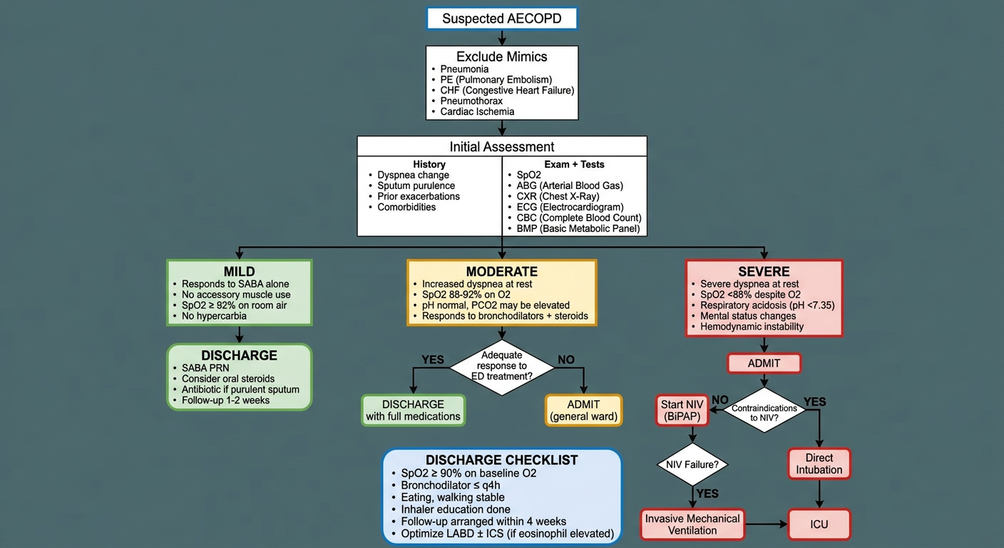
- Top section - Initial workup and exclusion of mimics (PE, pneumonia, CHF, pneumothorax)
- Three color-coded severity lanes:
- 🟢 Mild - SABA-responsive, SpO₂ ≥92% on room air → direct discharge pathway
- 🟡 Moderate - needs bronchodilators + steroids + antibiotics (if purulent) → re-assess response before discharge vs. ward admission
- 🔴 Severe - respiratory acidosis, altered mental status, hemodynamic instability → admit, trial NIV (BiPAP), escalate to invasive MV + ICU if NIV fails or is contraindicated
- Bottom discharge checklist - ensures safe discharge criteria are met before sending a patient home
management of postpartum hemorrhage
"postpartum hemorrhage" AND management
postpartum hemorrhage management guidelines 2024 2025 WHO ACOG
https://www.ncbi.nlm.nih.gov/books/NBK619236
Management of Postpartum Hemorrhage (PPH)
Definition
| Guideline | Definition |
|---|---|
| ACOG 2017 (reaffirmed 2024) | Blood loss ≥1,000 mL regardless of mode of delivery, OR blood loss with signs/symptoms of hypovolemia |
| WHO 2025 | Blood loss ≥500 mL after vaginal birth; ≥1,000 mL after cesarean |
| Practical definition | 10% drop in hematocrit, need for PRBC transfusion, or volume loss causing hemodynamic instability |
Note: Because pregnancy increases plasma volume by ~40% and RBC volume by ~25%, signs of hemorrhagic shock are masked. Up to 30% of total blood volume may be lost before BP drops. The first sign may be only mild tachycardia. - Tintinalli's Emergency Medicine, p. 3418
Classification
| Type | Timing | Common Causes |
|---|---|---|
| Primary PPH | Within first 24 hours of delivery | Uterine atony (75-90%), genital tract trauma (20%), retained placental tissue (10%), coagulopathy |
| Secondary PPH | 24 hours to 6 weeks postpartum | Subinvolution of placental site, retained placental tissue, genital tract wound, uterine infection |
The "4 Ts" - Causes of Primary PPH
| T | Cause | Details |
|---|---|---|
| Tone | Uterine atony | Most common cause (75-90%). Soft, boggy uterus on exam |
| Trauma | Genital tract lacerations, uterine rupture, uterine inversion | Second most common (~20%). Perineal, vaginal, cervical tears; episiotomy |
| Tissue | Retained placental fragments, placenta accreta spectrum | ~10% of cases |
| Thrombin | Coagulopathy | DIC, pre-existing clotting disorders, thrombocytopenia |
- Rosen's Emergency Medicine, p. 3419
Risk Factors
- Previous PPH or uterine surgery
- Placenta previa / abruption / accreta spectrum
- Multiple gestation, fetal macrosomia, polyhydramnios (uterine overdistension)
- Multiparity, anemia
- Coagulation disorders
- Prolonged labor (all stages)
- Chorioamnionitis, fever
- Oxytocin use during labor (receptor desensitization)
- Tocolytic use (magnesium sulfate, beta-mimetics)
- Operative delivery (forceps/vacuum), episiotomy
- Volatile anesthetic agents (halogenated - cause uterine relaxation)
- Preeclampsia
Initial Resuscitation (Simultaneous with Cause Identification)
- Call for help immediately - activate PPH response team (obstetrician, anesthesiologist, nurses, blood bank, interventional radiology)
- Two large-bore IV lines - establish promptly
- IV fluid resuscitation - isotonic crystalloids (lactated Ringer's preferred; isotonic crystalloids preferred over colloids per WHO 2025)
- Oxygen supplementation
- Uterine massage / bimanual compression - one fist in anterior fornix, other hand suprapubic compression of fundus
- Frequent vital signs monitoring
- Urinary catheter - monitor output
- Blood typing and crossmatch - send immediately; use O-negative unmatched blood in true emergencies
- Labs: CBC, coagulation panel (PT/PTT/fibrinogen), BMP, type and screen
- Creasy & Resnik's MFM, p. 947; Tintinalli's EM, p. 3419
Step-by-Step Management
Step 1: Identify and Treat the Cause
- Palpate uterus (atony = soft/boggy)
- Inspect cervix, vagina, perineum for lacerations
- Check for retained placental tissue (real-time ultrasound is helpful)
- Review coagulation labs
Step 2: Uterotonic Agents (For Atony - First-Line Medical Management)
First-Line: Oxytocin
| Route | Dose | Notes |
|---|---|---|
| IV infusion | 20-30 units in 1,000 mL crystalloid, rate ≤100 mU/min; up to 40 units total | Preferred; avoid large IV bolus |
| IM | 10 units after placental delivery | Standard prophylaxis dose |
-
Do NOT give as rapid IV bolus (>10 units) - causes hypotension, tachycardia, hemodynamic compromise, nausea, headache
-
Fluid overload risk if large volumes used (antidiuretic effect at high doses)
-
Per WHO 2025: IV oxytocin is the recommended uterotonic for treatment of PPH
-
Creasy & Resnik's MFM, p. 947; Miller's Anesthesia, p. 8906
Second-Line Uterotonics (If Oxytocin Fails)
| Drug | Dose | Route | Contraindications | Key Side Effects |
|---|---|---|---|---|
| Methylergonovine (Methergine) | 0.2 mg q2-4h | IM only (NOT IV) | Hypertension, cardiac disease, Raynaud's | Hypertension, CNS vasospasm, coronary spasm, nausea |
| Carboprost (PGF2α, 15-methyl) | 250 μg IM, repeat q15-90 min (max 8 doses = 2 mg) | IM or intramyometrial | Asthma | Bronchospasm, pulmonary HTN, diarrhea, nausea, desaturation |
| Misoprostol (PGE1) | 600-1000 μg; sublingual 800 μg OR rectal 1000 μg | Sublingual, rectal, oral, vaginal | - | Fever, chills, shivering, diarrhea |
| Ergometrine/Ergonovine | 0.2 mg IM | IM | Hypertension | Same as methylergonovine |
Important: Per WHO 2025, if IV oxytocin is unavailable or fails, use IV ergometrine, oxytocin-ergometrine combination, or a prostaglandin drug (including sublingual misoprostol 800 μg).
- Miller's Anesthesia, p. 8906; Rosen's EM, p. 3419; Creasy & Resnik's MFM, p. 947-948
Cochrane Network Meta-Analysis 2025 (PMID 40237648)
Step 3: Tranexamic Acid (TXA)
-
Dose: 1 g IV over 10 minutes; repeat 1 g IV if bleeding continues after 30 minutes, or if bleeding restarts within 24 hours
-
Timing: Must be given within 3 hours of birth - benefit diminishes rapidly after 3 hours
-
Mechanism: Antifibrinolytic - prevents plasmin-mediated clot breakdown
-
Per WHO 2025 (Recommendation 27): Early TXA in addition to standard care is recommended for PPH after vaginal birth OR cesarean section
-
Per WHO 2025 (Recommendations 14-15): TXA is NOT recommended for prevention of PPH (only treatment)
-
WOMAN trial (Lancet 2017) showed TXA given within 3 hours significantly reduced PPH death from bleeding
-
WHO Consolidated PPH Guidelines 2025
Step 4: Mechanical / Tamponade Measures
For Atony Persisting Despite Uterotonics:
| Technique | Description |
|---|---|
| Bimanual uterine compression | Continued manual massage |
| Uterine balloon tamponade (Bakri balloon, Rusch catheter, Jada System) | Intrauterine balloon inflated to apply counter-pressure. WHO 2025 recommends for atony not responding to first-line treatment. Jada System (vacuum-based) achieves control in >90% of patients at median 3 minutes |
| Uterine packing | Sterile gauze - WHO 2025 does NOT recommend plain or hemostatic gauze packing (insufficient evidence of benefit) |
| Non-pneumatic antishock garment (NASG) | Effective in remote settings to reduce blood loss and improve survival pending transfer |
- Miller's Anesthesia, p. 8906; Creasy & Resnik's MFM, p. 948; WHO 2025
Step 5: Interventional Radiology
- Uterine artery embolization (UAE): Recommended by WHO 2025 if other measures have failed AND resources/personnel are available. Allows uterine preservation.
- Best for hemodynamically stable patients
- Requires IR suite and experienced team
Step 6: Surgical Management
| Procedure | Details |
|---|---|
| Uterine compression sutures | B-Lynch "brace" suture - longitudinal sutures compress uterus and close blood supply. Hayman technique is a simpler variation. Uterine-conserving. |
| Uterine artery ligation (O'Leary sutures) | Bilateral ligation of uterine arteries. First surgical step. |
| Internal iliac (hypogastric) artery ligation | If uterine artery ligation fails. Reduces pulse pressure in pelvic vessels. No long-term consequences due to collateral circulation; subsequent pregnancies reported. |
| Peripartum hysterectomy | Definitive treatment for uncontrolled hemorrhage. Life-saving. Used when all other surgical measures have failed. |
Place patient in semilithotomy position to allow simultaneous vaginal exam to confirm cessation of bleeding.
- Creasy & Resnik's MFM, p. 948-949; Tintinalli's EM, p. 3419
Step 7: Blood Product Transfusion
-
Massive transfusion protocol (MTP) should be activated early for massive PPH
-
PRBCs: Transfuse to maintain Hb >8 g/dL in active hemorrhage
-
Fresh Frozen Plasma (FFP): For coagulopathy; standard MTP ratio FFP:PRBC ~1:1 (though exact ratios for obstetric hemorrhage are debated)
-
Platelets: Transfuse if <50,000/μL in active bleeding; aim >75,000-100,000
-
Cryoprecipitate / Fibrinogen concentrate: For hypofibrinogenemia (<2 g/L); fibrinogen level is an early predictor of PPH severity
-
O-negative unmatched blood: In true emergencies before crossmatch available
-
Miller's Anesthesia, pp. 8907-8908
Management by Specific Cause
Genital Tract Trauma (Lacerations)
- Inspect cervix, vagina, perineum systematically
- Repair with absorbable sutures (bedside or in OR)
- Contained hematomas: expectant if small/stable; vascular embolization or surgical drainage if expanding/large
Retained Placental Tissue
- Manual removal of placenta under anesthesia
- Uterine evacuation (curettage) - blunt preferred to avoid perforation
- Suspect placenta accreta if placenta fails to separate; may require hysterectomy
Placenta Accreta Spectrum (PAS)
- Multidisciplinary team essential
- Uterine-sparing approaches possible in selected cases
- Peripartum hysterectomy often required for percreta/increta
Coagulopathy / DIC
- Treat underlying cause
- Aggressive blood product replacement (FFP, cryoprecipitate, platelets)
- Recombinant factor VIIa (rFVIIa) as last resort in refractory coagulopathic bleeding
Uterine Inversion
-
Do NOT remove placenta before reinversion (may worsen hemorrhage)
-
Immediate manual reinversion with firm steady upward pressure on fundus
-
Tocolysis to relax uterus: IV magnesium sulfate, beta-mimetics (terbutaline), or nitroglycerin
-
General anesthesia with volatile agent if manual reinversion fails
-
After reinversion: uterotonics immediately
-
Creasy & Resnik's MFM, pp. 3290-3300; Tintinalli's EM
Prevention of PPH (Active Management of Third Stage of Labor)
| Intervention | Recommendation |
|---|---|
| Oxytocin 10 IU IM immediately after birth of baby | Recommended for all births - strongest recommendation |
| Controlled cord traction (CCT) | Recommended if skilled attendant present; small reduction in blood loss and third-stage duration |
| Uterine massage after placental delivery | Recommended as routine |
| TXA for prevention | NOT recommended (new 2024-2025 recommendation) |
| Delayed cord clamping (≥1 min) | Recommended for neonatal benefit; does not increase maternal PPH risk |
Escalation Algorithm Summary
PPH identified (blood loss ≥500 mL vaginal / ≥1000 mL cesarean, OR hemodynamic instability)
         ↓
Activate PPH team | 2 large-bore IVs | Type & crossmatch | Labs | Vitals monitoring
         ↓
ASSESS 4 Ts → identify primary cause
         ↓
TONE (atony) ──────────────→ Bimanual massage + Oxytocin (1st line)
         ↓ fails                    ↓ fails
         ↓              Methylergonovine / Carboprost / Misoprostol (2nd line)
         ↓                    ↓ all fail
Add TXA 1g IV (within 3h) → Balloon tamponade (Bakri/Jada)
         ↓ fails
         → UAE (if IR available + hemodynamically stable)
         → OR: Surgical (B-Lynch sutures → uterine artery ligation → hysterectomy)

TRAUMA ──────────────────→ Suture repair of lacerations / hematoma drainage
TISSUE ──────────────────→ Manual removal / curettage / hysterectomy (PAS)
THROMBIN ────────────────→ Blood products, treat DIC, rFVIIa (last resort)
fliud rescuscitation in postpartum hemorrhage
"postpartum hemorrhage" AND "fluid resuscitation"
fluid resuscitation postpartum hemorrhage crystalloid blood products massive transfusion protocol 2024 2025
PMID: 37535070
Fluid Resuscitation in Postpartum Hemorrhage
The Paradigm Shift: Away from Crystalloid-First Resuscitation
- Exacerbates dilutional coagulopathy (dilutes clotting factors, fibrinogen, and platelets)
- Causes tissue edema, including pulmonary edema
- Worsens metabolic acidosis
- Is associated with multiorgan failure
"Resuscitation with excessive crystalloid or colloid solutions should be avoided as it increases morbidity and mortality. Instead, hemostatic resuscitation including early administration of blood products should be instituted without delays." - AJOG 2025 (Hemostatic resuscitation during cesarean delivery)
- Barash's Clinical Anesthesia, p. 3519; Miller's Anesthesia, p. 8907
The "Lethal Triad" - What You Are Trying to Prevent
Hypothermia ──→ worsens coagulopathy
     ↑                    ↓
Acidosis  ←──── Coagulopathy ──→ more bleeding
| Derangement | Threshold | Effect |
|---|---|---|
| Hypothermia | Core temp <35°C | Every 1°C drop increases transfusion needs by ~20%; impairs enzyme function in coagulation cascade |
| Acidosis | pH <7.2 | Impairs thrombin generation; worsens coagulopathy |
| Coagulopathy | Fibrinogen <2 g/L, INR >1.5 | Prevents clot formation; fibrinogen falls early in PPH |
Phase 1: Immediate Resuscitation Measures
Vascular Access
- Two large-bore peripheral IVs (16G or larger) - immediate priority
- Arterial line - for continuous BP monitoring and frequent labs in major hemorrhage
- Central venous access - if peripheral access inadequate or vasopressors needed; also enables CVP monitoring
- Consider IO access if rapid IV access is not possible
Initial Volume Replacement
| Fluid | Recommendation |
|---|---|
| Lactated Ringer's (LR) | Preferred crystalloid - less hyperchloremic acidosis than normal saline |
| Normal Saline (0.9% NaCl) | Acceptable but risk of hyperchloremic acidosis with large volumes |
| Colloids (albumin, HES) | NOT recommended - no proven benefit over crystalloids and HES may worsen coagulopathy |
Phase 2: Blood Product Resuscitation
When to Trigger a Massive Transfusion Protocol (MTP)
- ≥4 units PRBC within 1 hour with ongoing bleeding
- ≥10 units PRBC within 24 hours
- Replacement of the patient's entire blood volume
- Loss of >50% blood volume in <3 hours
The MTP Protocol - Blood Product Ratios (1:1:1)
| Component | Ratio | Target |
|---|---|---|
| Packed Red Blood Cells (PRBCs) | 1 | Hb >7 g/dL (or >8 g/dL if ongoing active hemorrhage or cardiac disease) |
| Fresh Frozen Plasma (FFP) | 1 | INR <1.8; PT/PTT <1.5x normal |
| Platelets | 1 (apheresis unit ≈ 6 individual units) | Platelets >50,000/μL (>75,000-100,000 in ongoing severe hemorrhage) |
Note: The specific FFP:PRBC ratios from trauma settings "have been questioned by experts in obstetric hemorrhage in recent years" as obstetric physiology differs from trauma - Miller's Anesthesia, p. 8907
The Creasy & Resnik MFM Protocol (Serial Cooler Approach)
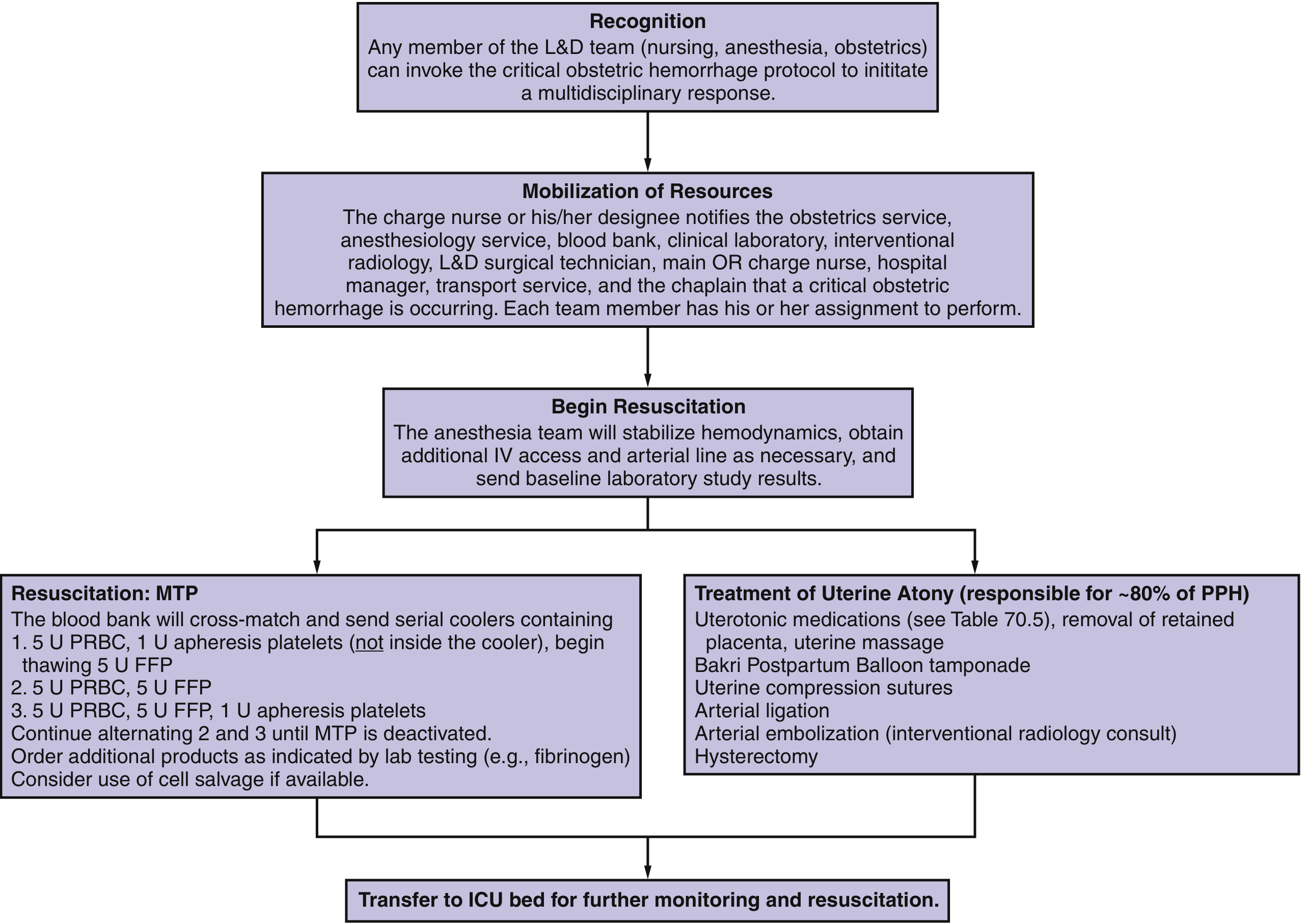
- Cooler 1: 5 U PRBCs + 1 U apheresis platelets (begin thawing 5 U FFP)
- Cooler 2: 5 U PRBCs + 5 U FFP
- Cooler 3: 5 U PRBCs + 5 U FFP + 1 U apheresis platelets
- Continue alternating coolers 2 and 3 until MTP deactivated
- Order additional products (fibrinogen, cryoprecipitate) as guided by labs
Phase 3: Fibrinogen - The Critical Obstetric-Specific Target
- Low fibrinogen is an early predictor of severe PPH and correlates with hemorrhage volume
- Obstetric hemorrhage causes rapid consumptive and dilutional coagulopathy - fibrinogen falls before PT/PTT become abnormal
- A normal fibrinogen at delivery is 4-6 g/L (much higher than non-pregnant normal of ~2-4 g/L) - so a "normal" fibrinogen level of 2 g/L in a postpartum woman indicates significant depletion
Fibrinogen Replacement Options
| Product | Fibrinogen Content | Dose | Notes |
|---|---|---|---|
| Cryoprecipitate | ~250 mg/unit | 10 units (pool) | Standard product; widely available; preferred over FFP for targeted fibrinogen replacement |
| Fibrinogen concentrate | 1 g/vial | 2-4 g IV | Pathogen-reduced; faster to prepare; increasing use |
| FFP | ~2-3 mg/mL | Large volumes needed | Less efficient for fibrinogen; risk of TACO/TRALI |
-
Obstetric hemorrhage at delivery: consider if fibrinogen <150-200 mg/dL (1.5-2 g/L)
-
Acute major bleeding: target fibrinogen ≥150 mg/dL
-
DIC: replace if fibrinogen <150 mg/dL
-
Barash's Clinical Anesthesia, p. 3519; Barash Table 17-8
Phase 4: Point-of-Care Viscoelastic Testing - TEG and ROTEM
| Test | Full Name | What It Measures |
|---|---|---|
| TEG | Thromboelastography | Clot formation time, clot strength, fibrinolysis |
| ROTEM | Rotational Thromboelastometry | Same domains; different reagent names |
- Diagnoses the specific coagulation defect (thrombocytopenia vs. low fibrinogen vs. factor deficiency vs. fibrinolysis)
- Guides targeted component therapy
- Reduces unnecessary blood product use
- Monitors fibrinolysis (helps guide TXA use)
- Can predict severe PPH early
The REFILL RCT (2023, PMID 37535070) found no clinically relevant difference between restrictive vs. liberal fluid strategies on ROTEM parameters in PPH <1,500 mL - suggesting fluid strategy may matter less than blood product strategy in moderate PPH, but viscoelastic testing remains valuable for guiding component therapy.
- Miller's Anesthesia, p. 8907; Creasy & Resnik's MFM, p. 1735; Barash's Clinical Anesthesia, p. 3519
Tranexamic Acid (TXA) - A Critical Resuscitation Adjunct
| Parameter | Details |
|---|---|
| Mechanism | Lysine analogue; binds plasminogen/plasmin → inhibits plasmin-mediated fibrin degradation → stabilizes clots |
| Dose | 1 g IV over 10 minutes; repeat 1 g if bleeding continues after 30 min or restarts within 24h |
| Timing | Must be given within 3 hours of birth - benefit is time-critical; no benefit if given >3 hours |
| Evidence | WOMAN Trial (20,060 women, RR for death from bleeding 0.69 if given within 3h, p=0.008) |
| Safety | No increase in thromboembolism. Give after cord clamping (crosses placenta into breastmilk) |
| Prevention | WHO 2025 does NOT recommend TXA for PPH prevention (prophylaxis) |
- Miller's Anesthesia, p. 8908; Barash's Clinical Anesthesia, p. 3519
Vasopressors and Hemodynamic Support
| Agent | Role |
|---|---|
| Norepinephrine | First-line vasopressor for vasodilatory/hemorrhagic shock |
| Phenylephrine | Alpha-agonist; raises SVR; useful adjunct |
| Epinephrine | For cardiac arrest or severe refractory shock |
| Vasopressin | Second-line adjunct for refractory shock |
Cell Salvage (Autotransfusion)
-
Historically avoided in obstetrics due to concern about amniotic fluid contamination
-
Now considered safe when used with a leukocyte reduction filter, which removes:
- Tissue factor
- Alpha-fetoprotein
- Fetal squamous cells
- Bacteria and other contaminants
-
Particularly valuable for patients who refuse allogenic blood products (e.g., Jehovah's Witnesses) or in massive hemorrhage
-
Cost-effective in cases of massive obstetric hemorrhage
-
In Rh-negative patients: give anti-D immunoglobulin after cell salvage with Kleihauer-Betke testing
-
Barash's Clinical Anesthesia, p. 3519; Miller's Anesthesia, pp. 8908-8909
REBOA (Resuscitative Endovascular Balloon Occlusion of the Aorta)
-
A balloon catheter is inserted via the femoral artery and advanced into the aorta
-
Inflation provides internal aortic occlusion to temporize hemorrhage while surgical control is achieved
-
Systematic review of 336 cases in abnormal placentation found it is feasible, safe, and effective for hemorrhage control in cesarean delivery
-
Placed by interventional radiologists or trauma surgeons
-
Creasy & Resnik's MFM, p. 1735
Monitoring Targets During Active Resuscitation
| Parameter | Target |
|---|---|
| Hemoglobin | >7 g/dL (>70 g/L) in stable patients; >8 g/dL if symptomatic or cardiac disease |
| Platelet count | >50,000/μL (>50 × 10⁹/L) in active bleeding |
| INR / PT | <1.8 |
| Fibrinogen | >1.5-2 g/L |
| Calcium (ionized) | Normal range - replace if low (massive transfusion depletes Ca²⁺ from citrate in blood products) |
| Temperature | ≥36°C (active warming: warm IV fluids, forced-air warming blanket) |
| pH / Lactate | pH >7.35; lactate <2 mmol/L (guides adequacy of perfusion) |
| Urine output | ≥0.5 mL/kg/hr |
Lab Frequency During Active MTP
- Hourly: CBC, INR, fibrinogen, calcium, lactate
- Continuous: arterial BP, SpO₂, heart rate
Quantitative Blood Loss (QBL) - Objective Assessment
- Weigh all sponges and drapes (1 mL blood = 1 g weight)
- Calibrated drapes/collection bags under the patient
- Visual aids for estimation training
- Creasy & Resnik's MFM, p. 6250
Summary: Resuscitation Framework
| Blood Loss (approx.) | Clinical State | Fluid Strategy |
|---|---|---|
| <500 mL | Normal / mild loss | Uterotonics; observe; no aggressive resuscitation usually needed |
| 500-1,000 mL | Alert, BP maintained | IV crystalloid 1-2 L; crossmatch; close monitoring; uterotonics + TXA |
| 1,000-1,500 mL | Mild tachycardia | PRBCs + FFP; activate MTP; TXA; serial labs; call for help |
| >1,500-2,000 mL | Hemodynamic instability | Full MTP (1:1:1 PRBC:FFP:Plt); cryoprecipitate/fibrinogen; TXA; vasopressors; consider ICU |
| >2,000 mL / ongoing | Shock | Damage control resuscitation; surgical/IR control; consider cell salvage, REBOA; ICU transfer |
hypertensive disorder in pregnancy in very detail
"hypertensive disorders of pregnancy" AND management
ACOG ISSHP hypertensive disorders pregnancy guidelines 2024 2025 preeclampsia classification management
1. Classification (ACOG / ISSHP)
| Disorder | Timing | Definition |
|---|---|---|
| Chronic hypertension | Before pregnancy or <20 weeks gestation | Preexisting or diagnosed before 20 weeks; persists >12 weeks postpartum |
| Gestational hypertension | ≥20 weeks, no proteinuria or organ damage | New HTN ≥140/90 mmHg; resolves by 12 weeks postpartum |
| Preeclampsia (de novo) | ≥20 weeks | New HTN + proteinuria OR end-organ damage |
| Preeclampsia superimposed on chronic HTN | Any gestational age | Worsening HTN + new proteinuria or organ damage in woman with chronic HTN |
| Eclampsia | Any time in pregnancy/postpartum | New-onset seizure in woman with preeclampsia |
| HELLP Syndrome | Usually 27-37 weeks | Hemolysis + elevated liver enzymes + low platelets; variant of preeclampsia |
| White-coat hypertension | Any | BP elevated in clinic, normal on HBPM |
| Masked hypertension | Any | BP normal in clinic, elevated on HBPM |
ISSHP recommends: preeclampsia should NOT be classified as 'mild' or 'severe' in an ongoing pregnancy. Instead, it is classified as "with" or "without" severe features.
- Creasy & Resnik's Maternal-Fetal Medicine; ISSHP 2021 Guidelines
2. Diagnosis of Hypertension in Pregnancy
Diagnostic Criteria for Preeclampsia
| Category | Criterion |
|---|---|
| Proteinuria | ≥300 mg/24-hour urine collection; protein:creatinine ratio ≥0.3; dipstick 2+ (only if quantitative testing unavailable) |
| Renal | Serum creatinine ≥1.1 mg/dL OR doubling of serum creatinine in absence of other renal disease |
| Hepatic | Elevated transaminases (AST/ALT) ≥2× upper limit of normal; severe right upper quadrant or epigastric pain |
| Hematologic | Platelet count <100,000/μL |
| Neurological | New-onset headache unresponsive to medication; visual disturbances |
| Pulmonary | Pulmonary edema |
| Uteroplacental | Fetal growth restriction (FGR) / abnormal umbilical artery Doppler |
Important: Proteinuria is NOT required for diagnosis of preeclampsia if end-organ involvement is present.
Severe Features of Preeclampsia (Any One):
-
Systolic BP ≥160 mmHg or diastolic ≥110 mmHg on two readings 15 min apart
-
Thrombocytopenia (platelets <100,000/μL)
-
Renal insufficiency (serum creatinine >1.1 mg/dL or doubling of baseline)
-
Impaired liver function (transaminases ≥2× normal, or severe RUQ/epigastric pain)
-
Pulmonary edema
-
New-onset headache unresponsive to medication, not accounted for by alternative diagnosis
-
Visual disturbances
-
Creasy & Resnik's MFM, pp. 1057-1070; ACOG Practice Bulletin 222
3. Epidemiology and Risk Factors
Risk Factors for Preeclampsia
| High Risk (≥2 moderate factors = high risk) | Moderate Risk |
|---|---|
| Prior preeclampsia | Nulliparity |
| Multifetal gestation | Obesity (BMI >30) |
| Chronic hypertension | Family history of preeclampsia |
| Type 1 or 2 diabetes mellitus | Low socioeconomic status |
| Renal disease | Age <18 or >35 years |
| Antiphospholipid syndrome | Black race (associated with more severe forms) |
| Autoimmune disease (SLE) | Interbirth interval >10 years |
| - | IVF conception |
-
White's Class B DM: 11-16% risk
-
Class C: 21-23% risk
-
Class D: 35-40% risk
-
Classes F and R: up to 70% risk
-
Creasy & Resnik's MFM, pp. 1057-1058
4. Pathophysiology
The Central Mechanism: Failed Spiral Artery Remodeling
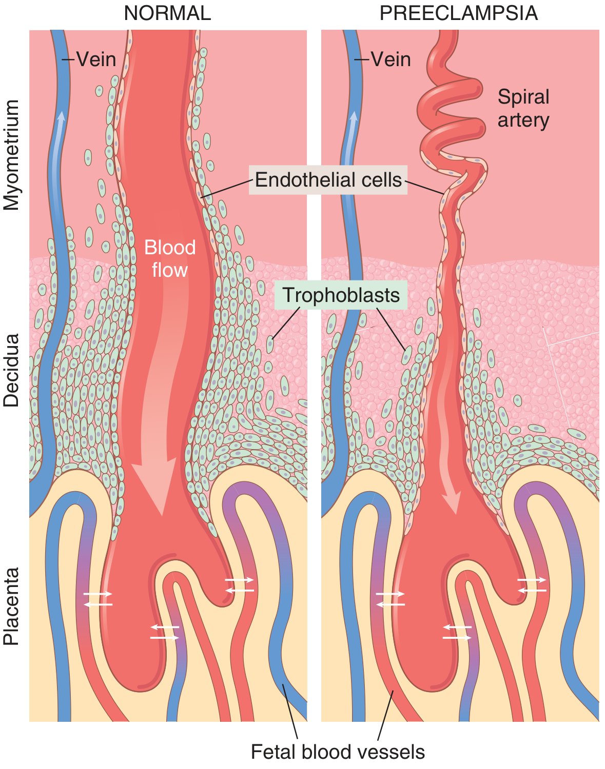
- Trophoblasts invade maternal spiral arteries deep into the myometrium
- Replace musculoelastic walls with fibrinoid material
- Transform into wide, low-resistance, high-flow vessels (6× wider)
- Placenta receives adequate perfusion
- Shallow trophoblast invasion - failure to remodel spiral arteries beyond the decidua
- Spiral arteries remain narrow, thick-walled, and high-resistance
- Placental ischemia and hypoxia result
- Ischemic placenta releases vasoactive factors into maternal circulation
Molecular Mechanisms
| Factor | Source | Effect |
|---|---|---|
| sFlt-1 (soluble fms-like tyrosine kinase-1) | Ischemic placenta - INCREASED | Binds and sequesters VEGF and PlGF → prevents them from reaching endothelial receptors |
| Soluble Endoglin (sEng) | Placenta - INCREASED | Antagonizes TGF-β → impairs endothelial nitric oxide production |
| PlGF (Placental Growth Factor) | Placenta - DECREASED | Normally maintains endothelial health in kidney and other organs |
| VEGF (Vascular Endothelial Growth Factor) | Various - DECREASED (sequestered by sFlt-1) | Normally maintains endothelial integrity |
- Decreased prostacyclin (vasodilator) production by endothelium
- Increased thromboxane A2 (vasoconstrictor)
- Reduced nitric oxide production → loss of vasodilation
- Increased sensitivity to angiotensin II (pressor)
- Widespread endothelial dysfunction
-
Preeclamptic women are hypersensitive to ALL endogenous pressors (vasopressin, epinephrine, norepinephrine, angiotensin II)
-
This can be detected as early as 14 weeks - weeks before clinical disease
-
Normal pregnant women require ~2.5× more angiotensin II to raise BP vs. non-pregnant; preeclamptic women are MORE sensitive than even non-pregnant women
-
Creasy & Resnik's MFM, pp. 494-503; Guyton & Hall Physiology, p. 1041
5. Systemic Pathologic Effects
Cardiovascular
- Generalized vasospasm → increased systemic vascular resistance
- Arterial tone elevated → hypertension
- Stroke: cerebral autoregulation is overwhelmed; risk highest when systolic BP >155 mmHg
Renal - "Glomerular Endotheliosis"
- Pathognomonic lesion: swelling of glomerular endothelial cells + fibrin deposition → capillary lumen constriction
- Decreased GFR and renal blood flow (opposite of normal pregnancy increase)
- Elevated serum uric acid (decreased excretion + oxidative stress)
- Oliguria in severe disease
- Proteinuria (up to 10-15 g/24h in severe cases)
Hepatic
- Periportal hemorrhagic necrosis
- Elevated AST/ALT (poor prognostic sign)
- Subcapsular hematoma → risk of hepatic rupture (rare but catastrophic)
- Hepatic enlargement + epigastric/RUQ pain = impending rupture
Neurological
- Cerebral vasospasm + edema (PRES - Posterior Reversible Encephalopathy Syndrome)
- Severe headache (frontal, unresponsive to analgesics)
- Visual disturbances (scotomata, blurred vision, cortical blindness)
- Hyperreflexia and clonus
- Eclampsia (seizures) - cerebral autoregulation overwhelmed
- Brain MRI: bilateral occipital white matter hyperintensities on T2 (PRES pattern)
Hematologic
- Consumptive thrombocytopenia (100,000-150,000/μL typical)
- Subtle DIC in many; overt DIC in <10% but present in 20% of severe cases
- Fibrin degradation products elevated; fibrinogen usually normal unless placental abruption
- Prolonged PT/PTT indicates procoagulant factor consumption
Pulmonary
- Pulmonary edema in ~2% of severe preeclampsia
- Causes: magnesium infusion-related fluid retention, circulatory overload, cardiogenic, decreased colloid oncotic pressure, pulmonary vascular leakage
- V/Q mismatch possible; PaO₂ usually maintained unless severe
- Airway edema: tongue, epiglottis, pharynx can be edematous - difficult intubation risk
Vascular Volume
- Despite edema, intravascular volume is contracted by 30-40% in severe preeclampsia
- Paradox: edematous woman who is intravascularly depleted
- Fluid shifts from intravascular → extravascular compartment
- Hemoconcentration (elevated Hct for preeclampsia)
Fetal/Placental
-
Reduced intervillous blood flow → placental insufficiency
-
Chronic fetal hypoxia and malnutrition
-
Fetal growth restriction (FGR)
-
Increased risk of preterm birth, stillbirth, abruption
-
Perinatal mortality substantially higher than in normal pregnancies
-
Barash's Clinical Anesthesia, pp. 3511-3512; Creasy & Resnik's MFM, pp. 220-245
6. Clinical Presentation
Symptoms of Preeclampsia
| Symptom | % With Symptom Preceding Eclampsia |
|---|---|
| Headache | 83% |
| Hyperreflexia | 80% |
| Proteinuria | 80% |
| Edema | 60% |
| Clonus | 46% |
| Visual signs | 45% |
| Epigastric pain | 20% |
Warning: 17% of eclamptic women had NO headache; 24% had NO proteinuria before seizure; 20% had NORMAL deep tendon reflexes. Eclampsia can occur without classic premonitory signs.
- Creasy & Resnik's MFM, Table 45.3, p. 1070
7. Investigation
| Test | Rationale |
|---|---|
| BP measurement (standardized) | Sitting, feet flat, correct cuff size, two readings |
| Urine protein | 24-hour urine (gold standard) or spot protein:creatinine ratio ≥0.3 |
| CBC | Thrombocytopenia; hemoconcentration (elevated Hct in severe disease) |
| LFTs (AST, ALT, LDH) | Hepatic involvement; elevated = poor prognosis |
| Serum creatinine | Renal impairment |
| Serum uric acid | Elevated; correlates with severity |
| Coagulation panel | If <100,000 platelets or DIC suspected |
| BNP/NT-proBNP | If cardiac dysfunction suspected |
| Blood smear / LDH / haptoglobin | Hemolysis (HELLP) |
| Fetal monitoring | NST, biophysical profile, umbilical artery Doppler |
| Fetal growth ultrasound | FGR assessment every 3-4 weeks |
| MRI brain | PRES in eclampsia (bilateral occipital T2 hyperintensities) |
| sFlt-1 / PlGF ratio | Biomarker for predicting near-term preeclampsia (investigational in US, available in some countries) |
Monitoring Frequency (Inpatient)
- Vital signs (BP, urine output, symptoms): at least every 8 hours
- Labs: daily when making diagnosis; decrease to 1-2x/week when stable; repeat with any clinical change
- Fetal well-being: daily NST
- Fetal growth: every 3 weeks
8. Prediction and Prevention
Screening (First Trimester)
- Uterine artery Doppler (pulsatility index)
- Mean arterial pressure (MAP)
- Placental Growth Factor (PlGF) - serum biomarker (decreased in preeclampsia weeks before clinical diagnosis)
- PAPP-A (pregnancy-associated plasma protein A)
- Maternal history/risk factors
Prevention
| Intervention | Recommendation | Evidence |
|---|---|---|
| Low-dose aspirin (81 mg/day) | High-risk women start at 12-16 weeks (ideally before 16 weeks), continue until delivery | ACOG strongly recommends; reduces preeclampsia by ~15-20% (ASPRE trial) |
| Calcium supplementation (1.5-2 g/day) | Women with low dietary calcium intake | WHO 2025 Cochrane review: reduces preeclampsia risk in low-calcium populations (PMID 41330480) |
| Weight management | Pre-pregnancy BMI optimization | Risk reduction |
| Antioxidant vitamins (C and E) | NOT recommended | Large RCTs showed no benefit; potential fetal risk |
| Pravastatin | Under investigation | Promising pilot data; not standard of care yet |
- Prior preeclampsia (especially early or severe)
- Multifetal gestation
- Chronic hypertension
- Type 1 or 2 diabetes
- Renal disease
- Autoimmune conditions (SLE, APS)
9. Management
Overview by Gestational Age and Severity
| Condition | Gestational Age | Management |
|---|---|---|
| Gestational HTN / Preeclampsia without severe features | ≥37 weeks | Delivery |
| Gestational HTN / Preeclampsia without severe features | 34-37 weeks | Delivery recommended |
| Gestational HTN / Preeclampsia without severe features | <34 weeks | Expectant management with close monitoring |
| Preeclampsia with severe features | ≥34 weeks | Delivery |
| Preeclampsia with severe features | 24-34 weeks | Expectant management after steroid course IF maternal/fetal status stable |
| Preeclampsia with severe features | <24 weeks (previable) | Delivery vs. expectant - individual counseling |
| Any preeclampsia with specific indications | Any age | Immediate delivery regardless of GA |
- Eclampsia
- Pulmonary edema
- DIC
- Uncontrollable severe-range hypertension
- Non-reassuring fetal status
- Placental abruption
- Reversed end-diastolic flow in umbilical artery (relative)
- Stillbirth
Antepartum Corticosteroids
- Administer betamethasone for fetal lung maturity if <34 weeks and delivery anticipated
- Monitor BP and blood glucose closely (steroids can worsen both)
- Do NOT delay delivery to complete a steroid course if immediate delivery is indicated
- Can administer during 24-hour monitoring window before delivery if appropriate
10. Antihypertensive Therapy
When to Treat
| BP Level | Action |
|---|---|
| <140/90 mmHg | No treatment in most cases (risk of uteroplacental hypoperfusion) |
| 140-159/90-109 mmHg | Oral antihypertensives for chronic treatment; threshold for starting varies by guideline |
| ≥160/110 mmHg (severe range) | Treat within 30-60 minutes to reduce risk of stroke/maternal morbidity |
Acute Severe Hypertension - First-Line Agents
| Drug | Dose | Notes |
|---|---|---|
| IV Labetalol | 20 mg IV bolus; repeat 20-80 mg q10-30 min to max 300 mg; OR infusion | Combined α+β blocker; preserves placental flow; well-tolerated. ACOG-endorsed |
| IV Hydralazine | 5 mg IV bolus; repeat 5-10 mg q20-40 min to max 20 mg | Direct vasodilator; increases uteroplacental blood flow; reflex tachycardia |
| Oral Nifedipine (immediate release) | 10-20 mg PO q20-30 min | Calcium channel blocker; effective and practical; monitor for hypotension |
| IV Nicardipine | 5-15 mg/hr infusion | Dihydropyridine CCB; more predictable than hydralazine |
| IV Clevidipine | 1-6 mg/hr | Ultra-short-acting CCB; rapidly titratable; may produce more profound decrease than labetalol |
Sodium nitroprusside: can be used in extreme refractory cases but risk of fetal cyanide toxicity limits use to very short term. ACE inhibitors, ARBs: absolutely contraindicated in pregnancy (fetotoxic).
- Rosen's Emergency Medicine, p. 2670; Barash's Clinical Anesthesia; ACOG/AAFP 2024
Chronic/Maintenance Antihypertensives in Pregnancy
| Drug | Class | Notes |
|---|---|---|
| Labetalol | Alpha-beta blocker | First-line; oral 100-400 mg q8-12h |
| Nifedipine XL | CCB | 30-120 mg/day; effective and well-studied |
| Methyldopa | Central alpha-2 agonist | Long safety record; sedating; less preferred |
| Hydralazine | Vasodilator | Second-line; used orally |
| Metoprolol/Atenolol | Beta blockers | Atenolol associated with FGR - prefer labetalol |
| ACE inhibitors / ARBs / renin inhibitors | - | ABSOLUTELY CONTRAINDICATED |
| Thiazide diuretics | - | Generally avoided; may reduce intravascular volume |
11. Magnesium Sulfate - Seizure Prophylaxis and Treatment
Dosing Protocol
| Phase | Dose | Route |
|---|---|---|
| Loading dose | 4-6 g over 15-20 minutes | IV |
| Maintenance | 1-2 g/hour continuous infusion | IV |
| Alternative (no IV access) | 5 g IM each buttock (total 10 g) | IM (Pritchard regimen) |
| If seizure occurs on MgSO₄ | Consider second 2-4 g bolus before adding second-line agent | IV |
Serum Magnesium Levels and Effects
| Serum Mg Level (mEq/L) | Effect |
|---|---|
| 4-7 | Therapeutic anticonvulsant range |
| 5-8 | Loss of deep tendon reflexes (DTRs) |
| 9-12 | Respiratory paralysis |
| >15 | Cardiac arrest |
Monitoring During MgSO₄
- Deep tendon reflexes - check before each dose; stop if absent
- Respiratory rate - must be ≥12/min; stop if <12
- Urine output - must be ≥25 mL/hr (Mg is renally cleared; accumulates in oliguria)
- Serum Mg level can be checked in oliguria/renal impairment
Mechanism of Action
- Decreases central nervous system irritability (anticonvulsant)
- Protects blood-brain barrier → limits cerebral edema formation
- Relaxes smooth muscle (partly through calcium antagonism)
- Reduces peripheral and cerebral vascular resistance
- Does NOT significantly lower BP - additional antihypertensives are needed
Antidote for Magnesium Toxicity
Duration of MgSO₄ Postpartum
-
Continue for 24 hours after delivery (most postpartum eclampsia occurs within 24 hours)
-
May discontinue earlier if diuresis occurs (indicates resolution of preeclamptic process)
-
Approximately one-third of all eclamptic seizures occur postpartum
-
Creasy & Resnik's MFM, pp. 647-668; Barash's Clinical Anesthesia, p. 3514
12. Eclampsia - Management
- Cerebral autoregulation overwhelmed at seemingly "low" pressures in young, previously healthy women
- Threshold for treatment lower than in other hypertensive emergencies (systolic >160 mmHg requires urgent treatment)
- Lateral decubitus position - prevent aspiration
- Secure airway, O₂ - maintain SpO₂ ≥95%
- Prevent injury - padded side rails
- MgSO₄ 4-6 g IV loading dose (if not already running); if seizure recurs, give second bolus of 2-4 g before adding alternative anticonvulsant
- Control BP if ≥160/110 with IV labetalol, hydralazine, or nifedipine
- Delivery - the definitive treatment after maternal stabilization
- Exclude other causes: hypoglycemia, drug overdose, CNS lesions (stroke, tumor)
- Rosen's EM, p. 2668-2670
13. HELLP Syndrome
Diagnostic Criteria
| Component | Laboratory Criterion |
|---|---|
| Hemolysis (microangiopathic) | Schistocytes on blood smear; elevated LDH (>600 U/L); elevated bilirubin; low haptoglobin |
| Elevated liver enzymes | AST >70 U/L (≥2× upper limit of normal); elevated ALT |
| Low platelets | <100,000/μL |
- Class 1: Platelets ≤50,000/μL (severe)
- Class 2: Platelets 50,000-100,000/μL (moderate)
- Class 3: Platelets 100,000-150,000/μL (mild)
Presentation
- Most present at 27-37 weeks gestation
- 11% may present before 27 weeks
- Can present postpartum (delayed presentation - important not to miss)
- Common presenting symptoms: malaise, right upper quadrant/epigastric pain, nausea/vomiting, often initially misdiagnosed as viral syndrome or gastroenteritis
Differential Diagnosis
| Feature | HELLP | TTP/HUS | AFLP |
|---|---|---|---|
| Hemolytic anemia | ++ | +++ | ± |
| Thrombocytopenia | ++ | +++ | ± |
| Coagulopathy | ± | - | + |
| Renal failure | + | +++ | ++ |
| Elevated AST | ++ | ± | +++ |
| Elevated ammonia | Normal | Normal | High |
| Hypertension | +++ | ± | ± |
| Effect of delivery | Recovery | None | Recovery |
| Management | Supportive + delivery | Plasma exchange | Supportive + delivery |
Management of HELLP
- Delivery - the definitive treatment; timing based on gestational age and disease severity
- Stabilize with MgSO₄ and antihypertensives
- Correct coagulopathy before delivery (platelets, FFP if needed)
- Corticosteroids (dexamethasone): historically used to increase platelet count; a randomized controlled trial showed no benefit from high-dose dexamethasone treatment
- Fetal lung maturity steroids if <34 weeks
- Consider plasmapheresis if HELLP persists postpartum >72 hours or if TTP/HUS cannot be excluded
- Brenner & Rector's The Kidney, pp. 2028-2040; Harrison's 22E; Sleisenger & Fordtran
14. Organ-Specific Complications and Their Management
Cerebral (PRES)
- Posterior Reversible Encephalopathy Syndrome: bilateral vasogenic edema in occipital/parietal lobes
- MRI shows T2 hyperintensities; enhancement indicates blood-brain barrier disruption
- Treatment: BP control, MgSO₄, delivery
Pulmonary Edema
- Occurs in only ~2% but carries high mortality
- Management: oxygen, diuretics (furosemide carefully), fluid restriction, possible mechanical ventilation
- Monitor carefully in postpartum diuresis phase when fluid remobilizes
Oliguria
- Often iatrogenic (excessive fluid restriction) or renal vasospasm
- Treat with cautious fluid challenge (500 mL bolus); monitor for pulmonary edema
- NOT a routine indication for aggressive IV fluids
Hepatic Rupture (Rare - <2%)
- Epigastric pain + hepatic enlargement = warning sign
- Management: emergent surgery; hepatic artery ligation, hepatic packing, or liver transplant in extreme cases
DIC
- Definitive therapy: delivery
- DIC alone is not an absolute indication for emergency delivery
- Acute coagulopathy: replace procoagulants before delivery (FFP, cryoprecipitate, platelets)
- Do NOT administer procoagulants to treat DIC without delivery - remove the inciting factor
15. Postpartum Management
- ~1/3 of all eclamptic seizures occur postpartum (most within 24 hours, almost all within 48 hours)
- Continue MgSO₄ for 24 hours postpartum (or until diuresis occurs)
- Continue BP monitoring and antihypertensives as needed
- Postpartum diuresis = resolution phase; can develop pulmonary edema paradoxically as fluid mobilizes
- Persistent HTN >12 weeks postpartum = reclassify as chronic hypertension
- Systolic ≥150 or diastolic ≥100 persistently postpartum: treat with oral antihypertensives
- Labetalol, nifedipine, enalapril (now safe - fetal concern no longer applies postpartum; minimal in breastmilk)
- A 2026 systematic review (PMID 41421750) supports active postpartum BP management to prevent eclampsia and readmission
16. Chronic Hypertension in Pregnancy
Diagnosis
- BP ≥140/90 before 20 weeks, or documented before pregnancy, or persisting >12 weeks postpartum
Baseline Workup (to detect superimposed preeclampsia later)
- Serum creatinine, BUN, CBC, 24-h urine protein or spot protein:creatinine
- Electrocardiogram (for cardiac hypertrophy)
- Ophthalmologic exam (for hypertensive retinopathy)
- Consider renal ultrasound to exclude secondary causes
Management
- CHDP Trial (NEJM 2022): Treating chronic HTN in pregnancy to target <140/90 mmHg significantly reduced severe adverse outcomes (stroke, preeclampsia with severe features) without affecting fetal growth
- Preferred agents: labetalol, nifedipine, methyldopa
- Aspirin 81 mg/day from early pregnancy to reduce superimposed preeclampsia risk (25% of women with chronic HTN develop superimposed preeclampsia)
- Monitor for superimposed preeclampsia: any new proteinuria, organ dysfunction, or sudden worsening of HTN
17. Anesthetic Considerations
- Preferred approach in preeclamptic women
- In volume-repleted patients: neuraxial analgesia does NOT cause unacceptable BP reduction and may improve placental perfusion by up to 75%
- Thrombocytopenia: check platelets before neuraxial; generally safe at >80,000-100,000/μL; below 70,000 is usually considered a contraindication
- Airway edema - tongue, epiglottis, pharynx; difficult laryngoscopy and intubation
- Hypertensive response to laryngoscopy - risk of cerebral hemorrhage; pretreat with antihypertensive
- Pulmonary edema with intubation/extubation response
- Avoid ketamine and ergot alkaloids in uncontrolled hypertension
- Magnesium potentiates neuromuscular blockers - use neuromuscular monitoring; reduce NMBD doses
- Barash's Clinical Anesthesia, pp. 3513-3514
18. Long-Term Implications
| Condition | Relative Risk |
|---|---|
| Chronic hypertension | 3.7× |
| Ischemic heart disease | 2.2× |
| Stroke | 1.8× |
| Venous thromboembolism | 1.8× |
| Diabetes mellitus | 2.0× |
| Renal failure | 5× |
Summary of Key Points
| Topic | Key Clinical Pearl |
|---|---|
| Pathophysiology | Shallow trophoblast invasion → placental ischemia → sFlt-1 and sEng release → endothelial dysfunction |
| Diagnosis | Proteinuria NOT required; end-organ damage is sufficient; severe features defined by BP ≥160/110 or organ damage |
| Prevention | Low-dose aspirin (81 mg/day) started before 16 weeks in high-risk women |
| Seizure prophylaxis | MgSO₄ 4-6 g IV load + 1-2 g/hr infusion; antidote is calcium gluconate 1 g IV |
| Severe HTN | Treat within 30-60 minutes with IV labetalol, IV hydralazine, or oral nifedipine |
| Delivery timing | ≥37 weeks (without severe features); ≥34 weeks (with severe features) |
| HELLP | Hemolysis + elevated LFTs + platelets <100,000; deliver; steroids show no benefit |
| Postpartum | 1/3 of seizures occur postpartum; continue MgSO₄ 24h; monitor BP |
| Contraindications | ACE inhibitors/ARBs absolutely contraindicated in pregnancy |
1. What the various deformities are present in a clubfoot at different zones of foot ? Just mention two clinical scoring system for the assessment of Clubfoot. How is a case of primary clubfoot managed in two years child presenting for the first time in OPD ? (2+2+2) 2. Write the Pathoanatomy of Clubfoot. What is the significance of 'Master knot of Henry'? Classify Clubfoot in general. (2+2+2) 3. There is a call from labour room for suspected case of right clubfoot. You are an attending doctor and after evaluating the case, you feel it is the case of Rt clubfoot. (10 = 2+2+2+4) Would you like to take any OBSTETRIC HISTORY, is there any relevance? (2) What other GENERAL / SYSTEMIC examinations relevant to this condition you may want to do? (2) What would be your plan of management for this neonate? (2) You happened to stay out hospital for six months and someone else was following the case. You happened to review the infant now at six months. How would you evaluate the case now (different possibilities) and manage accordingly?
clubfoot talipes equinovarus pathoanatomy zones deformities master knot Henry Ponseti management scoring systems
Clubfoot (Congenital Talipes Equinovarus) - Comprehensive Answers
QUESTION 1
(a) Deformities Present in Clubfoot at Different Zones of the Foot (2 marks)
| Zone | Deformity | Anatomical Description |
|---|---|---|
| Forefoot | Cavus (high arch) + Adductus | Pronation of the first ray; first ray plantar-flexed; forefoot appears supinated but is actually pronated relative to hindfoot. Metatarsals deviate medially at tarsometatarsal joints or shafts are adducted |
| Midfoot | Adductus (medial deviation) | Navicular displaced medially on the talar head; cuboid displaced medially on the calcaneus; deep medial skin crease |
| Hindfoot | Varus + Equinus | Calcaneus rotates medially beneath the talar neck; heel pulled superiorly by contracted Achilles tendon; calcaneal tuberosity moves toward fibular malleolus; deep posterior crease; "empty heel" sign |
| Ankle | Equinus | Fixed plantar flexion; ankle dorsiflexion restricted by contracted Achilles tendon, posterior joint capsule, and posterior ligaments |
| Global | Internal tibial torsion | Medial rotation of the entire limb below knee; often accompanies the foot deformity |
- Talus: talar neck is medially and plantarly deviated; talar body may be externally rotated in the ankle mortise
- Calcaneus: rotates in three planes - horizontally (beneath talus), coronally (varus), and sagittally (equinus); proximity to fibula due to horizontal rotation
- Navicular: extreme medial displacement around talar head (talonavicular subluxation)
- Cuboid: displaced medially on the calcaneus
-
Shortened/contracted muscles: Achilles tendon (gastrosoleus), tibialis posterior, FHL, FDL, intrinsic muscles
-
Contracted ligaments: calcaneonavicular (spring), calcaneofibular, deltoid (tibionavicular), posterior talocalcaneal, talonavicular capsule, Y-ligament, long plantar ligament
-
Muscle hypoplasia: gastrosoleus and tibialis posterior are smaller than normal with increased connective tissue and reduced myofibrils
-
Diminished/absent anterior tibial artery (associated)
-
Deep medial crease and single posterior crease present
-
Campbell's Operative Orthopaedics 15th Ed 2026, p. 1305; Bailey & Love's Short Practice of Surgery 28e
(b) Two Clinical Scoring Systems for Clubfoot Assessment (2 marks)
1. Pirani Scoring System
- Developed by Shafique Pirani
- Total score: 0 to 6 (higher = more severe)
- Comprises two subscores, each out of 3:
| Subscore | Signs Assessed (each 0, 0.5, or 1) |
|---|---|
| Midfoot Contracture Score (MCS) | 1. Curved lateral border (CLB) 2. Medial crease (MC) 3. Lateral head of talus coverage (LHT) |
| Hindfoot Contracture Score (HCS) | 4. Posterior crease (PC) 5. Empty heel (EH) 6. Rigid equinus (DF) |
2. Diméglio Scoring System
- Developed by Alain Diméglio
- Total score: 0 to 20 (higher = more severe; maximum 16 from 4 parameters + 4 additional points)
- Assesses 4 main parameters based on reducibility with gentle manipulation using a goniometer:
| Parameter | Scale | Max Points |
|---|---|---|
| Equinus in sagittal plane | 0 to 4 | 4 |
| Varus deviation in frontal plane | 0 to 4 | 4 |
| Derotation (calcaneoforefoot block around talus) | 0 to 4 | 4 |
| Adduction of forefoot on hindfoot (horizontal plane) | 0 to 4 | 4 |
| Extra points (1 each) | Posterior skin crease, medial skin crease, rigid cavus, poor muscle condition | Up to 4 |
-
Grade I (benign): score 1-5
-
Grade II (moderate): score 6-10
-
Grade III (severe): score 11-15
-
Grade IV (very severe): score 16-20
-
Campbell's Operative Orthopaedics 15th Ed 2026, p. 1306; PLOS One systematic review
(c) Management of Primary Clubfoot in a 2-Year-Old Child Presenting First Time to OPD (2 marks)
- Increased rigidity of deformity
- Denser soft tissues
- Greater bone maturity
- Less malleable cartilage
- Child is walking age, so casting is technically harder
Approach:
- Full clinical examination: confirm CAVE deformity, assess rigidity, check for signs of neglect or previous treatment, full systemic examination to rule out syndromic/neuromuscular causes
- Pirani and Diméglio scoring
- X-rays: weight-bearing AP and lateral views (talocalcaneal angles; Kite's AP angle normally 20-40°, reduced in clubfoot; Turco lateral angle normally 35°, reduced in clubfoot)
- Serial weekly manipulation and long-leg plaster-of-Paris casts
- Correction sequence: Cavus → Adductus → Varus → Equinus (CAVE)
- Longer casting period required compared to newborn (may need 8-10+ casts vs. 5-6 in newborn)
- More casts may be needed before tenotomy
- Required in ~90% of cases to correct hindfoot equinus
- Done under LA/GA; can be done as a day procedure
- Post-tenotomy cast in 15-20° dorsiflexion and 70° abduction for 3 weeks to allow Achilles healing
- Most critical step for preventing relapse
- Foot-abduction brace (Mitchell brace or Denis Browne bar): affected foot at 60-70° external rotation, unaffected foot at 30-40°
- Schedule: 23 hours/day for 3 months, then during sleep (12-14 hours/day) until age 4-5 years
- Non-compliance is the most common cause of relapse (~10-30% relapse rate)
- If dynamic supination deformity persists after casting (due to overpull of tibialis anterior)
- Transfer to third cuneiform (full transfer) after age 3-4 years when bone is ossified
- Resolves residual adductus and supination
- If conservative treatment fails or for rigid resistant deformity
- Posteromedial release (PMR / Cincinnati approach) or Ilizarov external fixator for very resistant/neglected cases
- Surgery is a last resort due to risk of stiffness, scarring, and inferior long-term outcomes compared to Ponseti method
Note: Multiple studies show Ponseti is successful even in children up to walking age and older; initial Pirani score and age influence number of casts required.
- Campbell's Operative Orthopaedics 15th Ed 2026, pp. 1307-1314; Miller's Review of Orthopaedics 9e, p. 238
QUESTION 2
(a) Pathoanatomy of Clubfoot (2 marks)
- The talar neck is medially and plantarly deviated (plantarmedial deviation)
- The talar body may be externally rotated within the ankle mortise
- This creates a fixed abnormal axis around which all other bones are misaligned
- Rotates abnormally in three planes around the interosseous talocalcaneal ligament:
- Horizontal (transverse) plane: rotates medially and slips beneath the talar head, calcaneal tuberosity approaches fibular malleolus
- Coronal (frontal) plane: rotates into varus
- Sagittal plane: equinus position
- Results in the calcaneus appearing to be "locked" beneath and medial to the talus
- Medially displaced around the talar head
- Talonavicular joint is in extreme inversion (subluxation)
- Navicular may contact the medial malleolus in severe cases
- Displaced medially on the calcaneus
- Calcaneocuboid joint rotated internally
- Adducted at tarsometatarsal joints, OR
- Shafts themselves are adducted
| Structure | Change |
|---|---|
| Gastrosoleus / Achilles tendon | Shortened; contracted; causes equinus |
| Tibialis posterior | Shortened; contracts; contributes to varus and adductus |
| FHL, FDL | Shortened; contribute to cavus and equinus |
| Intrinsic muscles | Contracted |
| Posterior tibial tendon sheath | Shortened and thickened; contains contractile myofibroblasts |
| Spring (calcaneonavicular) ligament | Contracted; resists talonavicular joint realignment |
| Deltoid ligament (tibionavicular) | Contracted |
| Calcaneofibular ligament | Resists talocalcaneal joint realignment |
| Y (bifurcated) ligament | Contracted at calcaneocuboid joint |
| Long plantar ligament | Shortened |
| Entire talonavicular capsule | Contracted on medial side |
- Campbell's Operative Orthopaedics 15th Ed 2026, pp. 1305-1306; Bailey & Love 28e, p. 8732
(b) Significance of the 'Master Knot of Henry' (2 marks)
- The tendon of Flexor Hallucis Longus (FHL) crosses superficial to
- The tendon of Flexor Digitorum Longus (FDL)
-
During posteromedial surgical release (McKay/Cincinnati approach): When correcting severe clubfoot surgically, the FHL is often tight after correction. The preferred technique to address FHL tightness is transection of FHL with formal tenodesis to FDL at the Master Knot of Henry. This sacrifices FHL function while preventing bowstringing and maintaining toe flexion through FDL.
-
As a surgical landmark: The medial plantar nerve and artery lie just deep and lateral to the FHL tendon at this level - must be protected during dissection.
-
Entrapment neuropathy: The medial plantar nerve can be entrapped at the knot of Henry between the navicular tuberosity superiorly and the abductor hallucis muscle belly inferiorly, causing heel and arch pain (jogger's foot / medial plantar nerve entrapment syndrome).
-
Relevance to tendon transfers: Any FHL harvest (e.g., for Achilles reconstruction, posterior tibial tendon reconstruction) must respect this anatomical chiasm to prevent complications.
- Campbell's Operative Orthopaedics 15th Ed 2026; Miller's Review of Orthopaedics 9e; Imaging Anatomy Vol. 3
(c) Classification of Clubfoot in General (2 marks)
Etiological Classification (Bailey & Love / General)
| Type | Description |
|---|---|
| Postural (Positional) | Soft, flexible deformity from intrauterine malposition. Passively correctable to neutral. Resolves by 3 months with physiotherapy/stretching. No true structural abnormality. |
| Idiopathic (Structural) | True structural deformity with bony and soft tissue changes. Most common type. Fixed, not passively correctable to neutral. Genetic (PITX1-TBX4 pathway). |
| Neuromuscular | Associated with spina bifida (myelomeningocele), cerebral palsy, arthrogryposis, poliomyelitis. More rigid; higher recurrence after treatment. |
| Syndromic | Associated with chromosomal abnormalities (Trisomy 18), diastrophic dysplasia, amniotic band syndrome (Streeter dysplasia), prune-belly syndrome, tibial hemimelia, VACTERL association. |
| Teratologic (in utero) | Arises from early intrauterine insult; may be associated with major systemic anomalies. |
Severity Classification - Diméglio's Grades
| Grade | Score (out of 20) | Description |
|---|---|---|
| I | 1-5 | Benign (postural) |
| II | 6-10 | Moderate (reducible) |
| III | 11-15 | Severe (partially reducible) |
| IV | 16-20 | Very severe (resistant/teratologic) |
By Response to Treatment
| Category | Description |
|---|---|
| Resolving | Responds to conservative treatment |
| Resistant | Does not respond adequately to conservative treatment; requires surgery |
| Relapsing | Initially corrected but deformity recurs (most common during brace non-compliance period) |
| Neglected | Untreated beyond 1-2 years; Ilizarov or surgery required |
- Campbell's Operative Orthopaedics 15th Ed 2026; Bailey & Love 28e; Miller's Review 9e
QUESTION 3 (10 marks)
Clinical Scenario: Neonatal Right Clubfoot
(a) Obstetric History - Relevance (2 marks)
| History | Relevance |
|---|---|
| Oligohydramnios | Reduced amniotic fluid → decreased fetal movement space → postural deformity; more commonly positional than structural clubfoot in this setting |
| Polyhydramnios | Suggests neuromuscular cause (fetal swallowing difficulty in NMD) associated with neuromuscular clubfoot |
| First-trimester amniocentesis (<11 weeks) | Associated with increased risk of clubfoot (limb positional effect) |
| SSRI use during pregnancy | Some SSRIs are associated with increased risk of structural clubfoot |
| Maternal smoking | Associated with higher frequency of clubfoot |
| Maternal obesity | Risk factor for clubfoot |
| Family history of clubfoot | Strong genetic component (PITX1-TBX4 pathway); sibling recurrence risk ~2-5%; if parent affected, higher risk |
| Multiple gestation | Restricted fetal movement can cause positional clubfoot |
| Antenatal USG findings | Prenatal diagnosis of clubfoot; bilateral involvement (higher risk of chromosomal anomaly/genetic syndrome); associated anomalies (neural tube defects, cardiac defects) |
| Mode of delivery | Breech presentation associated with postural deformities |
| Consanguinity | Risk of autosomal recessive syndromic conditions |
| Maternal drug history | Phenytoin, warfarin, alcohol, misoprostol associated with limb deformities |
(b) General / Systemic Examinations Relevant to Clubfoot (2 marks)
- Birth weight, gestational age, APGAR scores
- Dysmorphic features (low-set ears, micrognathia, cleft palate - suggest trisomies)
- Limb length inequality, limb hyper/hypoplasia
- Skin: hemangiomas, café-au-lait spots, dimples over spine
- Tone (hypotonia in neuromuscular causes)
- Deep tendon reflexes
- Moro, grasp, and other primitive reflexes
- Examine for myelomeningocele / spina bifida (sacral dimple, tuft of hair over spine, lipoma)
- Cranial nerve examination (facial palsy)
- Examine all four limbs (bilateral clubfoot? associated hand anomalies?)
- Arthrogryposis: multiple joint contractures, rigid joints, absent skin creases
- Hip examination: DDH is associated (perform Barlow and Ortolani maneuvers)
- Examine for tibial hemimelia (absent/hypoplastic tibia)
- Amniotic band syndrome (constriction rings on limbs)
- Limb girdle symmetry
- Heart auscultation: congenital heart disease (associated with chromosomal anomalies)
- Femoral pulses bilaterally
- Prune-belly syndrome: absent/deficient abdominal wall musculature + cryptorchidism + urinary tract abnormalities
- Scoliosis, kyphosis
- Posterior midline lesions (meningocele, lipomyelomeningocele)
- Chromosomal karyotype / chromosomal microarray if bilateral or associated anomalies
- Spinal USG or MRI if spinal abnormality suspected
- Hip USG at 6 weeks
(c) Plan of Management for the Neonate (2 marks)
Step 1: Confirm Diagnosis and Type
- Clinical examination: assess CAVE deformity severity
- Pirani and Diméglio scoring at baseline
- Distinguish structural (fixed, cannot passively correct) from postural (passively correctable)
Step 2: Ponseti Method - Treatment Phase
- Serial weekly manipulation and long-leg above-knee plaster casts
- Cast changes every 5-7 days (cartilaginous, pliable tissues in neonate - ideal for correction)
- Sequence of correction: Cavus first → Adductus + Varus (simultaneously) → Equinus last
- 1st cast: correct cavus by supinating forefoot and dorsiflexing first ray
- 2nd-4th casts: correct adduction and varus (apply pressure on lateral talar head as fulcrum)
- Final casts: address equinus
- Typically 5-6 casts required in a neonate
- Pirani score monitored at each visit
Step 3: Percutaneous Achilles Tenotomy
- Required in ~90% of cases for final hindfoot equinus correction
- Done at end of casting when all CAVE components are corrected except equinus
- Performed under local anesthesia as an outpatient
- Post-tenotomy cast applied for 3 weeks in maximum dorsiflexion and 70° abduction
Step 4: Foot Abduction Brace (FAB) - Maintenance Phase
- Most critical step - prevents relapse
- Dennis Browne / Mitchell shoe-bar brace
- Affected foot: 60-70° external rotation; Unaffected foot: 30-40° ER
- Schedule: Full-time (23 hours/day) for 3 months → nighttime/nap only (12-14 hrs/day) until age 4-5 years
- Parental counselling on compliance is essential
Step 5: Follow-Up
- Regular OPD reviews to monitor correction, brace compliance, Pirani scores
- Watch for relapse (commonest during brace-weaning period)
(d) Review at 6 Months - Evaluation and Management of Different Possibilities (4 marks)
Scenario A: Excellent / Full Correction - Brace Compliant
- Pirani score: 0 (all 6 signs normal)
- Full dorsiflexion ≥15°, good heel in neutral/slight valgus, plantigrade foot
- Brace worn correctly and consistently
- No signs of relapse
- Calf muscle: slightly smaller than opposite side (normal in treated unilateral CTEV)
- Continue brace (nighttime/nap-time) as per schedule until age 4-5 years
- Reassure parents; monthly review
- Counsel on relapse warning signs (loss of dorsiflexion, re-appearance of varus/adductus)
Scenario B: Relapse of Deformity (Dynamic or Structural)
- Most common cause: brace non-compliance - the MOST IMPORTANT cause of relapse
- Re-appearance of adductus, varus, equinus
- If walking age: dynamic supination when walking (foot internally rotates during swing phase = tibialis anterior overpull)
- Pirani score increased from baseline
- Assess rigidity of relapsed deformity
- Recasting: Repeat Ponseti casting (usually fewer casts needed than initial treatment)
- Repeat Achilles tenotomy if equinus has relapsed
- Resume brace wear with strict protocol; reinforce compliance counselling
- For dynamic supination deformity (overpull of tibialis anterior after age 3-4 years): Anterior Tibial Tendon Transfer (ATTT) to third cuneiform
Scenario C: Residual Deformity (Incomplete Correction)
- Partial correction achieved but foot not fully plantigrade
- May have isolated cavus, residual varus, or residual equinus remaining
- Pirani score: 1-3
- Continue Ponseti casting to achieve further correction
- Percutaneous tenotomy if equinus persists
- Address specific residual components:
- Residual cavus: plantar fascia release
- Residual varus: ATTT or tibialis posterior tendon lengthening
- Residual equinus: further tenotomy or posterior capsular release
Scenario D: Resistant / Failed Conservative Treatment
- Pirani score remains high despite appropriate casting
- Rigid, non-correcting deformity
- Check for underlying neuromuscular cause (spastic/paralytic clubfoot)
- Consider X-ray and MRI spine
- Posteromedial release surgery (if <2-3 years): Cincinnati approach; release of contracted posterior and medial structures with Achilles lengthening and posterior tibial tendon lengthening
- Ilizarov external fixator: for older neglected/relapsed cases, to achieve gradual correction
Scenario E: Possible Missed Neuromuscular Cause
- Foot softer or more rigid than expected
- Asymmetric muscle weakness, flaccid paralysis (spina bifida, polio)
- Spastic deformity (cerebral palsy)
- Poor brace compliance due to skin breakdown (anesthetic foot in spina bifida)
- Neurosurgical review if tethered cord / myelomeningocele
- Modified Ponseti for spina bifida clubfoot (higher recurrence, more casts needed)
- Long-term brace wear essential; custom orthoses