
Tamsulosin role in ureteric stone?
tamsulosin ureteral stone medical expulsive therapy
PMID: 37903004
Tamsulosin in Ureteric Stone (Medical Expulsive Therapy)
Mechanism of Action
- Smooth muscle relaxation in the ureter wall - reduces basal tone and decreases the frequency and force of ureteral peristalsis
- Ureteral dilatation particularly at the level of the obstructing stone, increasing the lumen diameter around the stone
- Reduced spasm at the site of stone impaction - spasmolytic effect reduces pain and facilitates passage
- Vesicoureteral junction relaxation - the lower ureter (intramural and juxtavesical portions) has the highest density of alpha-1 receptors, making tamsulosin especially effective for distal ureteric stones
Indications and Stone Size
| Guideline | Recommendation |
|---|---|
| AUA | Alpha-blocker MET for ureteric stones < 10 mm, observe 4-6 weeks |
| EAU | Alpha-blocker MET for stones > 5 mm (5-10 mm range) |
- Most effective for distal ureteric stones (where alpha-1 receptor density is highest)
- Best for stones 5-10 mm - smaller stones (≤5 mm) already have high spontaneous passage rates and benefit less
- Less evidence for proximal or mid-ureteric stones
Clinical Benefits
- Higher stone expulsion rate compared to conservative management alone
- Shorter time to stone expulsion
- Reduced analgesic requirement (fewer pain episodes)
- Better tolerance of ureteral stents when placed post-ESWL
- Adjunct to ESWL - tamsulosin improves expulsion of stone fragments post-lithotripsy
Dose
- Tamsulosin 0.4 mg orally once daily (typically taken at bedtime to reduce orthostatic hypotension)
- Duration: up to 4-6 weeks of observation
Comparison with Other Agents
- Tadalafil (PDE-5 inhibitor) showed a higher stone expulsion rate than tamsulosin for distal ureteric stones 5-10 mm (OR 0.55, 95% CI 0.38-0.80)
- No significant difference in time to expulsion or side effects (headache, dizziness, orthostatic hypotension) between the two drugs
- Tamsulosin remains superior to nifedipine (calcium channel blocker)
- Cochrane review (67 studies, 10,509 participants): alpha-blockers increase stone clearance and shorten expulsion time - effect was greater for stones >5 mm than ≤5 mm
Side Effects to Monitor
- Orthostatic hypotension / dizziness (most common)
- Retrograde ejaculation (due to alpha-1 blockade at bladder neck)
- Headache, rhinitis
Contraindications / Cautions
- Infection with obstruction (pyonephrosis, urosepsis) - requires urgent urologic drainage, not MET
- Stones >10 mm generally require ureteroscopy or ESWL rather than MET alone
- Failure to pass after 4 weeks is an indication for intervention (risk of ureteric stricture and renal deterioration increases beyond this point)
Summary
Find latest 2024-2025 clinical guidelines for ureteric stone management
ureteral stone management guidelines
EAU AUA ureteric stone management guidelines 2024 2025 recommendations
PMID: 40268592
PMID: 40583613
https://www.auanet.org/guidelines-and-quality/guidelines/sur…
https://www.auanet.org/documents/Guidelines/PDF/2025%20Guide…
EAU 2025 urolithiasis guidelines ureteral stone ureteroscopy ESWL MET alpha blocker specific recommendations
Latest 2024-2025 Clinical Guidelines: Ureteric Stone Management
- AUA 2025 Guideline: Surgical Management of Kidney and Ureteral Stones (published 2025, literature up to May 2025)
- EAU 2025 Guidelines on Urolithiasis (Skolarikos et al., Eur Urol, 2025 - PMID 40268592)
- EAU 2026 Pocket Guideline (updated version of 2025 recommendations)
1. Diagnosis
| Step | EAU 2025 | AUA 2025 |
|---|---|---|
| First-line imaging | Ultrasound | Low-dose CT (gold standard) |
| Gold standard | Low-dose CT (non-contrast) | Non-contrast CT |
| Biochemistry | Urine culture, serum creatinine, urine pH | Same |
| Stone analysis | Mandatory for all stone formers | Recommended |
2. Pain Management (Acute Colic)
- NSAIDs (e.g., diclofenac, ketorolac) are first-line for analgesia - EAU Strong recommendation
- Opioids reserved as secondary option only
- IV hydration for nausea/vomiting; forced diuresis is NOT recommended
3. Medical Expulsive Therapy (MET) - Updated 2025
AUA 2025 Guideline Statements:
| Statement | Stone location | Size | Recommendation | Evidence |
|---|---|---|---|---|
| #7 | Distal ureter | ≤10 mm | OFFER alpha-blocker MET for ~30 days | Strong; Grade A |
| #8 | Middle + proximal ureter | ≤10 mm | May offer alpha-blocker MET for ~30 days | Conditional; Grade B |
- 49 studies, >8,000 patients with distal ureteric stones ≤10 mm
- Alpha-blockers: RR 0.71 (95% CI 0.66-0.76) = 25 more passage events per 100 persons vs. control
- Significant reduction in pain episodes (MD: -0.59; 95% CI: -0.82 to -0.37)
- No significant benefit found for middle or proximal ureteric stones (RR: 0.84-0.88, CI crosses 1.0)
EAU 2025 Guideline:
- Offer alpha-blockers as MET for (distal) ureteric stones >5 mm - Strong recommendation
- MET most effective for stones 5-10 mm
- Silodosin appears more effective than tamsulosin as MET (new update vs. prior guidelines)
- Note: alpha-blockers remain off-label for this indication in most countries
- Combination therapy (phytotherapy + alpha-blockers) may be more beneficial than alpha-blockers alone
4. Indications for Active Stone Removal
EAU 2025 - Indications for intervention:
- Stones with low likelihood of spontaneous passage
- Persistent pain despite adequate analgesia
- Persistent obstruction
- Renal insufficiency (including solitary kidney)
- Infection with obstruction (emergency)
- Patient preference
5. Surgical Treatment - EAU 2025 Treatment Algorithm
URETERAL STONE (if removal indicated)
│
├── PROXIMAL URETERAL STONE
│   ├── >10 mm → 1st: URS (ante- or retrograde); 2nd: SWL
│   └── <10 mm → SWL or URS (equal options)
│
└── DISTAL URETERAL STONE
    ├── >10 mm → 1st: URS; 2nd: SWL
    └── <10 mm → SWL or URS (equal options)
AUA 2025 Guideline Statements:
| Statement | Setting | Recommendation |
|---|---|---|
| #10 | Distal stones ≤10 mm requiring surgery | May offer URS or SWL (Conditional; Grade B) |
| #11 | Proximal stones <2 cm requiring surgery | May offer URS or SWL (Conditional; Grade B) |
| #12 | SWL failure | May repeat SWL or proceed to URS |
| #13 | >2 cm ureteral stones / failed URS or SWL | Clinicians should offer PCNL or laparoscopic/robotic ureterolithotomy |
| (Special) | Morbid obesity | URS first-line (EAU Strong) |
6. URS vs. SWL - Key Comparison (2025 Guidelines)
| Parameter | URS | SWL |
|---|---|---|
| Stone-free rate (single procedure) | Higher | Lower |
| Complication rate | Higher | Lower |
| Repeat procedures | Less likely | More likely |
| EAU guidance | Informs patients URS = better SFR but higher complications | SWL feasible; repeat sessions within 1 day for ureteric stones |
| AUA guidance | Equal option to SWL for most ureteric stones | Equal option; up to 2 SWL sessions non-inferior to URS for secondary procedures |
7. Ureteroscopy Technical Recommendations (EAU 2025)
- Use Holmium:YAG (Ho:YAG) or Thulium Fiber Laser (TFL) for lithotripsy - Strong
- Stone extraction only under direct endoscopic visualisation - Strong
- Do NOT routinely insert a stent in uncomplicated cases - Strong
- Pre-treatment with alpha-blocker (1 week before URS) reduces need for active dilatation and improves stone-free rate
- Offer MET after laser lithotripsy to facilitate fragment passage - Strong
8. Observation / Conservative Management
- Observation is feasible in informed patients with no complications (EAU Strong)
- For newly diagnosed small stones: initial observation + periodic evaluation - EAU Strong
- AUA: confirm stone passage with follow-up imaging when passage is uncertain
- If stone not passed within 4-6 weeks - reassess for intervention (risk of stricture and renal deterioration)
- Emergency indications overriding observation: obstructed infected system (pyonephrosis/urosepsis) - urgent drainage mandatory
9. Recurrence Prevention
- Stone analysis mandatory for all formers (EAU/AUA)
- High fluid intake (urine volume >2.5 L/day) for all stone types
- Dietary modifications specific to stone composition
- Pharmacological prevention:
- Potassium citrate (calcium oxalate, uric acid, cystine stones)
- Thiazides (hypercalciuric stones)
- Allopurinol (hyperuricosuric stones)
- Theobromine (uric acid - new agent, ESD 2025)
- Genetic testing recommended for high-risk / hereditary stone formers (EAU 2025)
Quick Reference Summary
| Scenario | 2025 Guideline Action |
|---|---|
| Distal stone ≤10 mm, asymptomatic | Observe + alpha-blocker MET x30 days (AUA Strong/Grade A) |
| Distal stone ≤10 mm, needs surgery | URS or SWL (equal) |
| Proximal stone <10 mm | SWL or URS; MET conditional |
| Proximal stone <2 cm, needs surgery | URS or SWL |
| Stone >2 cm / failed URS+SWL | PCNL or laparoscopic ureterolithotomy |
| Obstructed + infected | Emergency drainage (nephrostomy/stent), IV antibiotics |
| Morbid obesity | URS first-line |
| Post-URS, uncomplicated | No routine stent |
| MET drug of choice | Silodosin > tamsulosin (EAU 2025 update) |
Opd management of renal colic and kidney stone.
renal colic outpatient management analgesia alpha blocker
OPD Management of Renal Colic and Kidney Stone
Step 1: Initial Assessment in OPD
History
- Character of pain: Sudden onset severe flank/loin pain radiating to groin/ipsilateral testis or labia - classic ureteric colic
- Associated symptoms: Nausea, vomiting, haematuria, dysuria, urinary frequency (suggests stone at UVJ)
- Fever/chills - red flag: suggests infected obstructed system
- Duration of symptoms (important - long obstruction = irreversible nephron loss)
- Previous stone history, family history, medications
Examination
- Costovertebral angle tenderness
- Abdominal tenderness
- Vital signs - fever, hypotension = emergency, admit immediately
- Rule out differential diagnoses (AAA, appendicitis, ectopic pregnancy)
Step 2: Investigations in OPD
| Investigation | Purpose | Notes |
|---|---|---|
| Urine dipstick / urinalysis | Haematuria (90%), pyuria, nitrites | Pyuria alone does not confirm UTI; leukocytosis from WBC demarginalisation |
| Urine culture | If infection suspected | Mandatory before any procedure |
| Serum creatinine / eGFR | Baseline renal function; obstruction impairs function | |
| Serum electrolytes | Metabolic assessment | |
| FBC | Leukocytosis may be from stress alone, not infection | |
| Serum calcium, uric acid | Metabolic stone work-up |
Imaging
| Modality | Sensitivity | Specificity | When to Use |
|---|---|---|---|
| Ultrasound (USG) | ~100% for hydronephrosis; up to 90% specific | First-line in OPD, pregnant women, children, and to limit radiation | EAU recommends as initial modality |
| Non-contrast CT (NCCT) | 95% | 98% | Gold standard; detects stones as small as 1 mm; reveals complications (hydronephrosis, ureteral edema, other pathology) |
| KUB X-ray | <70% specificity | Only for follow-up of a known radiopaque stone | Not reliable alone |
| IVP | 96% sensitive | Rarely used now; CT/USS have replaced it |
Practical OPD point: Patients with known nephrolithiasis, typical presentation, NO fever, NO infection on UA, NO solitary kidney - can skip CT and manage without imaging. NCCT if diagnosis is uncertain or complications suspected. - Rosen's Emergency Medicine
Step 3: Immediate Pain Management
NSAIDs - First Line
- Greater reduction in pain scores
- Less need for rescue analgesia
- Less nausea and vomiting
- Mechanism: reduce collecting system pressure by decreasing renal blood flow (PGE-mediated); COX-2 inhibitors also prevent downregulation of aquaporin channels
| Drug | Route | Dose |
|---|---|---|
| Diclofenac | IM/oral/suppository | 75 mg IM or 50 mg PO |
| Ketorolac | IM/IV | 15-30 mg IM |
| Ibuprofen | Oral | 400-600 mg TDS |
| Indomethacin | PO/PR | 50-75 mg |
- Renal insufficiency (exacerbates AKI by reducing RBF)
- Risk of GI bleeding (use COX-2 inhibitor or add PPI)
- Pregnancy (avoid in 3rd trimester)
- Cardiovascular disease (COX-2 inhibitors linked to MI/stroke risk)
Opioids - Second Line / Add-on
- Use when NSAIDs are contraindicated or pain is uncontrolled
- Options: tramadol, morphine, pethidine
- Side effects: nausea, vomiting, sedation, abuse potential
- Short prescription (3-5 days) if sent home with opioids
Antiemetics
- Add routinely for nausea/vomiting: metoclopramide, ondansetron, domperidone
- IV/IM route if vomiting prevents oral medication
Step 4: Decide - Admit or Discharge?
Criteria for ADMISSION (do not discharge):
| Red Flag | Action |
|---|---|
| Fever + ureteric obstruction (infected obstructed system) | Emergency - urgent urology referral, IV antibiotics, decompression (nephrostomy/stent) |
| Intractable pain not controlled with analgesia | Admit |
| Persistent vomiting - unable to tolerate oral medications | Admit |
| Solitary functioning kidney with obstruction | Admit - urgent decompression |
| Bilateral ureteric stones | Admit |
| Renal insufficiency / AKI worsening | Admit |
| Immunocompromised patient | Admit |
| Stone >10 mm (unlikely to pass) | Urology referral / plan intervention |
Suitable for OPD discharge if:
- Pain controlled with oral analgesia
- No fever or signs of infection
- Tolerating oral fluids
- Normal or near-normal renal function
- Small stone (≤10 mm), especially distal ureteric
- Informed and reliable patient who can return if needed
Step 5: OPD Medical Expulsive Therapy (MET)
| Stone size & location | Drug | Dose | Duration | Evidence |
|---|---|---|---|---|
| Distal ureteric stone ≤10 mm | Tamsulosin 0.4 mg once daily (at bedtime) | 0.4 mg OD | ~30 days | AUA 2025 Strong / Grade A |
| Distal ureteric stone ≤10 mm | Silodosin 8 mg OD | 8 mg OD | ~30 days | EAU 2025: silodosin > tamsulosin |
| Mid/proximal ureteric stone ≤10 mm | Alpha-blocker (conditional benefit) | Same | ~30 days | AUA 2025 Conditional / Grade B |
- Stones ≤5 mm: ~68% spontaneous passage
- Stones 6-10 mm: ~47% spontaneous passage
- Stones ≤10 mm at 4 weeks: ~80% in prospective studies
- Distal ureteric stones: 71% vs proximal stones: 22%
- MET for distal stones ≥5 mm: up to 57% increase in spontaneous passage
Step 6: Discharge Instructions to Patient
- Strain all urine - void into a glass jar or stone-strainer to capture the stone for analysis
- Maintain moderate fluid intake (~2-2.5 L/day) - forced over-hydration is NOT recommended
- Take analgesia as needed - NSAID + antiemetic at home
- Continue alpha-blocker (MET) daily
- Activity as tolerated - no bed rest required; normal ambulation is fine
- Return immediately if:
- Fever or chills develop
- Pain becomes severe and uncontrolled
- Difficulty urinating or urinary retention
- No stone passage after 4 weeks
Step 7: Follow-Up Plan
| Timepoint | Action |
|---|---|
| 1-2 weeks | Review pain control, stone passage, and urinalysis |
| 4 weeks | If stone NOT passed - confirm with imaging (USG or NCCT); refer urology for intervention |
| After stone passage | Send stone for composition analysis |
| Metabolic work-up | 24-hour urine collection (calcium, oxalate, uric acid, citrate, phosphate, sodium, creatinine) for recurrent or high-risk patients |
Step 8: Recurrence Prevention (Long-term OPD Management)
For ALL stone formers:
- High fluid intake - urine output >2 L/day (most important single intervention)
- Dietary sodium restriction - <2300 mg/day
- Moderate calcium intake - avoid low calcium diet (paradoxically increases oxalate absorption)
- Reduce animal protein intake
- Reduce oxalate-rich foods (spinach, nuts, tea, chocolate) if oxalate stones
Pharmacological prevention by stone type:
| Stone Type | Drug | Mechanism |
|---|---|---|
| Calcium oxalate / calcium phosphate | Hydrochlorothiazide 25 mg OD (if hypercalciuria) | Reduces urinary calcium |
| Calcium oxalate with low citrate | Potassium citrate 10-20 mEq TDS | Increases citrate (inhibitor), raises urine pH |
| Uric acid stones | Potassium citrate + Allopurinol (if hyperuricosuria) | Alkalinises urine (target pH 6-7), reduces uric acid |
| Struvite (infection stones) | Antibiotics + complete stone clearance | Treat underlying urea-splitting organism |
| Cystine stones | High fluid intake + potassium citrate + D-penicillamine/tiopronin | Alkalinise urine, reduce cystine crystallisation |
Summary Flowchart
Patient presents with renal colic
         ↓
Vitals, UA, Creatinine, USG/NCCT
         ↓
FEVER + OBSTRUCTION? → ADMIT → Emergency decompression + IV antibiotics
         ↓ No
Pain controlled? Tolerating oral? No infection? eGFR stable?
         ↓ Yes
DISCHARGE with:
  • NSAIDs ± antiemetic (oral)
  • Alpha-blocker (MET) if stone ≤10 mm
  • Urine straining instructions
  • Return precautions
  • Follow-up at 4 weeks
         ↓
4 weeks: Stone passed? → Stone analysis + metabolic work-up
         ↓ Not passed
Refer urology for URS / ESWL / PCNL
Opd Management of vercella zoster.
varicella zoster outpatient management antiviral treatment
herpes zoster postherpetic neuralgia treatment antiviral
PMID: 39029866
herpes zoster dermatomal rash skin lesions vesicles
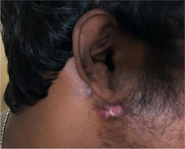
This clinical photograph displays a dermatomal skin rash on the right lateral face of a patient, consistent with herpes zoster (shingles). The lesions are primarily concentrated in the preauricular and mandibular regions, corresponding to the V2 (maxillary) and V3 (mandibular) branches of the trigeminal nerve. The rash is characterized by a cluster of erythematous papules and vesicles in varying stages of development. Key features include fluid-filled vesicles, some of which have ruptured to form shallow ulcers with a raw pink base, and others that have progressed to yellowish-white crusting and scabbing. There is evident perilesional erythema indicating localized inflammation. The distribution is strictly unilateral, stopping at the midline, which is a hallmark of varicella-zoster virus reactivation. This image serves as an educational example of the clinical presentation of herpes zoster facialis and its characteristic morphology of grouped vesicles on an erythematous base following a specific cranial nerve dermatome.
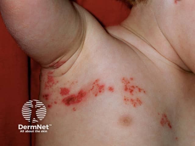
Clinical dermatology photograph of a pediatric patient shows an acute vesicular rash consistent with herpes zoster (shingles) in a dermatomal distribution along the left thorax/axillary region. The image demonstrates clustered vesicles on an erythematous base with surrounding mild edema and crusted lesions at several sites. Lesions appear arranged in a band-like pattern corresponding to a single spinal or cutaneous nerve dermatome, with unilateral involvement typical of varicella-zoster virus reactivation. The skin appears otherwise uninvolved beyond the affected dermatomes, and there is no obvious mucosal involvement in this view. Close-up, high-resolution photography captures the lesion morphology: discrete vesicles, some coalescing papules, thin crusts, and surrounding erythema. No purpura or ulceration is evident. The modality is noninvasive clinical photography, using standard illumination and close frontal view to document surface morphology without contrast or biopsy. Clinically, this image is relevant for differential diagnosis and educational discussions of zoster in children, including timing of antiviral therapy, pain management, and vaccination considerations. The finding's diagnostic significance lies in the dermatomal distribution, vesicular morphology, and temporal progression, aiding differentiation from disseminated varicella, contact dermatitis, and herpes simplex infection. Potential clinical uses include triage, medical education, and research on pediatric VZV reactivation patterns.
OPD Management of Varicella Zoster (VZV) Infections
Part A: PRIMARY VARICELLA (Chickenpox)
Clinical Diagnosis
- Prodrome: fever, malaise 1-2 days before rash
- Rash: Generalised, centripetal, pruritic - macules → papules → vesicles ("dewdrop on rose petal") → pustules → crusts, all stages simultaneously present (pathognomonic)
- Oral/mucosal involvement common
- Highly contagious (airborne + contact) from 2 days before rash until all lesions crusted
Investigations (usually clinical diagnosis)
- If confirmation needed: PCR from vesicle fluid - gold standard (highly sensitive and specific, can distinguish wildtype from vaccine strain)
- Tzanck smear (less specific - also positive in HSV)
- Direct fluorescent antibody - rapid but less sensitive than PCR
- Serology (ELISA) - for checking immune status / post-exposure prophylaxis need
Treatment Decision by Patient Group
1. Healthy Children (2-12 years) - OPD
- Antiviral not routinely recommended - varicella is benign and self-limited; acyclovir only modestly reduces symptoms by ~1 day
- Symptomatic treatment only:
- Antipyretics: Paracetamol/acetaminophen for fever
- AVOID ASPIRIN/SALICYLATES - risk of Reye syndrome
- Pruritus relief: Calamine lotion, cool compresses, tepid baths with baking soda or colloidal oatmeal (3 cups/tub)
- Oral antihistamines (e.g., chlorpheniramine, hydroxyzine) for itching
- Keep nails short; discourage scratching (prevents secondary bacterial infection)
- Do NOT use topical steroids or occlusive ointments
2. Adolescents, Adults, Pregnant (3rd trimester), Household contacts - OPD Antivirals Indicated
| Drug | Dose | Duration |
|---|---|---|
| Valacyclovir (preferred - higher bioavailability) | 1 g orally TDS | 7 days |
| Famciclovir (preferred) | 500 mg orally TDS | 7 days |
| Acyclovir (alternative) | 800 mg orally 5 times/day | 7 days |
| Children 2-18 yrs (valacyclovir) | 20 mg/kg TDS (max 1 g/dose, max 3 g/day) | 5 days |
Key point: Start within 24 hours of rash onset for maximum benefit. Treatment beyond 24 hours is not effective for routine healthy children. - Fitzpatrick's Dermatology
Valacyclovir and famciclovir are preferred over oral acyclovir for adults because of superior pharmacokinetics and higher drug levels. - Goldman-Cecil Medicine
3. Groups Requiring IV Acyclovir (ADMIT, not OPD)
- Immunocompromised patients (IV acyclovir 10 mg/kg every 8 hours, 7-10 days)
- Varicella pneumonitis
- Varicella encephalitis
- Neonates (mothers developed varicella within 5 days before/2 days after delivery): IV acyclovir 10 mg/kg or 500 mg/m² every 8 hours for 10 days
Secondary Bacterial Infection
- Most common complication - Staph. aureus and Group A Streptococcus
- Minor infections: warm soaks
- Cellulitis: systemic antibiotics - amoxicillin-clavulanate, cefalexin, or flucloxacillin
Isolation / Infection Control (OPD Counselling)
- Keep patient away from school/work until all lesions are crusted (usually 5-7 days after rash onset)
- Avoid contact with: pregnant women, newborns, immunocompromised individuals, non-immune healthcare workers
Part B: HERPES ZOSTER (Shingles) - Reactivation
Clinical Diagnosis
- Prodrome: Dermatomal pain, burning, allodynia 2-3 days before rash
- Rash: Unilateral, dermatomal, does not cross midline - clusters of vesicles on erythematous base
- Most common: thoracic (50-70%), trigeminal (ophthalmic branch most common)
- Diagnosis is clinical in typical cases
Special Forms (flag at OPD)
| Variant | Features | Action |
|---|---|---|
| Zoster ophthalmicus (V1 - ophthalmic branch) | Rash on forehead/nose tip (Hutchinson's sign), risk of keratitis, uveitis, blindness | Urgent ophthalmology referral + IV/oral antivirals |
| Ramsay Hunt syndrome (Geniculate ganglion) | Ear vesicles + facial palsy (CN VII) + vertigo, hearing loss | Oral antivirals + prednisolone; ENT referral |
| Disseminated zoster (>3 dermatomes) | Immunocompromised | ADMIT, IV acyclovir |
| Zoster meningitis / encephalitis | Headache, confusion, neck stiffness | ADMIT, IV acyclovir |
Who to Treat with Antivirals in OPD?
| Patient group | Antiviral? |
|---|---|
| Age <50 yrs, mild pain, uncomplicated | Often not needed |
| Age ≥50 yrs | Yes - reduces pain duration |
| Moderate-severe pain (any age) | Yes |
| Ophthalmic/facial zoster | Yes (urgently) |
| Immunocompromised | Yes (IV if severe) |
| Still forming new lesions beyond 72 hrs | Yes, still beneficial |
Key principle: Start within 72 hours of rash onset for best effect. However, treatment may still benefit if new lesions continue to appear after 72 hours. - Goldman-Cecil Medicine
Antiviral Dosing for Herpes Zoster
| Drug | Dose | Duration | Notes |
|---|---|---|---|
| Valacyclovir (preferred) | 1 g orally TDS | 7 days | Higher bioavailability than acyclovir |
| Famciclovir (preferred) | 500 mg orally TDS | 7 days | Equivalent efficacy to valacyclovir |
| Acyclovir (alternative) | 800 mg orally 5 times/day | 7 days | Less preferred due to lower bioavailability |
| IV Acyclovir (severe/immunocompromised) | 10 mg/kg every 8 hrs | 7-10 days or until all crusted | ADMIT |
Topical antivirals have NO role in treatment of varicella or herpes zoster. - Fitzpatrick's Dermatology
Pain Management in Zoster (OPD)
| Step | Drug | Notes |
|---|---|---|
| Step 1 | Paracetamol, NSAIDs | Mild pain |
| Step 2 | Tramadol, weak opioids | Moderate pain |
| Step 3 | Opioid analgesics (morphine, oxycodone) | Severe pain - use cautiously |
| Adjunct | Prednisolone 60 mg/day × 21 days (tapered) + acyclovir | Age >50 yrs; reduces acute pain and improves quality of life but does NOT prevent PHN |
A 2024 meta-analysis (Ding et al., PMID 39029866) confirmed that delayed initial treatment (≥3 days) is a significant risk factor for developing postherpetic neuralgia (PHN). Early treatment is the single most important preventive step for PHN.
Wound/Skin Care
- Keep lesions clean and dry
- Saline soaks or wet compresses for comfort
- Non-adherent dressings if needed
- Prevent scratching - secondary bacterial superinfection
Part C: POST-HERPETIC NEURALGIA (PHN) - OPD Management
Evidence-Based Pharmacotherapy (RCT-proven):
| Drug | Starting Dose | Target Dose | Notes |
|---|---|---|---|
| Gabapentin (first-line) | 300 mg at bedtime | Up to 1200 mg TDS | Titrate slowly; causes dizziness/sedation |
| Pregabalin (first-line) | 75 mg at bedtime | Up to 300 mg BD | Faster titration than gabapentin |
| Nortriptyline / Amitriptyline (TCA) | 25 mg at bedtime | Up to 150 mg/day | Caution in elderly (anticholinergic, cardiac) |
| 5% Lidocaine patch | Apply to affected area | Up to 12 hrs/day | Useful for localised allodynia |
| Capsaicin 8% patch (high concentration) | Applied by specialist | Single application | Pain from application itself; not tolerated by ~1/3 patients |
| Tramadol / Opioids | As needed | Lowest effective dose | Last resort; long-term safety concerns |
These agents provide adequate pain relief (pain <3/10 or ≥50% reduction) in 30-60% of patients. - Fitzpatrick's Dermatology
Part D: CRITERIA FOR ADMISSION (Do NOT manage OPD)
| Situation | Reason |
|---|---|
| Varicella pneumonia | Rapid deterioration risk; IV acyclovir |
| Varicella encephalitis (confusion, seizures) | IV acyclovir + ICU |
| Immunocompromised with varicella or disseminated zoster | IV acyclovir |
| Neonatal varicella | IV acyclovir |
| Zoster ophthalmicus with visual symptoms | IV antivirals + ophthalmology |
| Septic secondary bacterial infection | IV antibiotics |
| Pregnant with varicella pneumonia | High maternal mortality |
Part E: PROPHYLAXIS & PREVENTION (OPD Counselling)
Post-exposure Prophylaxis (PEP)
| Group | Intervention | Timing |
|---|---|---|
| Susceptible immunocompromised / pregnant / neonate | VZIG (Varicella-Zoster Immunoglobulin) | Within 10 days of exposure |
| Susceptible healthy adult/child (non-immune) | Varicella vaccine (if not contraindicated) | Within 3-5 days of exposure |
Vaccination
| Vaccine | Schedule | Target |
|---|---|---|
| Varicella vaccine (live attenuated Oka strain) | 2 doses: 12-15 months + 4-6 years (children); 2 doses 4-8 weeks apart (adults) | Prevention of primary varicella |
| Recombinant Zoster Vaccine (RZV/Shingrix) | 2 doses 2-6 months apart | Prevention of HZ in adults ≥50 yrs; immunocompromised from age 18+ |
| Live Zoster Vaccine (ZVL/Zostavax) | Single dose | Older recommendation; less effective than RZV |
Quick OPD Decision Summary
VARICELLA (Chickenpox)
├── Healthy child <12 yrs → Symptomatic only (calamine, paracetamol, NO aspirin)
├── Adolescent/Adult/Pregnant → Valacyclovir 1g TDS × 7d (start within 24h)
└── Immunocompromised/pneumonia/encephalitis → ADMIT, IV acyclovir

HERPES ZOSTER (Shingles)
├── Age <50, mild pain, uncomplicated → Symptomatic ± antiviral
├── Age ≥50 / moderate-severe pain → Valacyclovir 1g TDS × 7d + prednisolone
├── Ophthalmic/Ramsay Hunt → Urgent antiviral + specialist referral
└── Disseminated/encephalitis/immunocompromised → ADMIT, IV acyclovir

POST-HERPETIC NEURALGIA
└── Gabapentin/Pregabalin (first-line) + TCAs + Lidocaine patch