
Make me detailed pdf on topic thyroid gland with its anatomy , physiology, clinical examination, diseases , various surgical procedures with their details with pictures from scott and brown otorhinolaryngology and Cummings book
thyroid gland anatomy diagram lobes isthmus
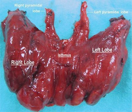
Anatomical specimen photograph of a human thyroid gland post-total thyroidectomy, set against a blue surgical background. The specimen is labeled to demonstrate a rare anatomical variation: bilateral pyramidal lobes. Centrally, the isthmus connects the voluminous right and left lateral lobes. Arising superiorly from the junction points between the isthmus and each lateral lobe are two distinct, elongated, conical structures identified as the right and left pyramidal lobes. The entire gland exhibits a reddish-brown, glistening parenchyma with a multinodular texture consistent with toxic multinodular goiter. The anatomical landmarks are labeled with white text: 'Right Lobe', 'Left Lobe', 'Isthmus', 'Right pyramidal lobe', and 'Left pyramidal lobe'. This clinical photograph is highly relevant for endocrine surgery and gross anatomy education, highlighting developmental remnants of the thyroglossal duct and the necessity of complete dissection during thyroidectomy to ensure total removal of all functional thyroid tissue.
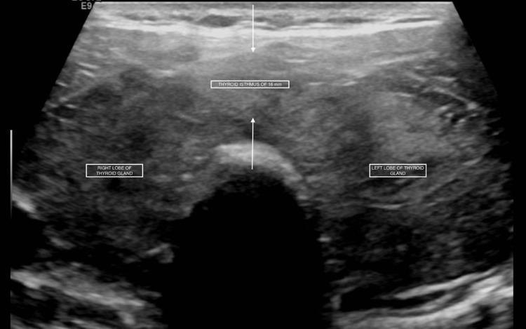
This diagnostic ultrasound image presents a transverse view of the thyroid gland, illustrating clinical signs of diffuse thyroid disease. The anatomy is clearly demarcated with labels for the 'RIGHT LOBE OF THYROID GLAND', 'LEFT LOBE OF THYROID GLAND', and the 'THYROID ISTHMUS'. The isthmus is significantly thickened, with a callout indicating a measurement of 16 mm, confirming thyromegaly. The parenchymal texture is notably heterogeneous and coarsely echogenic, showing a mixed pattern of hypoechoic and hyperechoic areas throughout both lobes. No discrete solid or cystic nodules are identified. The ultrasound findings are characteristic of diffuse thyroid enlargement, often associated with autoimmune conditions such as Graves' disease or Hashimoto's thyroiditis, particularly when correlating with the patient's reported history of hyperthyroidism. The image serves as an educational example of evaluating thyroid morphology, echotexture, and isthmic dimensions via sonography.
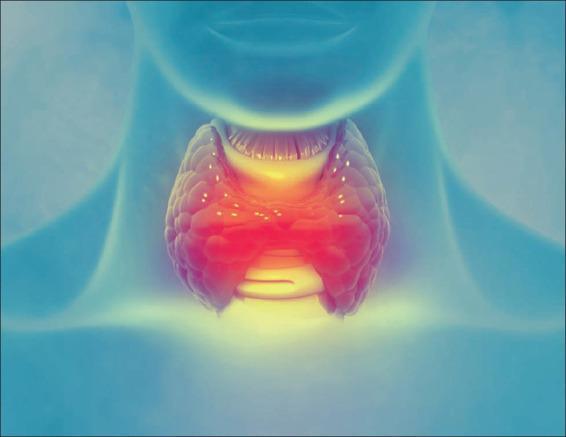
Anatomical diagram depicting the thyroid gland situated in the anterior neck, inferior to the larynx. The illustration utilizes a stylized, translucent blue silhouette of a human neck and torso as a background to provide anatomical context. The central focus is a bipedal, butterfly-shaped organ representing the thyroid gland, characterized by its right and left lobes connected by a central isthmus. The gland is rendered with a textured, lobulated surface and a vibrant color gradient ranging from deep red in the center to bright yellow at the superior and inferior margins, simulating metabolic activity or localized inflammation. Visible above the thyroid is the laryngeal prominence, and the tracheal rings are suggested beneath the isthmus. The image is designed for educational purposes to demonstrate the anatomical positioning of the thyroid gland within the endocrine system and its relationship to airway structures. It serves as a visual aid for discussions on thyroid disorders such as goiter, hyperthyroidism, or thyroiditis.
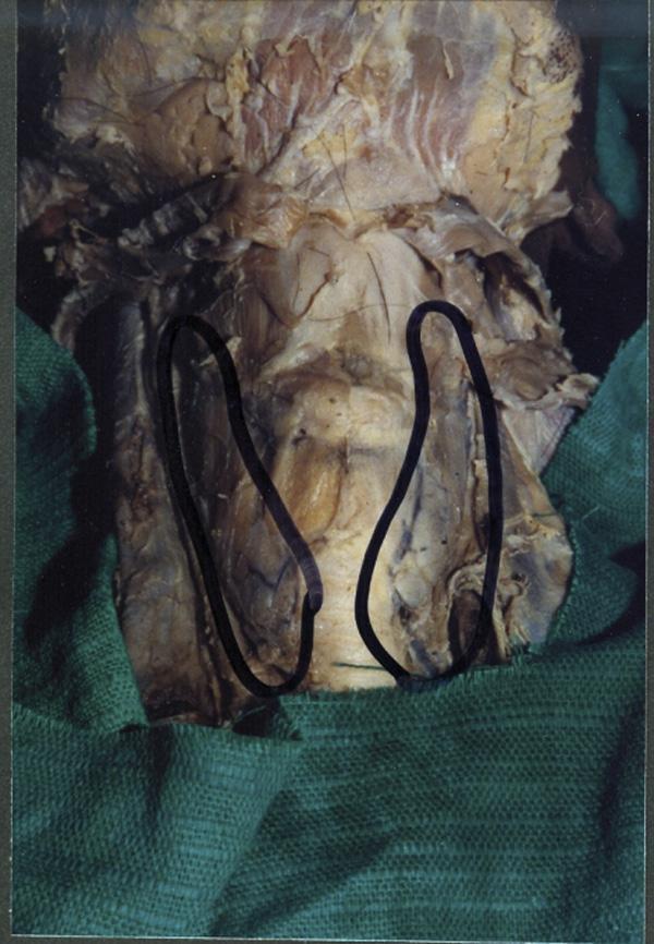
This clinical photograph of a gross anatomical specimen shows a midline dissection of the human neck, highlighting a congenital variation of the thyroid gland. The image depicts the thyroid gland consisting of two distinct, independent lateral lobes (outlined in black) positioned on either side of the trachea. Notably, there is a complete absence (agenesis) of the thyroid isthmus, which normally connects the two lobes across the second to fourth tracheal rings. In the area where the isthmus typically resides, only a thin, translucent layer of pre-tracheal fascia is visible covering the anterior surface of the trachea. The specimen demonstrates the anatomical relationship between the glandular lobes, the respiratory pathway, and the surrounding connective tissue structures. This visual is significant for medical education in endocrinology, anatomy, and surgery, as it illustrates a rare developmental anomaly where the median diverticulum of the thyroglossal duct failed to form the glandular bridge between the bifurcated lobes.
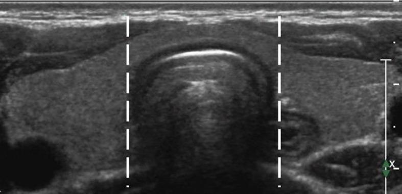
This diagnostic image is a transverse greyscale ultrasound of the anterior neck, specifically showing the thyroid gland. The image functions as an anatomical diagram to define the boundaries of the thyroid isthmus. The thyroid parenchyma appears homogeneous and moderately echogenic. Two vertical dashed white lines are superimposed on the image to delineate the lateral side-lines (lateral borders) of the trachea, which lies immediately posterior to the thyroid isthmus. The tissue situated between these two markers is identified as the isthmus, while the thyroid tissue lateral to these lines represents the left and right thyroid lobes. The trachea itself exhibits characteristic sonographic features, including an echogenic anterior border with posterior acoustic shadowing. This visual representation is used for clinical classification, particularly in determining the location of thyroid nodules or Papillary Thyroid Microcarcinomas (PTMC) for procedures like ultrasound-guided Radiofrequency Ablation (RFA).
thyroid cancer papillary follicular medullary ultrasound histology
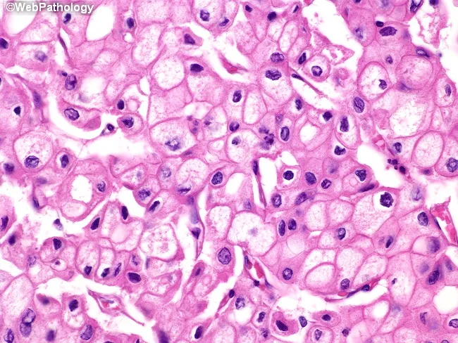
This is a histopathology slide of thyroid tissue prepared by hematoxylin and eosin staining, examined under brightfield light microscopy at high magnification. The specimen demonstrates epithelial neoplasm consistent with papillary thyroid carcinoma (PTC). The neoplastic cells are arranged in sheets and, where present, papillary fronds with fibrovascular cores are recognizable. The predominant cytoplasm is finely reticular and eosinophilic, with blander nuclear features. The nuclei show characteristic raisinoid appearance with nuclear grooves, giving an Orphan Annie eye look; the nuclear membranes are distinct and often thickened. Perinuclear halos, i.e., clear nuclear clearing around the chromatin, are evident. Some tumor cells exhibit voluminous cytoplasm, and in the plane of section, nuclei may be absent in those cells, a phenomenon of sectioning rather than true anuclear cells. The cytoplasmic clearing and grooves are diagnostic, aiding differentiation from benign nodules. The histology carries diagnostic significance, with PTC carrying generally favorable prognosis but potential for lymphatic metastasis; differential includes follicular carcinoma, medullary carcinoma, Hashimoto-related changes, and benign adenomas. Clinically, presence of these features correlates with occult metastasis risk and guides surgical planning, post-operative radioiodine therapy considerations, and molecular testing (e.g., BRAF V600E). This image is relevant for medical education, pathology teaching files, digital atlases, and research on thyroid cancer morphology.
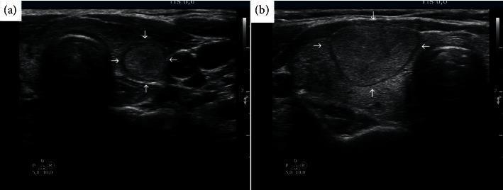
Diagnostic ultrasound comparison showing two distinct presentations of follicular thyroid cancer (FTC). Image (a) depicts a transverse ultrasound view of the thyroid gland containing a small (12 mm), relatively well-defined, circular, mildly hypoechoic nodule indicated by white arrows. This nodule represents a low-suspicion lesion (EU-TIRADS 3), which pathologically was confirmed as minimally invasive follicular cancer. Image (b) shows a significantly larger (28 mm), more heterogeneous, and lobulated nodule (arrows) in the thyroid gland of another patient. The larger mass in (b) represents a progressed case of follicular cancer that had metastasized to the lungs. The comparison illustrates the clinical challenge of diagnosing FTC using standard ultrasound-based Fine Needle Aspiration (FNA) criteria, as early-stage follicular cancers often lack the high-risk sonographic features (such as microcalcifications or irregular margins) seen in papillary thyroid carcinoma, potentially leading to diagnostic delays if triage is based solely on TIRADS size thresholds.
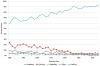
This line graph illustrates the epidemiological trends in the distribution of thyroid cancer cases by histological type from 1980 to 2012. The data, expressed as a percentage of all thyroid cancer cases, shows a prominent increase in papillary carcinoma, which rose from approximately 60% in 1980 to over 90% by 2012. Conversely, other histological subtypes show a steady decline in relative proportion over the same period. Follicular carcinoma, which initially accounted for nearly 25% of cases, decreased significantly. Medullary and anaplastic carcinomas, along with a category labeled as 'others,' remained at low percentages and trended downward toward the end of the study period. This visualization emphasizes the shifting landscape of thyroid pathology, highlighting the increasing dominance of papillary carcinoma in clinical diagnosis and incidence relative to non-papillary types. The graph serves as an educational resource for oncology and endocrinology, illustrating longitudinal changes in cancer subtypes within a specific population context.
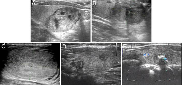
This composite diagnostic image features five B-mode and Doppler ultrasound scans (labeled A-E) of the thyroid gland, illustrating the sonographic variability of Bethesda IV thyroid nodules. Panel A demonstrates a follicular thyroid adenoma, characterized by an isoechoic to hypoechoic texture with internal cystic changes and a distinct hypoechoic halo. Panel B shows a follicular variant of papillary thyroid carcinoma (FVPTC), appearing as a hypoechoic nodule with well-defined margins. Panel C displays follicular thyroid carcinoma (FTC) as a predominantly isoechoic mass with noticeably irregular borders. Panel D illustrates multifocal papillary thyroid carcinoma (PTC) with multiple hypoechoic regions and irregular, poorly defined margins. Panel E shows medullary thyroid carcinoma (MTC) with color Doppler overlay indicating internal vascularity. This series serves as a clinical reference for comparing echogenicity (isoechoic vs. hypoechoic), margin characteristics (regular vs. irregular), and internal architecture (cystic vs. solid) across various benign and malignant thyroid pathologies identified during preoperative screening.
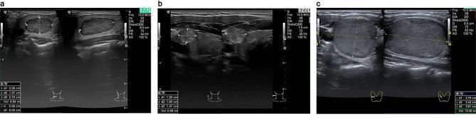
This diagnostic image displays three B-mode ultrasound panels illustrating different types of malignant thyroid nodules in the right lobe, categorized by the EU TI-RADS system. Panel (a) shows Medullary Thyroid Carcinoma (MTC) presenting as a nodule with mixed echogenicity, irregular contours, and internal microcalcifications. Panel (b) depicts Papillary Thyroid Carcinoma (PTC) characterized by an ill-defined, hypoechoic, and heterogeneous structure with scattered echogenic foci suggestive of microcalcifications. Panel (c) illustrates Follicular Thyroid Carcinoma (FTC), which appears as a relatively homogeneous, well-circumscribed nodule with a smooth contour, lacking obvious macrocalcifications. Each panel includes transverse and longitudinal views with calipers measuring nodule dimensions (d1, d2, d3) and calculated volume. These sonographic findings demonstrate key diagnostic features used to differentiate follicular and parafollicular thyroid malignancies, specifically focusing on echogenicity, border definition, and the presence of calcifications.
thyroidectomy surgical procedure neck incision
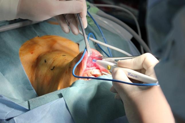
This clinical photograph captures a Transoral Video-Assisted Neck Surgery (TOVANS) for thyroidectomy. The patient is in a supine position with the neck extended to provide optimal access. The surgical field is established with sterile blue drapes, and the primary incision is located at the oral vestibule between the inferior lip and the gingiva, exposing the underlying vascular tissue and teeth. Two surgeons are actively engaged in the procedure; one uses a metal retractor to maintain the vestibular opening, while the other utilizes a white-handled Valleylab electrocautery device for tissue dissection. Surgical markings (black lines) are visible on the skin of the anterior neck to guide the subplatysmal tunneling. This approach is designed to provide a scarless outcome on the neck by accessing the thyroid gland through the oral cavity. Blue suction or irrigation tubing is present within the operative field, which is a key component of the sterile, controlled environment required for head and neck endocrine surgery.
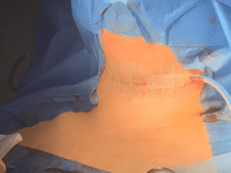
This clinical photograph shows a post-operative surgical site on the anterior neck, consistent with a completed total thyroidectomy and Sistrunk procedure. The primary incision is located in the lower anterior neck and is closed with medical adhesive strips (Steri-Strips) applied transversely across the horizontal wound line. A 15mm Blake aspirative drain is visible, exiting the lateral aspect of the surgical site to facilitate drainage and prevent hematoma formation. The drain is secured with additional adhesive dressing. The surgical field is surrounded by blue sterile drapes, exposing the area from the lower mandible to the clavicular region. The image demonstrates standard post-operative management of a thyroid surgery site, including primary skin closure techniques and active suction drainage. This visual is relevant for medical education concerning head and neck surgery, specifically thyroidectomy and thyroglossal duct cyst management.
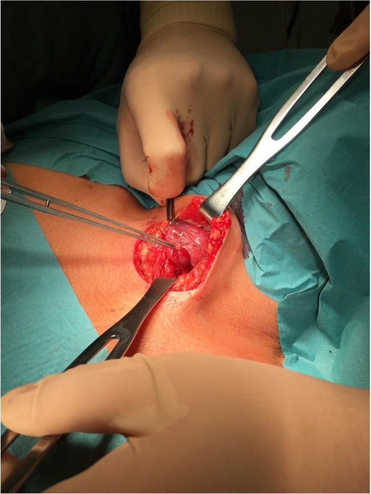
This clinical photograph shows an intraoperative view of a surgical procedure in the anterior neck region, likely a thyroidectomy or parathyroidectomy. The surgical field is exposed via a transverse cervical incision, with the underlying thyroid gland visible as a reddish, vascularized lobular structure. Multiple surgical instruments are in use for tissue manipulation and exposure: a pair of surgical forceps is retracting thyroid tissue to the left, while two metal retractors (superior and inferior) maintain the patency of the incision. A pointed probe or dissector is positioned in the center, directed toward a localized area on the thyroid surface to facilitate the identification of a parathyroid gland (PTG). The field demonstrates standard surgical anatomy and instrumentation for endocrine surgery of the neck, highlighting the careful dissection required to distinguish the PTG from surrounding thyroid parenchyma.
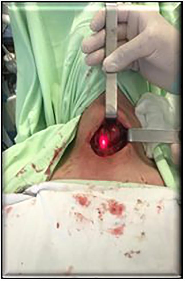
Clinical photograph of an intraoperative surgical setup during a neck procedure, likely a thyroidectomy or parathyroid surgery. The patient is in a supine position, draped with sterile green and white cloths showing minor blood staining around the surgical site. A gloved hand is using a metallic retractor to maintain exposure of the incision in the anterior neck. The surgical field is illuminated by a specialized near-infrared (NIR) spectrum light source, visible as a bright red-aiming beam, which is part of the SPY visualization system for indocyanine green (ICG) fluorescence angiography. This technology is utilized intraoperatively to identify parathyroid glands (PTG) and assess their perfusion before and after tissue resection. The image illustrates the integration of advanced fluorescence imaging into endocrine surgery for improved anatomical localization and preservation of delicate structures.
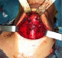
This clinical intraoperative photograph shows the anterior neck region during a surgical procedure, specifically following a total thyroidectomy. The operative field is exposed through a transverse cervical incision. Metallic surgical retractors are positioned laterally to maintain the retraction of the skin flaps and subcutaneous tissues, providing a clear view of the central neck compartment. The thyroid bed is visible, characterized by a raw, erythematous tissue surface with evidence of recent surgical dissection. No residual thyroid gland tissue is macroscopicially visible. The area shows expected post-surgical changes including minor fluid and heme presence on the exposed muscular and connective tissues. This image serves as a clinical example of the surgical site after the removal of the thyroid gland, typically performed for conditions such as thyroid carcinoma or thyroglossal duct cyst complications.
thyroid physiology hormone synthesis T3 T4 iodine
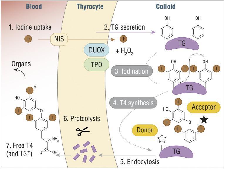
A pathophysiology diagram illustrating the seven-step process of thyroid hormone synthesis (T3 and T4) across three compartments: Blood, Thyrocyte, and Colloid. (1) Iodine uptake: Iodide (I-) enters the thyrocyte from the blood via the Sodium/Iodide Symporter (NIS). (2) TG secretion: Thyroglobulin (TG) is secreted into the colloid. (3) Iodination: Within the colloid, iodide is oxidized by Thyroid Peroxidase (TPO), dual oxidase (DUOX), and H2O2, leading to the attachment of iodine to tyrosine residues on TG. (4) T4 synthesis: A coupling reaction occurs between 'Donor' and 'Acceptor' iodinated tyrosine residues on the TG scaffold. (5) Endocytosis: The modified TG molecule is re-absorbed into the thyrocyte. (6) Proteolysis: Lysosomal enzymes break down TG, releasing individual hormones. (7) Release: Free T4 (thyroxine) and T3 (triiodothyronine) are secreted into the blood for systemic distribution to organs. The diagram includes detailed chemical structures of the phenolic rings and iodine substitutions characteristic of thyroid hormones.
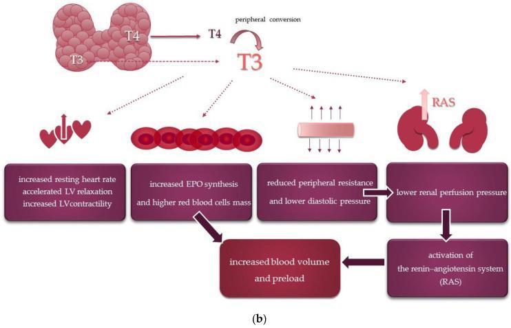
This medical flowchart illustrates the systemic and cardiovascular effects of thyroid hormones. The diagram begins with the thyroid gland releasing Thyroxine (T4) and Triiodothyronine (T3), highlighting the peripheral conversion of T4 to the more active T3. The downstream physiological impacts of T3 are categorized into four primary pathways: 1) Cardiac effects, including increased resting heart rate, accelerated left ventricular (LV) relaxation, and increased LV contractility. 2) Hematologic effects, specifically increased erythropoietin (EPO) synthesis leading to higher red blood cell mass. 3) Vascular effects, showing reduced peripheral resistance and lower diastolic pressure. 4) Renal effects, where reduced peripheral resistance leads to lower renal perfusion pressure. These combined pathways culminate in the activation of the renin-angiotensin system (RAS) and an overall increase in blood volume and preload. The illustration uses anatomical icons of the heart, blood cells, vessels, and kidneys to correlate physiological mechanisms with organ-level changes, demonstrating how hyperthyroidism or thyroid hormone signaling influences hemodynamic stability and cardiac output.
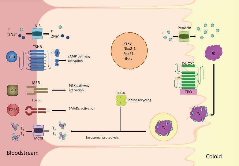
This medical schematic illustrates the pathophysiology and cellular mechanisms of a thyroid follicular cell, detailing the synthesis and secretion of thyroid hormones. The diagram is divided into three compartments: the bloodstream (basolateral side), the follicular cell cytoplasm, and the colloid lumen (apical side). Key transport proteins at the basolateral membrane include the Sodium-Iodide Symporter (NIS), which co-transports I- and Na+, and the Monocarboxylate Transporter 8 (MCT8), which facilitates the secretion of T3 and T4 into the blood. Signaling receptors TSHR, IGFR, and TGFBR are shown triggering cAMP, PI3K, and SMAD pathways respectively. At the apical membrane, Pendrin transports iodide into the colloid, where DUOX2 and Thyroid Peroxidase (TPO) facilitate the iodination of Thyroglobulin (Tg). The diagram further depicts the endocytosis of Tg, followed by lysosomal proteolysis to release T3 and T4. The enzyme DEHAL is shown recycling iodine from MIT and DIT. Centrally, the nucleus contains transcription factors Pax8, Nkx2-1, FoxE1, and Hhex, which regulate gene expression for thyroid differentiation.
goiter enlarged thyroid clinical examination neck
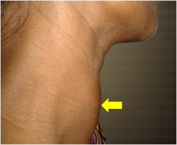
This clinical photograph shows a right lateral view of the neck of a 30-year-old female patient, demonstrating a diffusely enlarged thyroid gland, also known as a goiter. The anatomical region of interest is the anterior neck, where a significant convex protrusion is visible, indicated by a yellow arrow. The swelling appears symmetrical and smooth-surfaced, consistent with a diffuse rather than nodular enlargement of the thyroid lobes and isthmus. The skin overlying the mass is uniform in color without signs of inflammation, erythema, or ulceration. The neck is held in a slightly extended position to better visualize the anterior contour. This visual finding is characteristic of Graves' disease, as supported by the clinical context of thyrotoxicosis and diffuse radioactive iodine uptake mentioned in the patient's record. This image serves as an educational example of the physical examination findings associated with hyperthyroidism and autoimmune thyroid disease.
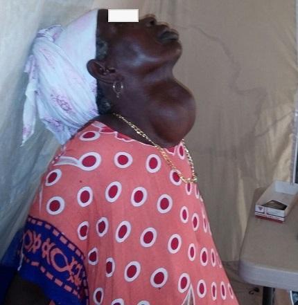
This clinical photograph shows a profile view of a 55-year-old female patient with a massive multinodular goiter (enlarged thyroid gland). The pathology is located in the anterior neck, presenting as a very large, rounded, and protuberant mass that extends symmetrically across both thyroid lobes. The overlying skin appears intact with a smooth texture and no visible ulceration, erythema, or inflammatory changes. The goiter is of such significant volume that it obscures the normal contours of the mandible and neck, and clinically, it was described as mobile without induration. This image illustrates a chronic, severe manifestation of iodine deficiency or benign thyroid disease, common in regions with limited surgical access. The clinical relevance focuses on the diagnostic physical examination of neck masses and the secondary effects of such enlargement, such as tracheal deviation or compressive symptoms. The patient is shown with a head covering and a patterned top, which provides cultural context but does not detract from the clear presentation of the anatomical abnormality.
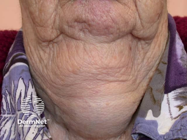
This clinical photograph shows the anterior neck of an adult patient with diffuse thyroid enlargement consistent with goiter. The image modality is clinical photography, captured in a frontal/anterior neck view with the patient seated or standing, natural lighting, and minimal magnification. The thyroid gland is visibly enlarged, producing a prominent cervical contour in the midline and bilateral lobes that extend laterally toward the sternocleidomastoid regions. Skin over the gland is wrinkled and thin, without evident ulceration or focal nodularity on the presented surface; there is modest venous prominence posteriorly. The enlargement appears diffuse rather than a single palpable nodule, suggesting a diffuse goiter or multinodular process that may be chronic. No tracheal deviation or airway compromise is clearly visible in this image, though clinical correlation is necessary to evaluate for compressive symptoms. Clinically, goiter etiologies include iodine deficiency, autoimmune thyroiditis (Hashimoto), Graves disease, or benign/multinodular thyroid neoplasms. Diagnostic significance: clinical confirmation of thyroid enlargement; subsequent assessment should include thyroid function tests (TSH, free T4), thyroid autoantibodies, and high-resolution neck ultrasound or CT/MRI as indicated. Differential considerations include diffuse toxic goiter, thyroiditis, and focal neoplasms. This image is educational for endocrine pathology, ENT, and radiology training, illustrating external thyroid enlargement and its gross appearance in situ.
recurrent laryngeal nerve thyroid surgery anatomy
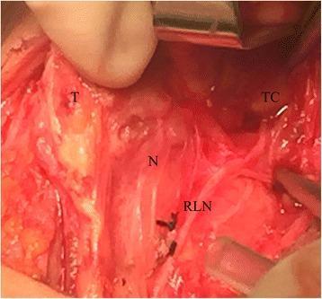
An intraoperative clinical photograph documenting an anatomical variation of the recurrent laryngeal nerve (RLN) during a thyroid surgery. The image shows the surgical field with the following landmarks labeled: the thyroid gland (T) medially retracted, the thyroid cartilage (TC) in the superior-lateral aspect, and the point of typical nerve entry (N). The recurrent laryngeal nerve (RLN) is visualized following an atypical trajectory, ascending along the medial margin of the superior thyroid pole and terminating beneath the superior cornu of the thyroid cartilage. This clinical case illustrates an anatomical variant that can lead to surgical complications if not properly identified. The surgical site displays typical tissue characteristics including erythematous vascularized structures and surgical retraction tools. This image serves as an educational resource for endocrine surgeons and medical students to understand neurovascular anatomy and the importance of intraoperative nerve monitoring in preventing iatrogenic vocal cord paralysis.
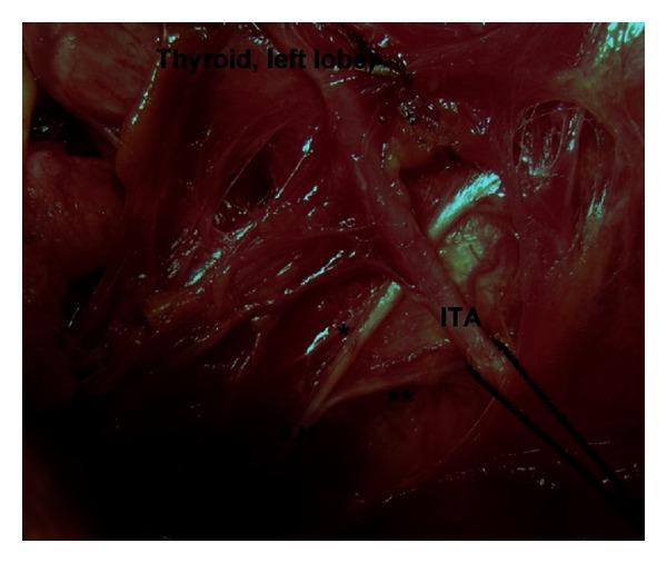
An intraoperative clinical photograph depicting the surgical anatomy of the left central neck compartment during thyroid surgery. The image highlights the anatomical relationship between the left inferior laryngeal nerve (ILN), also known as the recurrent laryngeal nerve, and the inferior thyroid artery (ITA). The photograph demonstrates a Type 4 variation, where the terminal division of the ILN into anterior (*) and posterior (**) branches occurs at the pre-arterial segment, proximal to its crossing with the ITA. The left lobe of the thyroid gland is visible in the upper portion of the surgical field. This visual is significant for endocrine surgery, illustrating extralaryngeal terminal bifurcation of the ILN, which is a critical landmark to identify to prevent iatrogenic nerve injury during thyroidectomy or parathyroidectomy. The image provides educational value regarding neurovascular variations and surgical risk regions in the cervical course of the laryngeal nerves.
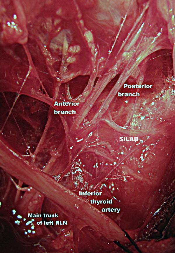
This magnified clinical operative photograph demonstrates the surgical anatomy of the left central neck compartment during thyroid surgery. The image highlights the variations in the course of the left recurrent laryngeal nerve (RLN) and its neurovascular relationships. The main trunk of the left RLN is visible in the lower quadrant, ascending and bifurcating into distinct anterior and posterior terminal branches. Medial to these branches, the inferior thyroid artery is identified, crossing the nerve path. A significant anatomical variation shown is the SILAB (Supra-Arterial Inferior Laryngeal Anastomotic Branch), a horizontal nerve anastomosis connecting the posterior branch of the RLN to adjacent neural structures near the laryngeal entry point. This photograph serves as an educational resource for identifying vulnerable neural structures during thyroidectomy and parathyroidectomy, emphasizing the clinical importance of meticulous dissection at the neurovascular crossing point to avoid iatrogenic nerve injury.
Graves disease hyperthyroidism exophthalmos thyroid eye disease

Clinical photograph of a 53-year-old male presenting with bilateral Graves' ophthalmopathy (thyroid eye disease). The image focuses on the periocular region, demonstrating bilateral exophthalmos (proptosis) characterized by the protrusion of the globes from the orbits. Significant scleral show is visible, particularly inferior to the iris, contributing to a characteristic 'staring' expression or Dalrymple's sign. The patient is wearing corrective eyeglasses and a surgical face mask, indicating a clinical setting. This visual illustrates the extrathyroidal manifestations of Graves' disease, an autoimmune condition causing hypertrophy of extraocular muscles and orbital adipose tissue. The image is an educational resource for medical professionals to recognize clinical signs of hyperthyroidism-associated orbitopathy and for calculating Clinical Activity Scores (CAS) in endocrinology and ophthalmology.
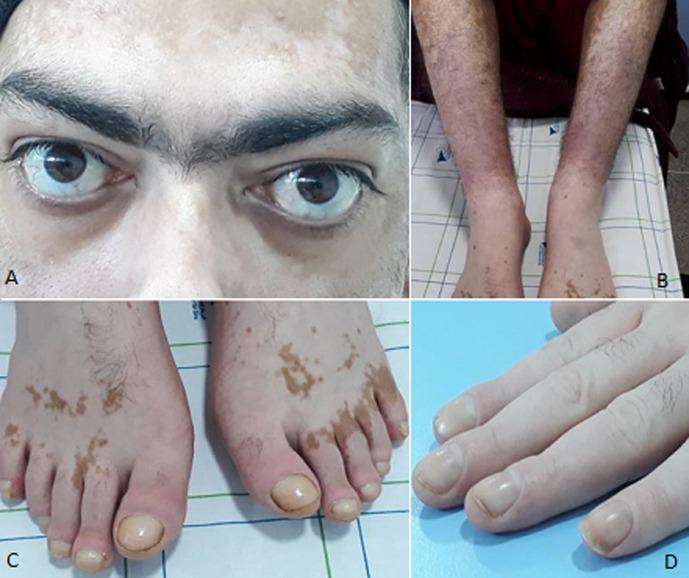
This composite clinical photograph illustrates the classic triad of extra-thyroidal manifestations associated with Graves' disease (Basedow's disease). Panel A shows bilateral exophthalmos (proptosis) and scleral show, along with periocular depigmentation consistent with vitiligo. Panel B demonstrates pretibial myxedema (thyroid dermopathy), characterized by diffuse, brawny, brownish-red induration and hyperpigmentation of the lower legs. Panels C and D display thyroid acropathy, a rare manifestation of Graves' disease. Notable features include digital clubbing (hippocratisme digital) of both fingers and toes, visible as bulbous swelling of the distal phalanges and increased convex curvature of the nail plates. Additionally, the skin on the dorsal aspect of the feet shows patchy, well-defined hyperpigmentation and hypopigmentation. This image serves as a comprehensive educational reference for the systemic dermatological and ophthalmological complications of autoimmune hyperthyroidism, highlighting the characteristic triad of exophthalmos, dermopathy, and acropathy.
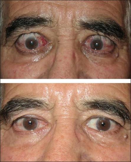
This comparison clinical photograph shows a 62-year-old male with active Graves' ophthalmopathy before and after medical intervention. The top image demonstrates hallmark signs of thyroid eye disease, including bilateral upper eyelid retraction (Dalrymple sign), exophthalmos (proptosis), and significant conjunctival hyperemia. Notably, the patient exhibits conjunctival chemosis and focal redness near the medial canthus of the right eye, accompanied by periorbital edema and increased forehead wrinkling likely due to compensatory frontalis muscle overactivity. The bottom image shows the clinical outcome following steroid treatment and botulinum toxin injection to the upper eyelids. There is a marked reduction in ocular surface inflammation and redness. The eyelid position has significantly improved with reduced scleral show, and the periorbital region appears less edematous with a more relaxed brow position. This visual illustrates the management of ocular manifestations in autoimmune thyroid disease, focusing on the improvement of lid retraction and clinical activity scores (CAS).
mkdir -p /tmp/workspace/thyroid-pdf && ls /tmp/workspace/thyroid-pdf/
Make me detailed pdf on topic thyroid gland with its anatomy , physiology, clinical examination, diseases , various surgical procedures with their details with pictures from scott and brown otorhinolaryngology and Cummings book