
Severe alcohol withdrawal management
"alcohol withdrawal" AND management
alcohol withdrawal CIWA-Ar scale delirium tremens management
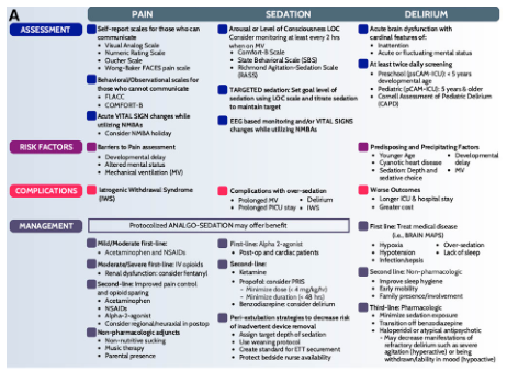
Summary : This figure presents a side-by-side comparison of pain, sedation, and delirium assessment and management in pediatric intensive care, organized into four main categories: Assessment, Risk Factors, Complications, and Management. Each category is subdivided for pain, sedation, and delirium, with specific tools, risk factors, complications, and management strategies listed for each. flowchart/table hybrid: # Overall Structure : • The figure is divided into three vertical columns: Pain, Sedation, and Delirium. • Four horizontal sections: Assessment, Risk Factors, Complications, and Management. • Each cell contains bulleted lists of tools, factors, or strategies. # Pain : ## Assessment : • Self-report scales for communicative children: Visual Analog Scale, Numeric Rating Scale, Oucher Scale, Wong-Baker FACES pain scale. • Behavioral/observational scales for non-communicative children: FLACC, COMFORT-B. • Acute vital sign changes with NMBA use; consider NMBA holiday. ## Risk Factors : • Barriers to pain assessment: Developmental delay, altered mental status, mechanical ventilation (MV). ## Complications : • Iatrogenic Withdrawal Syndrome (IWS). ## Management : • Mild/Moderate: Acetaminophen, NSAIDs. • Moderate/Severe: First-line IV opioids, consider fentanyl for renal dysfunction. • Second-line: Improved pain control and opioid sparing (acetaminophen, NSAIDs, alpha-2 agonist). • Non-pharmacologic: Non-nutritive sucking, music therapy, parental presence. # Sedation : ## Assessment : • Arousal/Level of Consciousness: Monitor every 2 hours when on MV. • Comfort-B scale, State Behavioral Scale (SBS), Richmond Agitation-Sedation Scale (RASS). • Targeted sedation: Set goal, titrate sedation. • EEG-based monitoring and vital sign changes with NMBA use. ## Risk Factors : • Complications with over-sedation: Prolonged MV, prolonged PICU stay, delirium, IWS. ## Complications : • Over-sedation: Prolonged MV, delirium, prolonged PICU stay, IWS. ## Management : • Protocolized analgo-sedation may offer benefit. • First-line: Alpha-2 agonist (post-op, non-cardiac). • Second-line: Ketamine, minimize dose (4 mg/kg/h), minimize duration (<48h), benzodiazepines for delirium. • Peri-extubation strategies: Assign wean target, use extubation protocol, create standard for ETT securement, protect bedside nurse availability. # Delirium : ## Assessment : • Acute brain dysfunction: Inattention, fluctuating mental status. • Acute daily screening: Preschool (psCAM-ICU-5), school-age (pCAM-ICU-5), Cornell Assessment of Pediatric Delirium (CAPD). ## Risk Factors : • Predisposing/precipitating: Young age, developmental delay, congenital heart disease, MV, sedative/analgesic use. ## Complications : • Worse outcomes: Longer ICU/hospital stay, greater cost. ## Management : • First-line: Treat medical disease (labs, brain MAPS). • Hypoactive: Over-sedation, lack of sleep. • Hyperactive: Infections. • Second-line: Non-pharmacologic (sleep hygiene, early mobilization, family presence). • Third-line: Minimize sedation exposure, avoid benzodiazepines, consider alpha-2 agonist, may need dexmedetomidine for withdrawal/mood (hypoactive). # Design Encodings : • Blue, purple, and pink colour coding for Pain, Sedation, and Delirium columns. • Section headers highlighted with background shading. • Bulleted lists for clarity. • Icons for assessment, risk factors, complications, and management. # Analysis : • The figure provides a comprehensive, parallel overview of pain, sedation, and delirium management in pediatric critical care. • Each domain is addressed with specific assessment tools, risk factors, complications, and management strategies. • There is emphasis on protocolized approaches, minimizing complications, and integrating non-pharmacologic interventions. • The structure facilitates rapid comparison and highlights the interconnectedness of pain, sedation, and delirium in clinical practice.
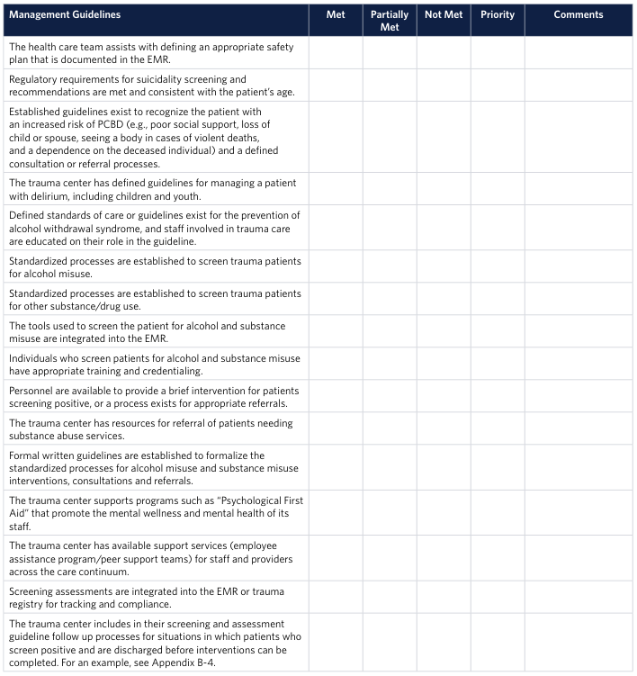
<table><thead><tr><th>Management Guidelines</th><th>Met</th><th>Partially Met</th><th>Not Met</th><th>Priority</th><th>Comments</th></tr></thead><tbody><tr><td>The health care team assists with defining an appropriate safety plan that is documented in the EMR.</td><td></td><td></td><td></td><td></td><td></td></tr><tr><td>Regulatory requirements for suicidality screening and recommendations are met and consistent with the patient's age.</td><td></td><td></td><td></td><td></td><td></td></tr><tr><td>Established guidelines exist to recognize the patient with an increased risk of PCBD (e.g., poor social support, loss of child or spouse, seeing a body in cases of violent deaths, and a dependence on the deceased individual) and a defined consultation or referral processes.</td><td></td><td></td><td></td><td></td><td></td></tr><tr><td>The trauma center has defined guidelines for managing a patient with delirium, including children and youth.</td><td></td><td></td><td></td><td></td><td></td></tr><tr><td>Defined standards of care or guidelines exist for the prevention of alcohol withdrawal syndrome, and staff involved in trauma care are educated on their role in the guideline.</td><td></td><td></td><td></td><td></td><td></td></tr><tr><td>Standardized processes are established to screen trauma patients for alcohol misuse.</td><td></td><td></td><td></td><td></td><td></td></tr><tr><td>Standardized processes are established to screen trauma patients for other substance/drug use.</td><td></td><td></td><td></td><td></td><td></td></tr><tr><td>The tools used to screen the patient for alcohol and substance misuse are integrated into the EMR.</td><td></td><td></td><td></td><td></td><td></td></tr><tr><td>Individuals who screen patients for alcohol and substance misuse have appropriate training and credentialing.</td><td></td><td></td><td></td><td></td><td></td></tr><tr><td>Personnel are available to provide a brief intervention for patients screening positive, or a process exists for appropriate referrals.</td><td></td><td></td><td></td><td></td><td></td></tr><tr><td>The trauma center has resources for referral of patients needing substance abuse services.</td><td></td><td></td><td></td><td></td><td></td></tr><tr><td>Formal written guidelines are established to formalize the standardized processes for alcohol misuse and substance misuse interventions, consultations and referrals.</td><td></td><td></td><td></td><td></td><td></td></tr><tr><td>The trauma center supports programs such as "Psychological First Aid" that promote the mental wellness and mental health of its staff.</td><td></td><td></td><td></td><td></td><td></td></tr><tr><td>The trauma center has available support services (employee assistance program/peer support teams) for staff and providers across the care continuum.</td><td></td><td></td><td></td><td></td><td></td></tr><tr><td>Screening assessments are integrated into the EMR or trauma registry for tracking and compliance.</td><td></td><td></td><td></td><td></td><td></td></tr><tr><td>The trauma center includes in their screening and assessment guideline follow up processes for situations in which patients who screen positive and are discharged before interventions can be completed. For an example, see Appendix B-4.</td><td></td><td></td><td></td><td></td><td></td></tr></tbody></table>
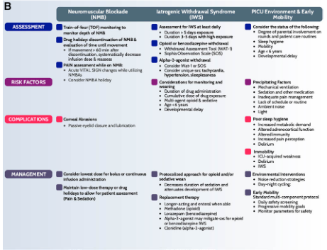
Summary : This figure presents a side-by-side comparison of three key domains in pediatric intensive care: Neuromuscular Blockade (NMB), Iatrogenic Withdrawal Syndrome (IWS), and the Pediatric Intensive Care Unit (PICU) Environment & Early Mobility. For each domain, the figure outlines assessment strategies, risk factors, complications, and management approaches. flowchart: # Domains : • Neuromuscular Blockade (NMB) • Iatrogenic Withdrawal Syndrome (IWS) • PICU Environment & Early Mobility # Assessment : • NMB: – Train-of-four (TOF) monitoring for depth of NMB – Drug holiday/discontinuation and movement evaluation – Reassessment and titration of dose – Pain assessment while on NMB – Monitor acute vital sign changes • IWS: – Assess for IWS at least daily – Duration: 5+ days exposure, 3–5 days with weaning – Use opioid/benzodiazepine withdrawal tools (WAT-1, Sophia Observation Scale) – Consider WAT-1 or SOS • PICU Environment & Early Mobility: – Consider parental involvement, delirium, sleep, mobility, and developmental delay # Risk Factors : • NMB: – Prolonged NMB use – Consider NMB holiday • IWS: – High cumulative opioid/benzo dose – Multi-agent opioid/sedative use – Age <6 months, developmental delay • PICU Environment & Early Mobility: – Precipitating factors: mechanical ventilation, sedation, infection, acute illness, inadequate pain management, delirium, developmental delay # Complications : • NMB: – Corneal abrasions (due to incomplete eyelid closure/lubrication) • IWS: – Signs/symptoms of withdrawal (not detailed in this section) • PICU Environment & Early Mobility: – Poor sleep hygiene, increased metabolic demand, altered immune function, delirium, immobility, ICU-acquired weakness, IWS # Management : • NMB: – Use lowest effective dose/bolus or continuous infusion – Maintain low-dose therapy or drug holidays for patient assessment (pain & sedation) • IWS: – Protocolized opioid/benzo weaning – Decrease duration/sedation to prevent IWS – Replacement therapy: long-acting/enteral wean, methadone, lorazepam, clonidine (alpha-2 agonist) • PICU Environment & Early Mobility: – Environmental interventions: day/night cycling, noise reduction – Early mobility: multi-component protocol, daily safety screening, monitor safety parameters # Layout : • Three vertical columns, each representing a domain (NMB, IWS, PICU Environment & Early Mobility) • Each column subdivided horizontally into Assessment, Risk Factors, Complications, and Management • Colour-coded sections: blue (Assessment), purple (Risk Factors), red (Complications), dark blue (Management) # Analysis : • The figure systematically compares the three domains, highlighting both unique and overlapping considerations (e.g., risk factors and management strategies). • Emphasis is placed on regular assessment, minimizing risk, and protocolized management to improve patient outcomes in the PICU. • The structure facilitates quick reference for clinicians to identify best practices and potential complications in each domain.
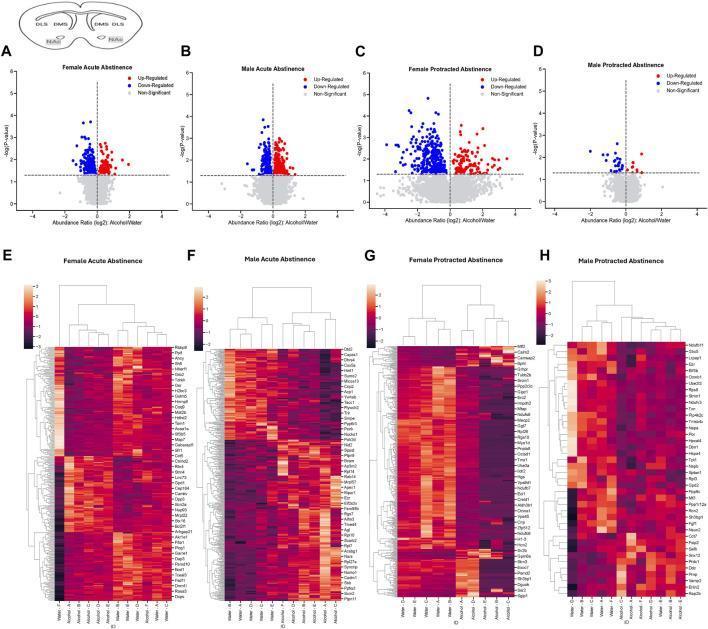
This composite figure presents proteomic analysis of the mouse nucleus accumbens (NAc) following alcohol exposure and subsequent abstinence. The image is organized into two rows. The top row (A-D) features four volcano plots mapping protein abundance changes (Alcohol/Water ratio) against statistical significance (-log10 p-value). Red dots signify up-regulated proteins, blue dots indicate down-regulated proteins, and gray dots represent non-significant changes across four experimental groups: Female Acute Abstinence (A), Male Acute Abstinence (B), Female Protracted Abstinence (C), and Male Protracted Abstinence (D). The bottom row (E-H) displays corresponding heatmaps and hierarchical clustering dendrograms for the same four groups. These clusterplots visualize the differential protein expression profiles, with rows labeled by specific GeneIDs and columns representing individual samples. A color scale indicates intensity of protein abundance. This visual data illustrates sex-based and temporal differences in the striatal proteome, highlighting significant protein alterations during different stages of alcohol withdrawal.
Severe Alcohol Withdrawal Management
Pathophysiology
- ROSEN's Emergency Medicine, p. 2883
- Harrison's Principles of Internal Medicine 22E, p. 3723
Clinical Spectrum and Timeline
| Phase | Timing after last drink | Features |
|---|---|---|
| Minor withdrawal | 6-12 hours onset, peaks 24-48 h | Tremor, anxiety, nausea/vomiting, diaphoresis, tachycardia, hypertension, insomnia |
| Alcoholic hallucinosis | 8-48 hours | Visual/auditory hallucinations or illusions; clear sensorium (distinguishes from DTs) |
| Withdrawal seizures | 12-48 hours (peak ~24-36 h) | Generalized tonic-clonic; usually 1-2 brief seizures; may occur without other withdrawal signs |
| Delirium tremens (DTs) | 72-96 hours (range 48 h to 5 days) | Agitated delirium, marked tremor, hyperthermia, severe autonomic dysregulation, visual/tactile hallucinations, paranoid delusions |
- Maudsley Prescribing Guidelines 15ed, p. 505-506
- Washington Manual of Medical Therapeutics, p. 998
Severity Assessment - CIWA-Ar
- < 8 - Mild; rarely requires medication
- 8-15 - Moderate; benzodiazepines indicated
- > 15 - Severe; high risk for seizures and DTs; close monitoring mandatory
- Maximum score: 67
Delirium Tremens - Recognition and Risk
-
History of prior DTs or withdrawal seizures (most important predictor)
-
Severe alcohol dependence / high daily consumption
-
Self-detoxification without medical input
-
Multiple prior admissions for withdrawal
-
Concurrent medical illness (liver disease, infection, GI bleeding)
-
Electrolyte abnormalities: hypokalemia, hypomagnesemia
-
Thiamine deficiency
-
Concurrent benzodiazepine or other sedative use
-
Older age
-
Maudsley Prescribing Guidelines, p. 506
-
Harrison's, p. 3723
Management
1. Immediate Stabilization (ALL patients)
- Thiamine 100-500 mg IV/IM BEFORE any glucose - Wernicke's encephalopathy can be precipitated by glucose in thiamine-deficient patients
- Multivitamins including folic acid
- Correct electrolytes: Mg²⁺, K⁺, glucose (hypomagnesemia and hypokalemia are common and worsen seizure risk)
- Balanced diet/nutrition as tolerated
- Avoid IV fluids unless hemodynamically compromised, significant vomiting/diarrhea, or recent bleeding - most patients are normally hydrated or mildly overhydrated
- Harrison's, p. 3723; Washington Manual, p. 998
2. Benzodiazepines - First-Line for ALL Severe Withdrawal
| Drug | Route | Half-life | Notes |
|---|---|---|---|
| Diazepam | PO/IV | Long (20-100 h + active metabolites) | Rapid IV onset (1-3 min); preferred for fast loading; avoid in severe liver failure |
| Chlordiazepoxide | PO | Long | Standard oral agent for mild-moderate; avoid in liver disease |
| Lorazepam | IV/IM/PO | Medium (12 h, no active metabolites) | Preferred in liver disease and elderly; good IM absorption; safe in renal failure |
| Oxazepam | PO | Short (5-15 h) | Hepatically safe (renal excretion); use in liver failure |
- Diazepam IV: 5 mg IV every 5-10 min; escalate to 10 mg/dose if 5 mg is insufficient; titrate to light somnolence. Up to 800 mg/day of chlordiazepoxide equivalent reported in severe DTs.
- Lorazepam IV: 2-4 mg IV every 15-20 min until symptom control; can repeat 1-4 mg IV every 5-15 min for severe withdrawal
- Symptom-triggered (preferred): give benzodiazepine when CIWA-Ar ≥8-10; reassess every 1-2 hours
- Fixed-taper (alternative if CIWA-Ar not feasible): e.g., chlordiazepoxide 25-50 mg or diazepam 10 mg PO every 4-6 hours day 1, tapered to zero over 5 days
- ROSEN's Emergency Medicine, p. 2884; Harrison's, p. 3723; Washington Manual, p. 998-999
3. Management of Delirium Tremens (ICU Setting)
- High-dose benzodiazepines are the cornerstone - there is no maximum dose limit in severe refractory DTs; titrate to control agitation
- For benzodiazepine-refractory DTs: closely monitored propofol or phenobarbital can be used
- A 2025 systematic review (PMID 39415533) supports propofol as a rescue agent in ICU for refractory AWS
- Dexmedetomidine is generally not recommended as monotherapy - it does not act on GABAergic/glutamatergic systems, may mask autonomic signs without preventing seizures, and has not improved patient-centered outcomes in trials - Washington Manual, p. 999
- DTs typically run a self-limiting course of 3-5 days regardless of therapy; treatment goal is controlling agitation and preventing complications
- Antipyretics for hyperthermia; cardiac monitoring; airway protection if needed
- Harrison's, p. 3723; Maudsley Prescribing Guidelines, p. 506
4. Seizure Management
- Alcohol withdrawal seizures are usually brief, generalized, self-limiting; antiepileptic drugs (AEDs) are NOT routinely indicated
- Long-acting benzodiazepines (diazepam) are first-line for seizure prophylaxis in high-risk patients (prior seizure history)
- Phenytoin does not prevent alcohol withdrawal seizures - do not use as monotherapy or adjunct
- Carbamazepine loading is an option in patients with untreated epilepsy or seizures despite adequate benzodiazepine loading
- If seizures occur, exclude structural/metabolic causes (hypoglycemia, trauma, electrolyte disturbance, meningitis)
- If hypoglycemia is present, give thiamine before glucose
- Recent evidence (2024): phenobarbital shows promise in the ED for refractory withdrawal seizures - two systematic reviews (PMID 37923363, PMID 37589203) support its use as an adjunct or alternative to benzodiazepines in the emergency setting
- Washington Manual, p. 998; Maudsley, p. 505
5. Adjuncts and Supportive Care
| Intervention | Indication |
|---|---|
| Thiamine 100-500 mg IM/IV | ALL patients before glucose; prevents Wernicke's |
| Magnesium supplementation | Hypomagnesemia (lowers seizure threshold) |
| Potassium replacement | Hypokalemia |
| Antipsychotics (haloperidol) | Adjunct for hallucinations only - never as monotherapy in DTs; use with caution |
| Propofol infusion | Refractory DTs not responding to benzodiazepines (ICU only) |
| Phenobarbital | Adjunct/alternative to BZDs, especially in ED (emerging evidence) |
| Fluid resuscitation | Only if clinically dehydrated, bleeding, or significant vomiting |
Indications for Inpatient / ICU Admission
- Regular alcohol >30 units/day or SADQ >30
- History of seizures or prior DTs
- Concurrent benzodiazepine or polydrug use
- Comorbid medical illness (liver disease, cardiac, infection)
- Pregnancy
- Elderly or minor
- No social support / homeless
- Cognitive impairment or psychiatric comorbidity
- Failed community detoxification previously
Differential Diagnosis (Especially for DTs)
Recent Evidence Updates (2024-2026)
- Phenobarbital has strong emerging evidence as a first-line alternative or adjunct to benzodiazepines for AWS in the ED, with two 2024 systematic reviews supporting its safety and efficacy (PMID 37923363, PMID 37589203)
- Propofol is supported as a rescue agent for refractory AWS in ICU settings (PMID 39415533, 2025 systematic review)
- Ethanol infusion for AWS management remains controversial - a 2025 systematic review (PMID 39559850) found insufficient evidence to recommend it; not standard of care