
prepare a pdf ofHistolgical images andImmunofluorescence patterns Images of all types of vasculitis with key description
vasculitis histology microscopy necrotizing inflammation vessel wall
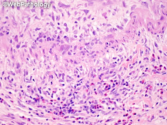
This histopathology image depicts a gallbladder wall section showing classic necrotizing vasculitis of medium-sized muscular arteries consistent with polyarteritis nodosa (PAN). The salient feature is transmural inflammation of the vessel wall with fibrinoid necrosis and disruption of the tunica media and internal elastic lamina, best seen with elastic stains but evident on routine H&E as pale, eosinophilic necrosis surrounding a dense inflammatory infiltrate. In active lesions, the infiltrate comprises neutrophils and eosinophils admixed with histiocytes; skip areas indicate discontinuous involvement with uninvolved interposed segments. The inflammatory process may spare adjacent gallbladder parenchyma, defining a bystander phenomenon rather than true parenchymal inflammation. In healing stages, the vessel shows fibrointimal proliferation and progressive luminal narrowing, reflecting irreversible remodeling. The image underscores the need to distinguish true necrotizing vasculitis from secondary vascular inflammation seen with acute or chronic cholecystitis. Clinically, PAN-related gallbladder involvement signals a systemic vasculitis that may accompany renal and gastrointestinal manifestations; diagnosis rests on histology, correlation with serology (often ANCA-negative in classic PAN), and integration with radiographic or clinical vasculitis features. This specimen exemplifies how vascular lesions can be segmental and heterogenous within a single gallbladder biopsy, influencing prognosis and therapeutic planning (steroids, immunosuppressants). Guiding patient management decisions.
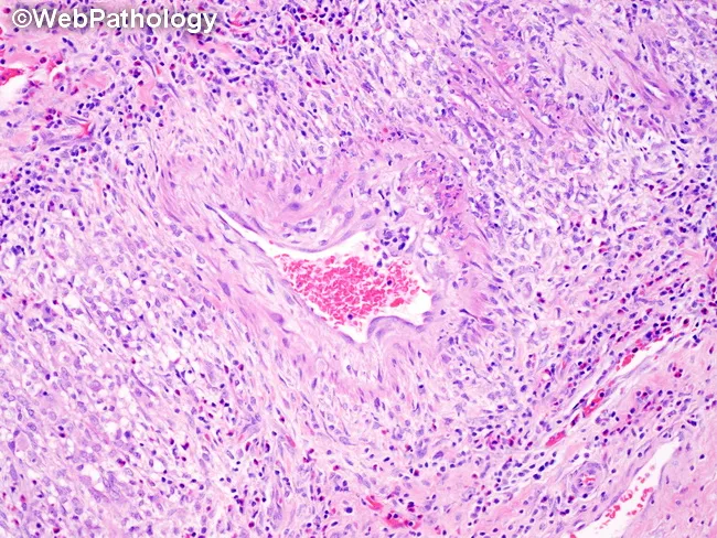
Histopathology of gallbladder tissue reveals necrotizing transmural vasculitis of medium-sized muscular arteries within the gallbladder wall. The vessel wall exhibits fibrinoid necrosis and inflammatory infiltrates that are initially neutrophilic and eosinophilic, with histiocytes; the surrounding tissue typically shows minimal inflammatory reaction. Lesions are discontinuous, with skip areas, and multiple vessels commonly display varying stages of evolution within a single specimen. In active lesions, transmural inflammation encroaches on all vascular layers, culminating in destruction of the tunica media and internal elastic lamina; elastic stains most clearly demonstrate lamina destruction. In healed or healing stages, fibrointimal proliferation produces luminal narrowing or occlusion. The inflammatory infiltrate may transition to a lymphocytic predominance during resolution. Importantly, bystander inflammation related to adjacent cholecystitis must be distinguished from true vasculitis. The grossly preserved surrounding gallbladder parenchyma contrasts with the targeted vascular injury. These features are characteristic of polyarteritis nodosa and may reflect systemic vasculitis when observed in the gallbladder, prompting clinical correlation for systemic symptoms and laboratory markers (ANCA status is often negative in classic PAN). This histology underlines the need for immunosuppressive therapy and vigilance for renal, gastrointestinal, and vascular complications. Correlation with clinical data guides targeted treatment and prognosis assessment in systemic vasculitis workup.
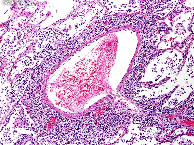
This is a light microscopy image of a tissue biopsy prepared for histopathology and stained with Hematoxylin and Eosin (H&E). The section displays a prominent blood vessel at the center with a lumen containing erythrocytes and a thickened, eosinophilic wall. Surrounding the vessel is dense perivascular inflammatory infiltrate composed of small, dark-staining nuclei (lymphocytes and macrophages) with occasional plasma cells. The adjacent stroma shows interstitial edema and mild congestion. The vascular intima may be focally thickened, and there is mild adventitial inflammation. The background parenchyma appears fragmented with dispersed inflammatory cells, making any definitive parenchymal pattern (e.g., granulomas, necrosis) not distinctly visible. No obvious malignant cells are evident in this field. The image highlights features consistent with an inflammatory or vasculitic process, such as endarteritis or perivasculitis, though infection cannot be excluded without ancillary studies. Diagnostic significance: In a patient with systemic inflammatory symptoms, this pattern supports a vasculitic or immune-mediated vascular injury diagnosis and prompts serologic testing (ANCA), infectious workup, and clinical correlation. Potential clinical use cases include evaluation of suspected vasculitis, inflammatory lung or soft-tissue lesions, and research into vascular inflammatory pathology. This description emphasizes terminology suitable for indexing, including vasculitis, perivasculitis, endarteritis, and H&E histology cues.
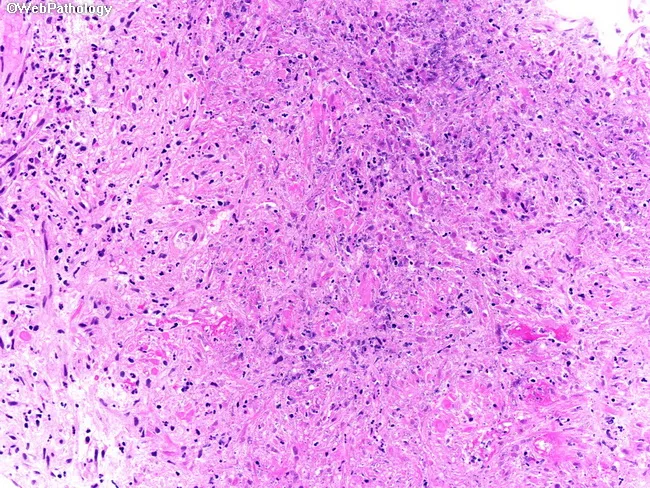
This is a high-magnification histopathology image of prostatic tissue examined by light microscopy after hematoxylin and eosin staining. The specimen is a core biopsy obtained from the prostate gland in a patient with known granulomatosis with polyangiitis (Wegener) and recent pulmonary involvement. The tissue shows necrotizing granulomatous inflammation characterized by aggregates of epithelioid histiocytes and multinucleated giant cells surrounding central areas of eosinophilic or caseous-like necrosis. A dense inflammatory infiltrate, including neutrophils and lymphocytes, permeates the stroma and disrupts normal prostatic architecture, with partial preservation of acini in some regions. Small- to medium-sized vessel involvement compatible with vasculitis can be identified within the inflammatory infiltrate. The palisading arrangement of histiocytes and necrotic foci are hallmark features. No definite malignancy is evident in the sampled cores. The appearance is typical for prostatic GPA involvement, although infectious etiologies or sarcoidosis may mimic granulomatous prostatitis; correlation with microbiologic studies and serology is advised. Clinically, prostatic GPA, though uncommon (estimates 2.5–7.5%), carries significance for disease activity assessment and therapeutic decision making, as immunosuppressive regimens are tailored to systemic vasculitis burden. This image supports multidisciplinary evaluation of genitourinary involvement in GPA. The image is a teaching reference for recognizing GPA-associated granulomatous prostatitis on prostatic biopsy and to illustrate the importance of correlating clinical history with histology.
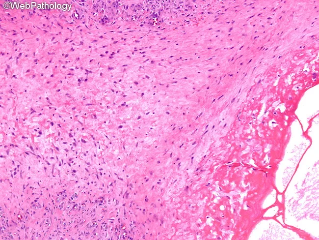
High magnification light microscopy of a formalin-fixed tissue section stained with hematoxylin and eosin (H&E) reveals a focal area of fibrinoid necrosis within the vessel wall (lower right) accompanied by cystic change. Surrounding this necrotic core is a prominent zone of vascular tissue characterized by marked fibroblastic proliferation and remodeling, with spindle-shaped cells and thickened intima forming a dense, fibrous stroma (upper left). The necrotic area displays eosinophilic, amorphous material consistent with fibrin deposition, often seen in necrotizing vasculitis. The adjacent parenchyma shows a relatively preserved architecture with scattered inflammatory cells, a pattern that may reflect an early or localized vasculitic process rather than a fully developed granulomatous lesion. Overall, the image demonstrates a cascade from acute fibrinoid injury toward progressive fibrous scarring, typical of angiitis with fibroproliferative response. The combination of fibrinoid necrosis and fibroblastic proliferation within a vascular niche has diagnostic relevance for necrotizing vasculitis syndromes (e.g., polyarteritis nodosa, granulomatosis with polyangiitis) and related connective tissue diseases. Clinically, these features are relevant for differential diagnosis, prognosis, and therapeutic decisions, as they inform decisions about immunosuppressive therapy, vascular risk assessment, and correlation with serology such as ANCA. This micrographic pattern is essential for pathology education and case documentation.
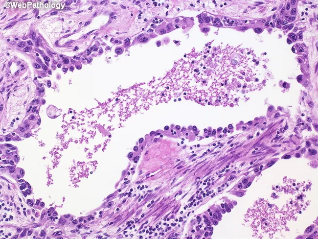
This is a high-power brightfield light microscopy image of a formalin-fixed tissue section stained with Hematoxylin and Eosin. The specimen reveals a small- to medium-sized vessel with luminal content dominated by eosinophilic, granular material compatible with fibrin or organized thrombus. The vessel wall shows minimal intimal thickening and surrounding perivascular tissue contains a mixed inflammatory infiltrate, including scattered neutrophils and lymphocytes, with occasional spindle-shaped fibroblasts indicative of early fibrous remodeling. The mucosal or epithelial architecture surrounding the vessel is not clearly defined, but adjacent stroma demonstrates increased cellularity and edema. There is no overt malignant epithelial proliferation evident in this field. The overall pattern is suggestive of a vascular inflammatory or thrombotic process; features could be compatible with vasculitis, thrombotic microangiopathy, or infectious endovascular involvement, depending on clinical context. No granulomas or necrosis are conspicuously present in this field, though a small focus of macrophage-like cells is seen in the perivascular region. This image supports educational discussions of vascular pathology, thrombus formation, inflammatory vasculopathy, and differential diagnoses in patients with acute or chronic vascular inflammation. The illustrated features emphasize correlation with clinical presentation and laboratory data to distinguish vasculitis from simple thrombosis.
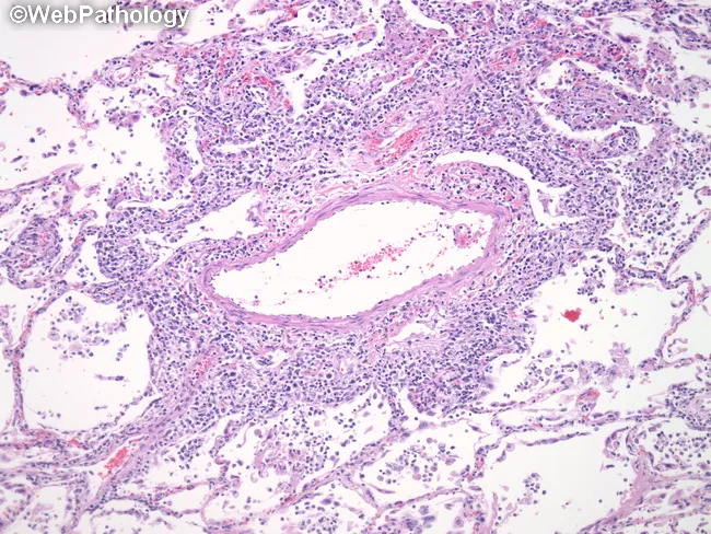
Imaging modality and technique: light microscopy of a formalin-fixed, paraffin-embedded lung biopsy stained with Hematoxylin and Eosin (H&E). The panel reveals an angiocentric lymphoid infiltrate surrounding a small blood vessel within lung parenchyma. The infiltrate is composed predominantly of small lymphocytes with scant cytoplasm and dark, round to slightly irregular nuclei, consistent with T-cell lineage; rare accompanying histiocytes may be present. Perivascular cuffs encroach on the vascular wall and create an unmistakable angiocentric pattern that is characteristic of lymphomatoid granulomatosis (LG). Overall lung architecture in the field is variably preserved, with focal interstitial inflammatory changes near vessels. In LG the lesion often contains endothelial damage and can progress to necrotizing vasculitis and, with time, clonal B-cell proliferation that is frequently EBV-associated; this exact panel does not provide EBV status. Clinically, LG presents with pulmonary symptoms such as cough, dyspnea, occasional chest pain, and may be accompanied by systemic symptoms like fever and weight loss. The cited histology is pivotal for differential diagnosis against granulomatosis with polyangiitis and infectious vasculitides. This image serves as an educational exemplar for pulmonary pathology, hematopathology, and EBV-associated lymphoproliferative disorders, illustrating the critical angiocentric, T-cell–predominant pattern. Correlation with imaging studies and clinical data is essential for accurate diagnosis and management.
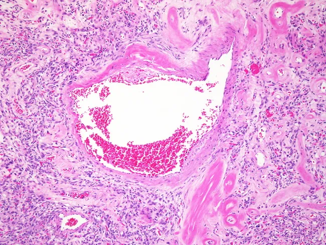
This histology slide is a hematoxylin and eosin stained cross-section of vascular tissue showing an occlusive intraluminal thrombus within a small vessel. The thrombus is erythrocyte-rich with scattered pale eosinophilic fibrin strands, producing a heterogeneous red-pink to pale-white appearance within the lumen. The vessel wall displays mild intimal thickening and subtle edema of the surrounding media. A perivascular inflammatory infiltrate, comprising neutrophils and small lymphocytes, is embedded in loose fibrous stroma surrounding the vessel. There is no overt necrosis, granulomatous inflammation, or malignant cells detected in the observed field. Adjacent extracellular matrix appears relatively loose with modest collagen deposition, compatible with chronic inflammatory change. Overall, the image demonstrates a thrombotic vasculopathy pattern with secondary inflammatory response. Clinically relevant to thrombosis risk assessment, hypercoagulable states, inflammatory vasculitis, or vascular injury, this finding requires correlation with patient history and laboratory tests. The staining highlights cellular detail, fibrin deposition, and vessel-wall integrity, supporting differential diagnoses such as acute thrombosis, embolic occlusion, and vasculopathic processes, and aiding pathologic correlation and therapeutic decision-making.
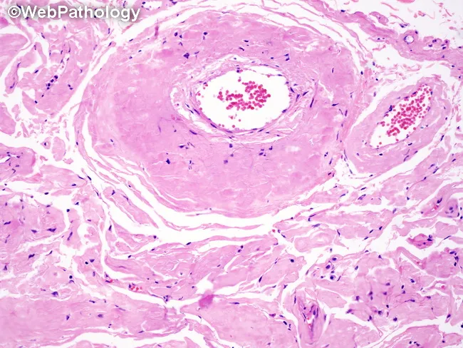
Comprehensive Description: This is a light microscopy histopathology image of a small artery/arteriole in hematoxylin and eosin (H&E) stained tissue, captured at a transverse cross-section. The vascular wall shows pronounced onion-skin hyperplasia with concentric, laminated intimal and medial cell layering, leading to marked luminal narrowing. The wall is thickened by smooth muscle cell proliferation and basement membrane reduplication, producing alternating dense and pale bands around the lumen. The surrounding parenchyma reveals fibrous connective tissue and sparse inflammatory cells; endothelial cell details are not distinct at this magnification. The appearance is characteristic of hyperplastic arteriolosclerosis, classically associated with malignant hypertension or accelerated hypertension. In contrast to hyaline arteriolosclerosis, which produces homogeneous eosinophilic thickening, the onion-skin pattern reflects proliferative remodeling of the vessel wall. Notable features may include mild perivascular fibrosis and variable luminal caliber, with potential ischemic susceptibility in involved territories. Clinically, this finding supports a hypertensive emergency in context with acute end-organ injury (kidney, brain, retina) and guides aggressive blood pressure management. Differential considerations include polyarteritis nodosa and other vasculitides when necrotizing inflammation is present; however, this image lacks transmural fibrinoid necrosis and significant inflammatory infiltrate. This slide is educational for pathology, histology teaching, and radiology-pathology correlation. Practice today together.
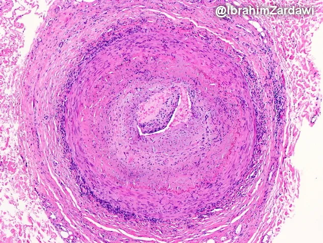
This histology image captures a cross-sectional temporal artery biopsy illustrating the hallmarks of giant cell arteritis. Prepared as a paraffin-embedded tissue section and stained with Hematoxylin and Eosin, the slide reveals a transmural inflammatory infiltrate that traverses all arterial wall layers. At the center, a narrowed lumen is surrounded by concentric intimal hyperplasia with marked medial disruption. Multinucleated giant cells and mononuclear macrophage clusters are present within the media, accompanied by lymphocytic infiltrate along the vessel wall. The elastic lamina appears disrupted, and fragmentation of the media is evident, consistent with robust granulomatous vasculitis. These features produce near-occlusive narrowing of the artery, explaining the ischemic risk associated with temporal arteritis. Clinically, such histopathology confirms a diagnosis of GCA and helps differentiate from atherosclerotic disease, infectious vasculitis, or other large-vessel vasculitides. The image underscores the systemic nature of the disease, as inflammation can extend from cranial vessels to the aorta. The diagnostic significance lies in correlating histology with patient presentation, including age-related cranial symptoms and risk of vision loss; timely immunosuppressive therapy can prevent irreversible ischemic complications. This specimen exemplifies classic angiitis with granulomatous inflammation and intimal hyperplasia driving luminal compromise.
immunofluorescence vasculitis IgA deposits ANCA pauci-immune kidney glomerulonephritis
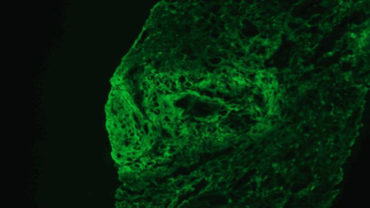
This diagnostic image shows direct immunofluorescence (DIF) of a renal biopsy specimen, specifically highlighting fibrinogen deposition. The image reveals intense, apple-green fluorescence with a heterogeneous distribution. The staining pattern is prominently seen within the walls of a large-caliber renal artery and associated perivascular spaces, appearing as bright, irregular, and somewhat fibrillar or linear deposits. This visual finding is characteristic of necrotizing vasculitis, where fibrinoid necrosis leads to the entrapment of fibrinogen within the damaged vascular walls. The surrounding interstitial tissue displays fainter, more diffuse staining patterns. In the clinical context of ANCA-associated pauci-immune necrotizing vasculitis, this fibrinogen-positive staining serves as a critical marker for active vascular injury, contrasting with the absence of significant immunoglobulin or complement (C3) deposits that would otherwise suggest immune-complex mediated glomerulonephritis. The image is a primary educational resource for nephropathology, illustrating the visual manifestations of vascular necrosis at a microscopic level.
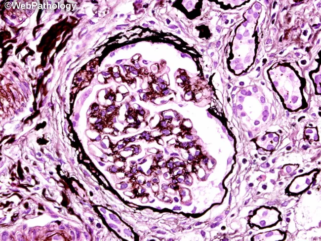
This is a high-fidelity histopathology image obtained from a human kidney biopsy, prepared as a routine H&E stained section and viewed under bright-field illumination at high magnification. The focal area centers on a renal glomerulus within the renal cortex; the cortical architecture surrounds the glomerulus with adjacent tubules and interstitium. The glomerular tuft exhibits marked cellularity with apparent endocapillary proliferation and crowding of capillary loops. Mesangial regions appear expanded with increased cellular elements. Intraluminal deposits and fibrillar material are suggested by variably dense purple staining within capillary lumina, though higher-resolution immunofluorescence would be needed for definitive immune complex characterization. The basement membranes appear thickened in places, but overall normal-appearing capillary wall architecture is not consistently evident due to the dense cellular infiltrate. Overall, the image displays a pathologic proliferative glomerulonephritis pattern, with luminal narrowing and potential early crescents in extreme cases not clearly resolved in this single field. The observed changes have clinical significance because they correlate with nephritic presentations such as hematuria, proteinuria, and reduced eGFR, and they guide differential diagnoses including post-infectious GN, IgA nephropathy, membranoproliferative GN, or pauci-immune GN. Confirmatory studies (IF/EM) and correlation with serology are recommended. Clinical history and imaging further refine prognosis and treatment planning for patients.
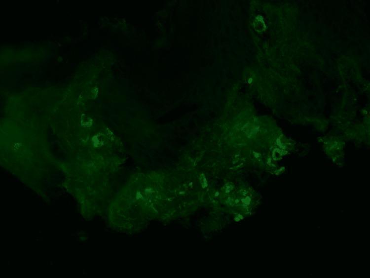
This diagnostic image demonstrates direct immunofluorescence (DIF) of a skin punch biopsy specimen. The micrograph reveals a characteristic granular green fluorescent staining pattern against a dark background. These fluorescent deposits are primarily concentrated in a perivascular distribution, localized around the superficial dermal blood vessels. The morphology of the staining is punctate and clumped, typical of immune complex deposition. Clinically, this visual evidence of perivascular IgA deposition is a hallmark diagnostic feature of IgA vasculitis (formerly Henoch-Schönlein purpura). The image illustrates the classic immunopathologic findings associated with leukocytoclastic vasculitis involving the small vessels of the dermis. This material is suitable for dermatology and rheumatology education, specifically for teaching the diagnostic criteria of systemic small-vessel vasculitides.
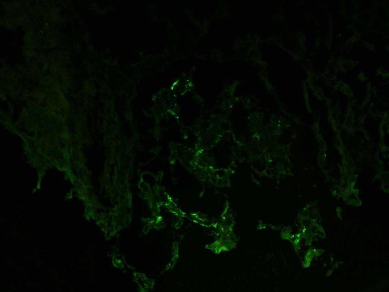
This diagnostic image is a direct immunofluorescence (DIF) micrograph of a renal biopsy specimen. It shows positive staining for IgA antibodies within a glomerulus. The fluorescence pattern is characterized by granular, segmental deposits primarily localized in the mesangium, creating a scattered apple-green signal against a dark, non-reactive background. The distribution is discontinuous and varies in intensity, which is a hallmark finding in IgA nephropathy (Berger's disease). The image illustrates the specific immunological deposition that leads to glomerular injury, highlighting the importance of immunofluorescence in differentiating various types of glomerulonephritis. Key educational concepts include the recognition of mesangial immune complex deposition and the diagnostic utility of antisera specific for immunoglobulin subclasses in nephropathology.
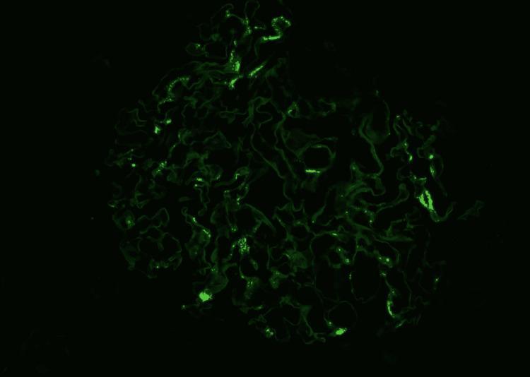
This diagnostic immunofluorescence image demonstrates a renal glomerulus exhibiting a characteristic staining pattern for IgA nephropathy. Against a dark background, a bright green fluorescent signal is localized primarily within the mesangial regions. The signal presents as granular, irregular deposits concentrated in the mesangial matrix, reflecting the entrapment of immunoglobulin A (IgA) immune complexes. The overall structure of the glomerulus shows a complex, lobulated architecture with visible capillary loops. While some areas demonstrate intense, discrete granular fluorescence, others show a more diffuse or attenuated signal, which may correlate with underlying morphological changes such as mesangial hypercellularity or segmental sclerosis. This imaging modality is essential in nephropathology for differentiating between various forms of glomerulonephritis based on the specific distribution (mesangial vs. capillary wall) and nature (granular vs. linear) of immune deposits.
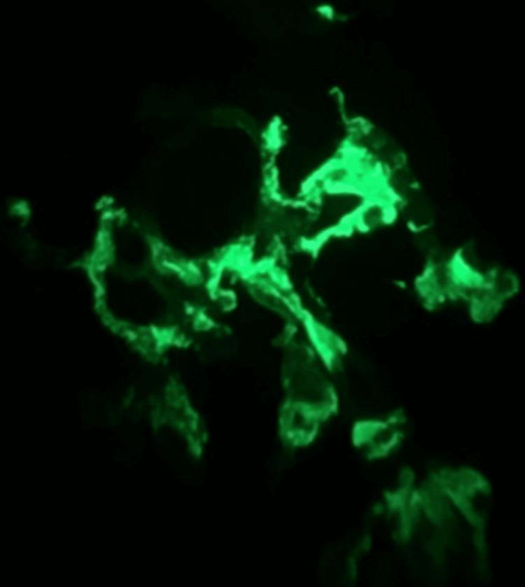
This diagnostic image shows immunofluorescence microscopy of a renal biopsy specimen, specifically demonstrating glomerular IgA staining. Set against a dark background, the image reveals intense apple-green fluorescence within the mesangium and along some capillary loops of a glomerulus. The staining pattern is granular and predominantly mesangial, characterized by irregular clumps and lobulated deposits of IgA immune complexes. This visual finding is a hallmark diagnostic feature of IgA nephropathy (Berger disease) or IgA vasculitis (Henoch-Schönlein purpura) in a clinical context of palpable purpura and subnephrotic proteinuria. The distribution illustrates the localization of immunoglobulin A deposits within the glomerular tuft, which is essential for differentiating immune-mediated glomerulonephritides.
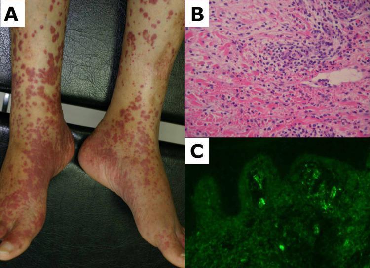
This composite image illustrates the clinical, histological, and immunopathological hallmarks of IgA vasculitis (Henoch-Schönlein purpura). Panel A is a clinical photograph of the bilateral lower limbs showing extensive palpable purpura. The lesions are erythematous to purplish-red macules and papules, ranging from pinpoint petechiae to confluent patches, predominantly involving the feet, ankles, and shins. Panel B presents a skin biopsy specimen stained with hematoxylin and eosin (H&E) at high magnification, demonstrating leukocytoclastic vasculitis. Key features include a dense perivascular neutrophilic infiltrate, leukocytoclasis (nuclear dust), and extravasated erythrocytes within the dermis. Panel C shows a direct immunofluorescence (DIF) micrograph of the dermis, highlighting characteristic granular immunoglobulin A (IgA) deposits within the superficial dermal capillary walls, appearing as bright apple-green fluorescence. This educational sequence correlates the gross dermatological presentation of small-vessel vasculitis with its underlying microscopic vascular damage and specific immune complex deposition pattern.
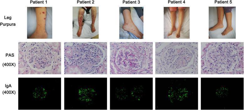
A multi-panel medical figure displaying clinical and histopathological findings for five pediatric patients with IgA vasculitis (Henoch-Schönlein purpura). The top row presents clinical photographs of the lower extremities, showing characteristic non-thrombocytopenic palpable purpura. Presentation ranges from sparse, discrete petechiae and ecchymoses (Patient 1, 3, 5) to extensive, confluent, and necrotic-appearing vasculitic lesions (Patient 2). The middle row shows renal biopsy specimens stained with Periodic Acid-Schiff (PAS) at 400X magnification, revealing varying degrees of mesangial hypercellularity and matrix expansion consistent with IgA nephropathy. The bottom row demonstrates direct immunofluorescence (DIF) microscopy at 400X magnification, highlighting bright, granular IgA deposits primarily within the glomerular mesangium. The intensity and distribution of the green fluorescence indicate the severity of immune complex deposition. This comparison illustrates the clinical and pathological spectrum of Henoch-Schönlein purpura nephritis (HSPN) prior to treatment.
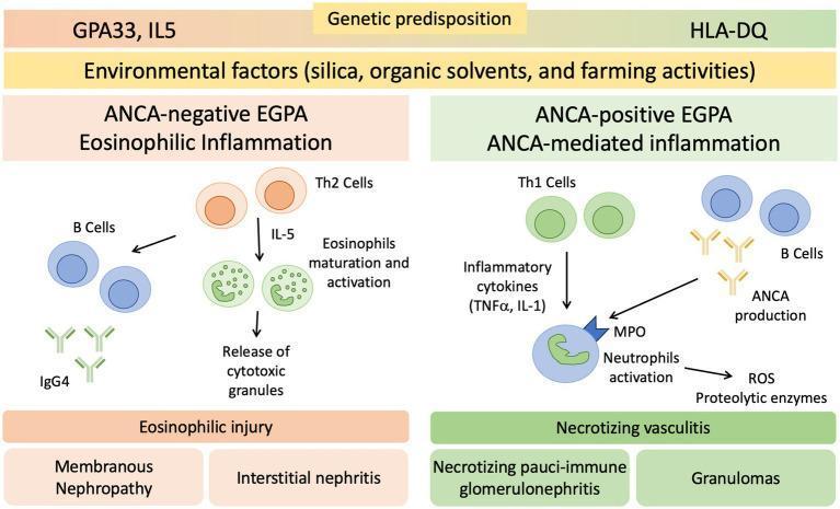
A medical pathophysiology diagram illustrating the dual-pathway pathogenesis of Eosinophilic Granulomatosis with Polyangiitis (EGPA) based on ANCA status. The diagram is divided into two primary sections: ANCA-negative EGPA (Eosinophilic Inflammation) and ANCA-positive EGPA (ANCA-mediated inflammation). Both pathways are preceded by genetic predispositions (GPA33, IL5 for ANCA-negative; HLA-DQ for ANCA-positive) and environmental triggers like silica and organic solvents. The ANCA-negative pathway shows Th2 cells secreting IL-5, leading to eosinophil maturation, activation, and release of cytotoxic granules, resulting in eosinophilic injury such as membranous nephropathy and interstitial nephritis; B cells contribute via IgG4 production. The ANCA-positive pathway depicts Th1 cells producing inflammatory cytokines (TNFα, IL-1) and B cells producing ANCA. These mediators activate neutrophils via myeloperoxidase (MPO), leading to reactive oxygen species (ROS) and proteolytic enzyme release. This results in necrotizing vasculitis, manifesting as necrotizing pauci-immune glomerulonephritis and granulomas.
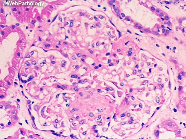
Imaging modality: Light microscopy (Hematoxylin and Eosin stain) of a renal cortical biopsy. Primary subject: renal glomerulus captured at high magnification (≈400x) showing a single glomerular tuft within the renal cortex. Visual features include crowded cellularity within the glomerular capillary loops and increased mesangial cell population, with indistinct capillary lumina due to cellular proliferation. The surrounding parenchyma shows mild tubulointerstitial involvement with scattered inflammatory cells; however, the main focus remains the glomerulus. The mesangial matrix appears variably expanded; there is no obvious crescents, fibrin deposition, or global sclerosis at this field of view. Background stroma is eosinophilic consistent with hematoxylin-eosin processing. Notable features such as endocapillary proliferation may indicate an inflammatory glomerulonephritis, though the absence of definitive immune complex deposits cannot be assessed on H&E alone. Diagnostic significance: these findings raise suspicion for an acute proliferative or mesangial proliferative glomerulonephritis; correlation with immunofluorescence, electron microscopy, and clinical data is essential. Potential clinical use cases include evaluation of proteinuria, hematuria, reduced glomerular filtration rate, or nephritic syndrome; differential diagnoses include IgA nephropathy, post-infectious GN, lupus nephritis, membranoproliferative GN. This image is valuable for medical education and research on renal pathology, glomerular architecture, and inflammatory patterns in the kidney.
giant cell arteritis temporal artery biopsy granuloma giant cells histology
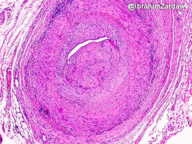
Histology image obtained from a temporal artery biopsy, prepared with Hematoxylin and Eosin stain and viewed under light microscopy at low magnification. The specimen demonstrates classic giant cell arteritis with transmural, granulomatous inflammation encompassing the full thickness of the arterial wall (intima, media, and adventitia) in a segmental distribution. Multinucleated giant cells are interspersed with a dense lymphohistiocytic infiltrate, and fragmentation or disruption of the internal elastic lamina is evident. The inflammatory process extends to the vasa vasorum and small venules within the adventitia, indicating widespread perivascular involvement. Accompanying features include marked intimal hyperplasia producing marked luminal narrowing and, in this field of view, near-occlusive lumen occlusion. The arterial media may show loss of elastic fibers and necrosis is not conspicuous here. These findings are diagnostic of GCA and help differentiate from other large-vessel vasculitides and nonspecific atherosclerotic changes. Clinically, such histology supports a diagnosis of temporal arteritis in patients with new headaches, visual symptoms, jaw claudication, and elevated inflammatory markers, guiding urgent corticosteroid therapy to prevent ischemic complications. This image exemplifies the radiologic-pathologic correlation typical of vasculitic arteritis. The slide highlights diagnostic relevance, research utility, and educational value for trainees and clinicians in practice. For medical education and research.
This histology image captures a cross-sectional temporal artery biopsy illustrating the hallmarks of giant cell arteritis. Prepared as a paraffin-embedded tissue section and stained with Hematoxylin and Eosin, the slide reveals a transmural inflammatory infiltrate that traverses all arterial wall layers. At the center, a narrowed lumen is surrounded by concentric intimal hyperplasia with marked medial disruption. Multinucleated giant cells and mononuclear macrophage clusters are present within the media, accompanied by lymphocytic infiltrate along the vessel wall. The elastic lamina appears disrupted, and fragmentation of the media is evident, consistent with robust granulomatous vasculitis. These features produce near-occlusive narrowing of the artery, explaining the ischemic risk associated with temporal arteritis. Clinically, such histopathology confirms a diagnosis of GCA and helps differentiate from atherosclerotic disease, infectious vasculitis, or other large-vessel vasculitides. The image underscores the systemic nature of the disease, as inflammation can extend from cranial vessels to the aorta. The diagnostic significance lies in correlating histology with patient presentation, including age-related cranial symptoms and risk of vision loss; timely immunosuppressive therapy can prevent irreversible ischemic complications. This specimen exemplifies classic angiitis with granulomatous inflammation and intimal hyperplasia driving luminal compromise.
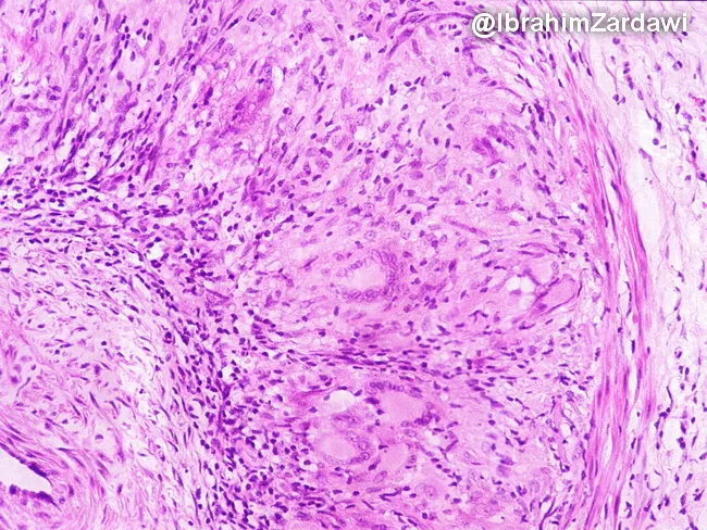
Imaging modality and technique: Brightfield light microscopy of a temporal artery biopsy stained with Hematoxylin and Eosin (H&E). Specimen: arterial wall from the temporal region; cross-sectional vascular biopsy showing inflammatory arteritis. Key features include granulomatous inflammatory infiltrate surrounding the media and adjacent to the internal elastic lamina, composed of lymphocytes, plasma cells, epithelioid histiocytes, and multinucleated giant cells. Giant cells are often present but may be absent; their presence supports diagnosis but is not required. The inflammatory infiltrate disrupts the media and may extend into the adventitia; fragmentation and phagocytosis of the internal elastic lamina are characteristic. Medial necrosis is minimal to absent; there is usually no well-formed granuloma or extensive necrosis. Neointimal hyperplasia and luminal narrowing can be observed as secondary consequences of chronic vasculitis. The image demonstrates a granulomatous arteritis pattern typical of giant cell arteritis. Diagnostic significance: classic histopathology includes granulomatous inflammation of medium-sized arteries with IEL fragmentation; biopsy positivity confirms temporal arteritis; however, skip lesions can yield false negatives; clinical correlation with headache, scalp tenderness, jaw claudication, visual symptoms and elevated inflammatory markers (CRP/ESR) is essential. Differential considerations include Takayasu arteritis, atherosclerosis, infectious vasculitis, and other granulomatous vasculitides. This slide is used in educational settings to illustrate GCA pathology and IEL involvement.
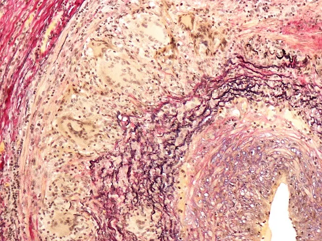
This histopathology image shows a temporal artery biopsy stained with an elastic stain (e.g., Verhoeff-Van Gieson) to highlight elastic laminae within the arterial wall. The specimen demonstrates classic giant cell arteritis features: fragmentation and partial duplication of the internal elastic lamina with loss of elastic fibers, and prominent intimal hyperplasia producing marked luminal narrowing. Inflammatory infiltrates, including mononuclear cells and multinucleated giant cells, surround the media in a granulomatous vasculitis pattern. Elastic lamina disruptions create irregular vascular borders and eccentric thickening of the intima. Some sections reveal medial edema and preserved outer adventitia, without definite necrosis. The overall morphology supports inflammatory arteritis affecting cranial arteries, particularly the superficial temporal artery. Clinically this histology aligns with temporal headache, jaw claudication, decreased vision risk, and elevated ESR/CRP in patients over 50. The diagnostic significance is high when elastic lamina fragmentation is present with compatible clinical symptoms. Differential considerations include atherosclerotic changes with intimal thickening and luminal narrowing, other large-vessel vasculitides such as Takayasu arteritis in younger patients, and other granulomatous diseases. Correlation with serology, imaging, and clinical presentation is essential for accurate diagnosis and timely glucocorticoid therapy. This image supports education, research, and diagnostic case reviews in rheumatology and pathology practice today.
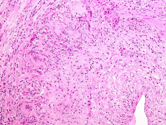
Imaging modality: Light microscopy of a temporal artery biopsy stained with Hematoxylin and Eosin. Precise anatomical localization: cranial temporal artery wall (temporal artery) with transmural involvement of the tunica intima and media. Visual features include a transmural lymphohistiocytic infiltrate with granulomatous inflammation and multinucleated giant cells; the internal elastic lamina is often disrupted with elastin debris that may be phagocytized. The architecture may be segmental, reflecting skip lesions; biopsy sensitivity can be limited, with a proportion of clinically suspected cases yielding negative results. Serial sectioning and submission of the entire specimen increase diagnostic yield. Clinical relevance centers on giant cell arteritis (temporal arteritis) in elderly patients presenting with new headaches and risk of irreversible vision loss if untreated. Differential considerations include Takayasu arteritis and polyarteritis nodosa. This histology image exemplifies canonical features: granulomatous vasculitis, giant cells, IEL disruption, and elastin fragmentation near the wall, reinforcing the importance of correlating histology with clinical syndrome and treatment urgency.
granulomatosis with polyangiitis Wegener lung kidney histology ANCA c-ANCA immunofluorescence
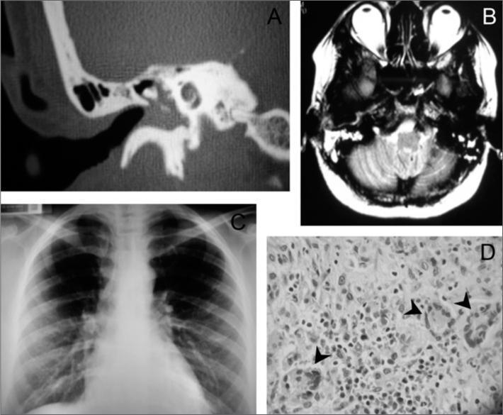
A multi-panel clinical figure presenting diagnostic findings for granulomatosis with polyangiitis (Wegener's). Panel A is a right-side temporal bone CT scan showing opacification or blurring of the tympanic cavity and mastoid air cells, suggesting inflammatory involvement. Panel B shows a T2-weighted axial skull MRI with a hyperintense signal in the right mastoid region, confirming fluid or soft tissue replacement of normal aeration. Panel C is a posterior-anterior chest X-ray demonstrating bilateral reticulonodular infiltrates, predominantly distributed in the lung bases. Panel D is a high-power histopathology micrograph (H&E stain) showing non-caseating granulomas, marked by arrows, characterized by localized clusters of epithelioid histiocytes and multinucleated giant cells within a background of dense inflammatory cell infiltration. Collectively, these images illustrate the multi-systemic nature of ANCA-associated vasculitis, involving the upper respiratory tract (ear/mastoid), lower respiratory tract (lungs), and microscopic tissue changes.
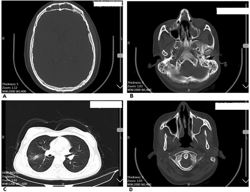
This composite of non-contrast Computed Tomography (CT) scans illustrates the multisystem manifestations of Granulomatosis with Polyangiitis (GPA), formerly known as Wegener’s granulomatosis. Panel A (axial head) shows mucosal thickening in the frontal sinuses. Panel B (axial head) reveals bilateral opacification of the maxillary sinuses and fluid within the mastoid air cells, diagnostic of bilateral sinusitis and mastoiditis. Panel C (axial chest) demonstrates pulmonary involvement characterized by focal ground-glass opacities and infiltrates in the lung parenchyma, features common in GPA-related vasculitis. Panel D (axial head) provides a detailed view of the nasal cavity showing significant destruction and perforation of the nasal septum alongside unilateral maxillary sinus opacification. These findings highlight the characteristic triad of upper respiratory tract, lower respiratory tract, and potentially renal involvement associated with this small-vessel vasculitis. The images serve as an educational tool for identifying typical radiological markers such as sinonasal destruction and pulmonary parenchymal disease.
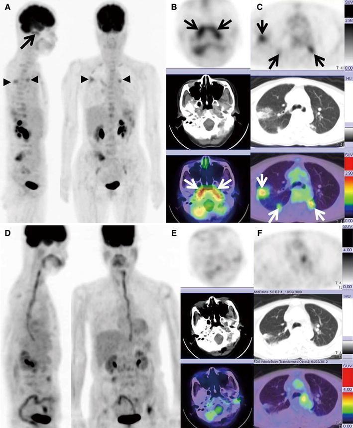
Diagnostic imaging series comprising FDG PET/CT scans of a 65-year-old patient with Granulomatosis with Polyangiitis (GPA/Wegener's). Panel A (MIP image) and Panels B-C (axial PET, CT, and fused PET/CT) show the baseline state with marked metabolic activity. Key findings include intense FDG uptake in the nasopharynx and bilateral auditory tubes (max SUV 4.30), and hypermetabolic nodular shadows in both lungs (max SUV 3.41). These findings correlate with elevated MPO-ANCA and PR3-ANCA titers. Panels D, E, and F display follow-up imaging five months post-treatment, illustrating a complete metabolic response. The repeat MIP and axial fused images show the resolution of previously seen hypermetabolism in the nasal mucosa and pulmonary nodules (max SUV reduced to 1.11–1.41), coinciding with the normalization of ANCA titers. This comparison illustrates the utility of FDG PET/CT in monitoring treatment response and disease activity in systemic vasculitis affecting the upper and lower respiratory tracts.
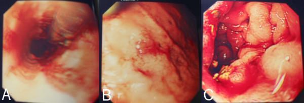
This set of three endoscopic images (A, B, and C) illustrates severe gastrointestinal manifestations of systemic vasculitis, specifically granulomatosis with polyangiitis (Wegener's). Panel A displays an esophageal view characterized by circumferential erythema, mucosal friability, and active longitudinal erosions. Panel B focuses on the gastric mucosa, showing diffuse, patchy inflammation with focal areas of hemorrhage and superficial ulcerations. Panel C provides a view of the duodenum, demonstrating significant mucosal edema and structural distortion. The duodenal surface exhibits intense confluent erythema, scattered petechiae, and adherent fibrinous exudates or sloughing tissue, particularly visible in the foreground. Collectively, these diagnostic images represent systemic inflammatory involvement of the upper gastrointestinal tract, leading to diffuse esophagitis, gastritis, and duodenitis. These findings are clinically significant as rare but severe complications of ANCA-associated vasculitis, highlighting the risk of mucosal ischemia and gastrointestinal hemorrhage.
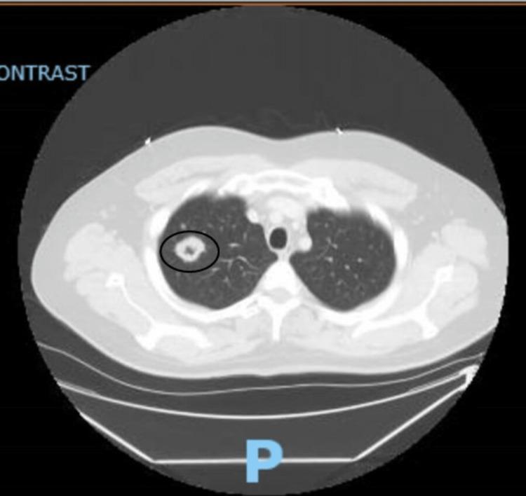
This diagnostic image is an axial non-contrast computed tomography (CT) scan of the upper chest. The primary finding is a solitary, well-defined cavitary nodule located in the right upper lobe of the lung, highlighted by a black circular annotation. The lesion exhibits a relatively thick, irregular wall with a lucent, air-filled central cavity, measuring approximately 2.9 cm in maximum diameter. The surrounding lung parenchyma appears largely unremarkable without evidence of significant consolidation or pleural effusion. Anatomical landmarks including the trachea, apical mediastinum, and ribs are visible and intact. Clinically, this radiographic presentation in a patient with positive c-ANCA titers is highly characteristic of Granulomatosis with Polyangiitis (GPA), formerly known as Wegener's granulomatosis. This image serves as a classic educational example for differential diagnosis of cavitary lung lesions, which includes GPA, tuberculosis, fungal infections, and malignancy.
Takayasu arteritis aorta histology granulomatous inflammation large vessel
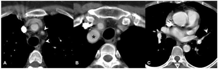
Diagnostic axial computed tomography (CT) scans of the chest in a patient with Takayasu arteritis, demonstrating large and medium-vessel vasculitis manifestations. Image A shows significant wall thickening and complete occlusion of the left common carotid and left subclavian arteries, indicated by white arrowheads, where contrast enhancement is absent within the lumen. Image B reveals aneurysmal dilation of the brachiocephalic trunk (marked with a black asterisk), characterized by an abnormal increase in vessel diameter. Image C illustrates involvement of the pulmonary circulation, specifically showing stenosis of a left pulmonary segmental artery (white arrowhead). This narrowing is attributed to granulomatous inflammation and subsequent fibrous thickening of the arterial wall. These images collectively highlight the diverse morphological changes—occlusion, dilation, and stenosis—typical of Takayasu arteritis, an idiopathic chronic inflammatory disease predominantly affecting the aorta and its primary branches.

This set of four axial Computed Tomography Angiography (CTA) scans demonstrates the thoracic aorta in a patient with Takayasu arteritis, highlighting the diagnostic features of large-vessel vasculitis. Panels (a) and (b) show the pre-treatment phase, characterized by significant circumferential wall thickening of the aortic arch (arrows) with associated mural contrast enhancement. This 'halo' or 'macaroni' sign indicates active vessel wall inflammation. Panels (c) and (d) present follow-up imaging of the same anatomical regions after medical intervention. These post-treatment scans show a marked reduction in both mural thickness and contrast enhancement, signifying a positive therapeutic response and decreased disease activity. The imaging modality utilizes intravenous iodinated contrast to differentiate between the arterial lumen and the pathological vessel wall. This comparison is clinically significant for monitoring disease progression and the efficacy of immunosuppressive therapy in vasculitis patients.
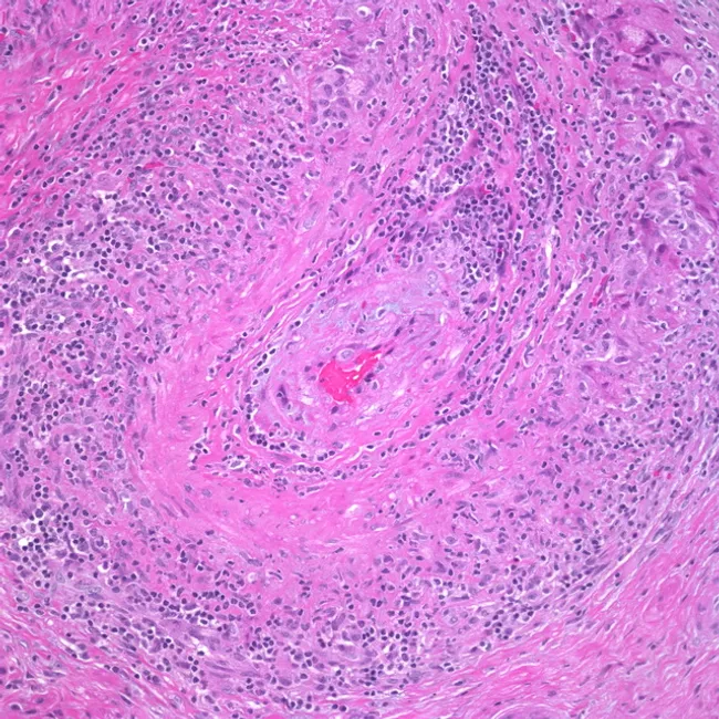
High-power hematoxylin and eosin stained transverse section of a medium-sized artery demonstrating granulomatous arteritis. The lumen is narrowed by edema and fibrosis of the intima, while the tunica media is uniformly expanded by a mixed inflammatory infiltrate. Lymphocytes, plasma cells, and histiocytes predominate, with several multinucleated giant cells visible in the upper right quadrant. The internal elastic lamina is variably destroyed, with fragmentation evident at the media- adventitia interface. Transmural inflammation produces concentric thickening of the vessel wall and marked luminal compromise. Admixed eosinophils are sparse; occasional foamy histiocytes emphasize granulomatous architecture. The adventitia shows mild edema and vascular remodeling. Overall features are classic for granulomatous vasculitis, most consistent with giant cell arteritis (temporal arteritis) or other large-vessel vasculitides such as Takayasu arteritis. Diagnostic significance: histology provides definitive confirmation when clinical suspicion is high and ESR/CRP elevations are present. Differential considerations include polyarteritis nodosa, infectious vasculitis, and eosinophilic granulomatosis with polyangiitis depending on clinical context. Clinically relevant correlations include age >50 years, new headaches, jaw claudication, visual symptoms, and constitutional signs. This image is valuable for pathology education, residency training, and radiologic-pathologic correlation studies in vasculitis. Useful for teaching differential diagnosis and correlating clinical features with histologic patterns in practice.
microscopic polyangiitis pauci-immune crescentic glomerulonephritis necrotizing capillaritis
A medical pathophysiology diagram illustrating the dual-pathway pathogenesis of Eosinophilic Granulomatosis with Polyangiitis (EGPA) based on ANCA status. The diagram is divided into two primary sections: ANCA-negative EGPA (Eosinophilic Inflammation) and ANCA-positive EGPA (ANCA-mediated inflammation). Both pathways are preceded by genetic predispositions (GPA33, IL5 for ANCA-negative; HLA-DQ for ANCA-positive) and environmental triggers like silica and organic solvents. The ANCA-negative pathway shows Th2 cells secreting IL-5, leading to eosinophil maturation, activation, and release of cytotoxic granules, resulting in eosinophilic injury such as membranous nephropathy and interstitial nephritis; B cells contribute via IgG4 production. The ANCA-positive pathway depicts Th1 cells producing inflammatory cytokines (TNFα, IL-1) and B cells producing ANCA. These mediators activate neutrophils via myeloperoxidase (MPO), leading to reactive oxygen species (ROS) and proteolytic enzyme release. This results in necrotizing vasculitis, manifesting as necrotizing pauci-immune glomerulonephritis and granulomas.
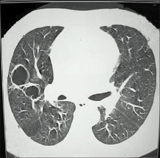
This diagnostic image is an axial computed tomography (CT) scan of the thorax, specifically focused on the lung parenchyma using a lung window setting. The image reveals bilateral pulmonary pathology characterized by multiple thin-walled, air-filled cavities. These cavities are variably sized, with a prominent cluster of large, well-defined circular and oval lucencies located in the right lung field. The surrounding parenchyma shows some evidence of increased interstitial markings and potential ground-glass opacities, which may indicate inflammation or hemorrhage. In the clinical context of pauci-immune crescentic glomerulonephritis and recurrent hemoptysis, these radiological findings are highly suggestive of pulmonary involvement in small-vessel vasculitis, such as Granulomatosis with Polyangiitis (GPA). The image demonstrates the hallmark features of cavitary lung disease, serving as a critical teaching tool for medical students and clinicians specializing in pulmonology, nephrology, and radiology to recognize the thoracic manifestations of systemic vasculitides and their potential for life-threatening complications like massive pulmonary hemorrhage.
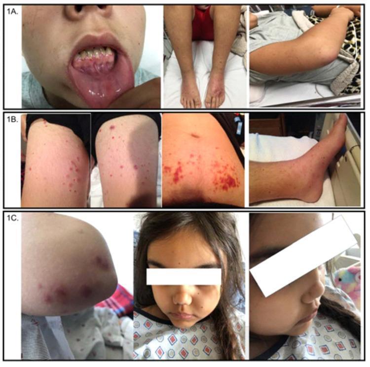
A composite of clinical photographs (labeled 1A, 1B, 1C) showcasing the dermatologic and oral manifestations of pediatric Granulomatosis with Polyangiitis (GPA). Panel 1A illustrates significant gingival hyperplasia with associated mucosal bleeding and friability, alongside diffuse purpura and pitting edema on the bilateral lower extremities and right arm. Panel 1B displays a diffuse, erythematous papular rash distributed across the bilateral thighs, lower legs, and anterior trunk. Panel 1C presents scattered, palpable purpuric lesions on the upper extremities and a characteristic saddle nose deformity involving the collapse of the nasal bridge. Collectively, these images demonstrate the multisystemic nature of ANCA-associated vasculitis in children, highlighting key clinical signs such as small-vessel vasculitic skin lesions, characteristic midface skeletal changes, and upper respiratory tract involvement. The visual evidence supports medical education on the physical examination findings essential for diagnosing systemic autoimmune diseases and pauci-immune crescentic glomerulonephritis.
This diagnostic image shows direct immunofluorescence (DIF) of a renal biopsy specimen, specifically highlighting fibrinogen deposition. The image reveals intense, apple-green fluorescence with a heterogeneous distribution. The staining pattern is prominently seen within the walls of a large-caliber renal artery and associated perivascular spaces, appearing as bright, irregular, and somewhat fibrillar or linear deposits. This visual finding is characteristic of necrotizing vasculitis, where fibrinoid necrosis leads to the entrapment of fibrinogen within the damaged vascular walls. The surrounding interstitial tissue displays fainter, more diffuse staining patterns. In the clinical context of ANCA-associated pauci-immune necrotizing vasculitis, this fibrinogen-positive staining serves as a critical marker for active vascular injury, contrasting with the absence of significant immunoglobulin or complement (C3) deposits that would otherwise suggest immune-complex mediated glomerulonephritis. The image is a primary educational resource for nephropathology, illustrating the visual manifestations of vascular necrosis at a microscopic level.
Kawasaki disease coronary artery histology aneurysm eosinophilic granulomatosis polyangiitis Churg-Strauss eosinophils
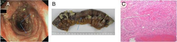
A three-panel figure documenting colonic involvement in Eosinophilic Granulomatosis with Polyangiitis (EGPA), also known as Churg-Strauss syndrome. Panel A is an endoscopic image of the transverse colon showing severe mucosal inflammation, erythema, and irregular ulcerations (blue arrows) indicative of vasculitis. Panel B is a macroscopic clinical photograph of a resected 15-20 cm segment of the transverse colon; the specimen exhibits transmural ischemia, necrotizing lesions, and areas of dark, necrotic tissue with adherent fibrinous exudate. Panel C is a histopathological micrograph (H&E stain) of the resected colon demonstrating characteristic features of EGPA, including dense eosinophilic infiltration within the tissue and thrombotic vessel occlusion. This collection illustrates the progression from endoscopic signs of vasculitis to clinical bowel perforation and the underlying microscopic vascular damage.
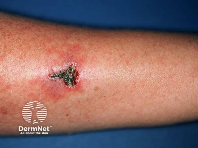
Clinical photography of a solitary forearm lesion demonstrates an irregular ulcer with a dark necrotic crust and surrounding erythema. The image depicts cutaneous involvement that can accompany eosinophilic granulomatosis with polyangiitis (Churg-Strauss syndrome), a small-vessel vasculitis characterized by eosinophilic infiltrates and granulomatous inflammation. Visual features include a punched-out appearance, eschar formation, and a tender, inflamed rim. This macroscopic dermatologic finding is non-specific but clinically significant when integrated with systemic symptoms such as asthma, eosinophilia, neuropathy, and vasculitic organ involvement. The diagnostic value lies in documenting skin involvement, guiding biopsy targeting, and assisting differential diagnosis against leukocytoclastic vasculitis, pyoderma gangrenosum, infectious ulcers, or other cutaneous vasculitides. In clinical practice, this image would be used in dermatology, rheumatology, and pathology workflows to correlate histology with morphology, monitor treatment response, and educate learners about cutaneous manifestations of EGPA and related eosinophilic vasculitides. These attributes enhance cross-disciplinary teaching, case-based learning, and AI-enabled retrieval in diagnostics practice.
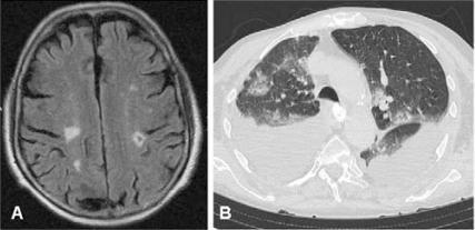
This dual-panel diagnostic image displays multi-organ involvement in a patient with Eosinophilic Granulomatosis with Polyangiitis (Churg-Strauss syndrome). Panel A is an axial MRI scan of the brain (FLAIR sequence) showing multiple hyperintense foci within the subcortical white matter and cortex, primarily in the frontal and parietal lobes, consistent with multiple embolic or vasculitic infarcts. Panel B is an axial CT scan of the thorax in a lung window, demonstrating bilateral, patchy ground-glass opacities and pulmonary infiltrates. Significant fluid accumulation is noted in the pleural spaces bilaterally, representing bilateral pleural effusion. Together, these diagnostic images illustrate the systemic nature of small-to-medium vessel vasculitis, specifically highlighting the neurologic (central nervous system ischemia) and pulmonary (eosinophilic infiltrates and exudative effusion) manifestations of the disease. This content is suitable for medical education regarding rheumatological emergencies and systemic vasculitides.
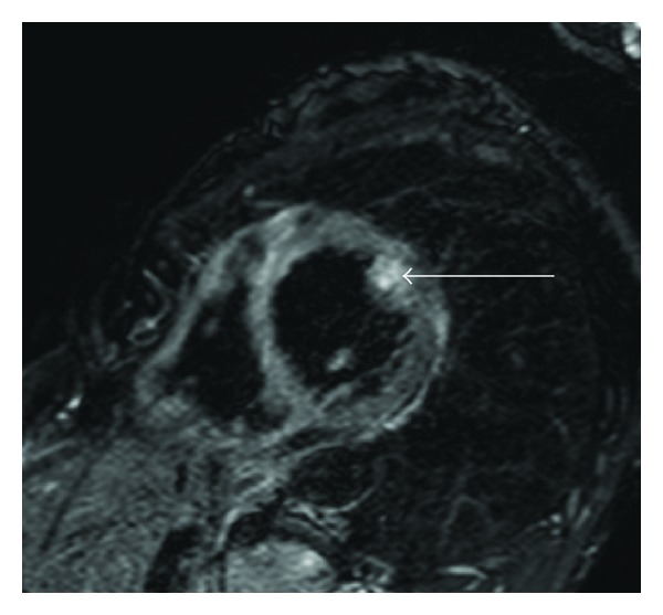
**Imaging Modality:** Cardiac Magnetic Resonance Imaging (MRI), T2-weighted sequence. **Anatomical Region:** Heart, mid-ventricular short-axis view. **Observed Pathology:** Eosinophilic myocarditis secondary to Churg-Strauss syndrome (Eosinophilic Granulomatosis with Polyangiitis). **Characteristic Visual Features:** The image demonstrates focal areas of increased signal intensity (hyperintensity) within the myocardium. A prominent, hyperintense patchy area is localized to the papillary muscle (indicated by the white arrow), consistent with localized myocardial edema. Additional subtle, patchy intramural T2 hyperintensities are visible within the left ventricular wall. **Key Diagnostic Features:** T2-weighted hyperintensity is a primary radiologic marker for myocardial edema and active inflammation. In the clinical context of systemic vasculitis, this distribution pattern—specifically involving the papillary muscles and exhibiting a non-coronary, patchy distribution—is characteristic of eosinophilic infiltration and associated inflammatory response. **Clinical Significance:** This finding is essential for diagnosing cardiac involvement in systemic hypereosinophilic syndromes and monitoring the inflammatory phase of myocarditis.
Goodpasture anti-GBM linear immunofluorescence kidney basement membrane
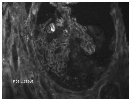
This diagnostic image shows a direct immunofluorescence (DIF) microscopy of a percutaneous kidney biopsy specimen at 20X magnification. The image demonstrates a hallmark pathognomonic finding: strong, continuous, linear immunofluorescence staining along the glomerular basement membrane (GBM). The staining outlines the capillary loops of the glomerulus with high intensity and uniformity, indicating the deposition of immunoglobulin G (IgG) antibodies directly against the basement membrane. The surrounding mesangial regions and Bowman's space show significantly less fluorescence, creating a sharp contrast that emphasizes the linear pattern. This visual presentation is characteristic of anti-glomerular basement membrane (anti-GBM) antibody disease, often associated with Goodpasture syndrome or rapidly progressive (crescentic) glomerulonephritis. The clinical significance of this finding is the confirmation of autoimmune-mediated renal damage where autoantibodies target Type IV collagen within the GBM.
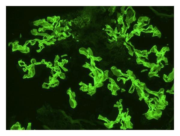
This diagnostic image is a direct immunofluorescence (DIF) micrograph of human renal tissue. The image demonstrates a characteristic linear staining pattern along the glomerular basement membrane (GBM). The fluorescent signal, appearing as a bright green (FITC) stain, reveals continuous, ribbon-like deposits of IgG that follow the complex, convoluted contours of the glomerular capillaries. This specific linear distribution is a hallmark of anti-GBM disease, such as Goodpasture syndrome, distinguishing it from the granular deposits typically seen in immune-complex-mediated glomerulonephritis. The clusters of highlighted structures represent multiple glomerular segments within the biopsy sample, showing uniform intensity of antibody binding. This finding is critical for the diagnosis of rapidly progressive glomerulonephritis (RPGN) and guides urgent clinical intervention like plasmapheresis and high-dose corticosteroid therapy.
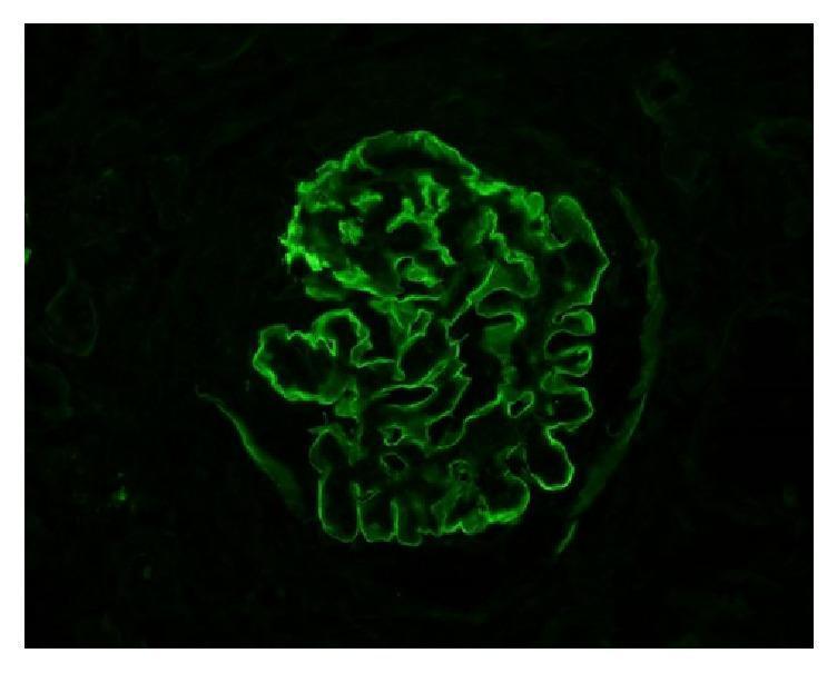
This diagnostic image is a high-power immunofluorescence (IF) microscopy of a human renal glomerulus. The image demonstrates a hallmark diagnostic pattern for anti-glomerular basement membrane (anti-GBM) disease. There is intense, continuous, and highly smooth linear staining for IgG (immunoglobulin G) along the glomerular capillary loops. Unlike the granular appearance typical of immune complex deposition (e.g., in lupus nephritis or post-infectious glomerulonephritis), this linear ribbon-like fluorescence highlights the entire circumference of the capillary basement membranes. The background is appropriately dark, emphasizing the bright green fluorescein isothiocyanate (FITC) signal. This visual finding is a key educational feature for identifying Goodpasture syndrome or anti-GBM mediated crescentic glomerulonephritis in a clinical pathology context. The pattern reflects the direct binding of autoantibodies to the non-collagenous domain of the alpha-3 chain of type IV collagen within the basement membrane.
cryoglobulinemic vasculitis leukocytoclastic vasculitis skin biopsy lupus full-house immunofluorescence
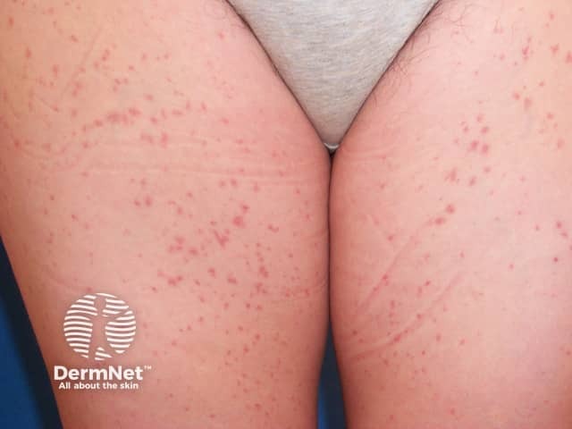
This dermatology clinical photograph demonstrates bilateral anterior thighs with a scattered eruption of small, non-blanching red-to-purple macules and papules consistent with cutaneous vasculitis. The lesions are primarily pinpoint to palm-sized clusters, with some faint purpuric coalescence along the inguinal folds. The appearance is compatible with palpable purpura from leukocytoclastic vasculitis (small-vessel vasculitis) affecting dermal capillaries. The distribution is symmetrical and non-exudative, without overt necrosis. No ulceration. The image is acquired as a close-up frontal view using digital photography in standard diagnostic lighting. There is a labeling watermark. The clinical significance: cutaneous vasculitis may be primary or secondary (infection, medications, autoimmune disease). Diagnostic significance: must correlate with symptoms, complete history, and laboratory tests; confirm by skin biopsy with immunofluorescence showing leukocytoclastic vasculitis and possibly IgA deposition in IgA vasculitis. Differential diagnoses include Henoch-Schonlein purpura (IgA vasculitis), drug-induced leukocytoclastic vasculitis, cryoglobulinemic vasculitis, essential thrombocytopenia-associated purpura, pityriasis lichenoides et varioliformis acuta. Potential clinical use cases: dermatology teaching, triage in suspected systemic vasculitis, biopsy planning, case series documentation, AI-enabled image retrieval for vasculitis phenotypes. Limitations of a single-view image include potential underestimation of lesion depth and evolution. For documentation, standardize lighting, include multiple angles, and capture high-resolution regional views. Serial photographs can help track progression and response to therapy and monitor systemic involvement.
This diagnostic image demonstrates direct immunofluorescence (DIF) of a skin punch biopsy specimen. The micrograph reveals a characteristic granular green fluorescent staining pattern against a dark background. These fluorescent deposits are primarily concentrated in a perivascular distribution, localized around the superficial dermal blood vessels. The morphology of the staining is punctate and clumped, typical of immune complex deposition. Clinically, this visual evidence of perivascular IgA deposition is a hallmark diagnostic feature of IgA vasculitis (formerly Henoch-Schönlein purpura). The image illustrates the classic immunopathologic findings associated with leukocytoclastic vasculitis involving the small vessels of the dermis. This material is suitable for dermatology and rheumatology education, specifically for teaching the diagnostic criteria of systemic small-vessel vasculitides.
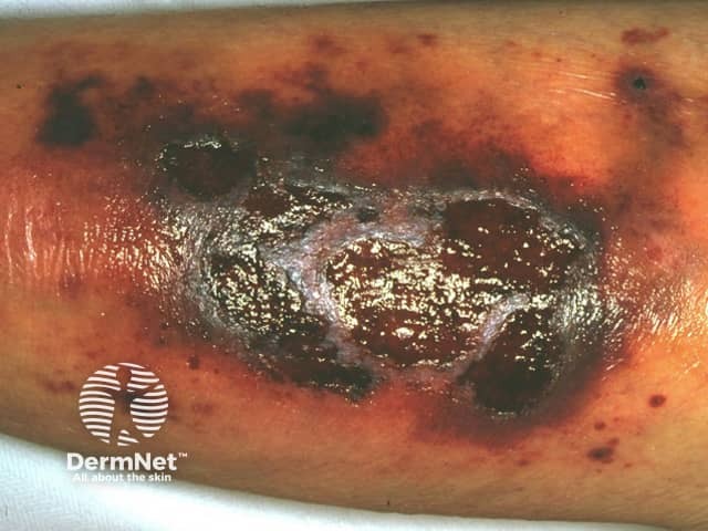
Clinical photography of a lower leg skin lesion showing a large necrotic ulcer with a dark eschar, surrounding erythema, edema, and crusting. The ulcer involves superficial to potentially deep dermal tissue with moist slough and irregular, undermined borders. The presentation is characteristic of cutaneous necrotizing vasculitis or severe vasculitic ulceration, with necrosis and pigment changes around the lesion. The image emphasizes key features such as necrotic center, black/brown eschar, vascular-mediated tissue injury, and inflammatory halo. Differential diagnoses include cutaneous leukocytoclastic vasculitis, polyarteritis nodosa with skin involvement, cryoglobulinemic vasculitis, granulomatosis with polyangiitis, and secondary infection risk. Clinically, this pattern may reflect systemic vasculitis or isolated cutaneous vasculitis and warrants correlation with patient history, systemic examination, and laboratory testing. Next steps typically include targeted skin biopsy with histopathology and direct immunofluorescence, serologic evaluation for ANCA, complements, cryoglobulins, and infectious workup to exclude septic etiology. Management implications involve immunomodulatory therapy consideration and infection control. This image is valuable for dermatology, rheumatology, and infectious disease education, case-based discussions, and AI-driven search datasets. It illustrates the importance of recognizing necrotizing vasculitic skin lesions and guiding diagnostic pathways. Standardized captions support multi-terminology indexing for clinicians, researchers, and trainees across dermatology and rheumatology domains in educational repositories.
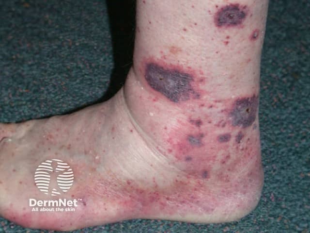
Clinical photography of the skin demonstrates multiple non-blanching, violaceous purpuric patches and plaques concentrated over the distal leg, ankle, and surrounding soft tissue. The lesions are irregular in shape, vary in size from a few millimeters to several centimeters, and appear coalescent in places with mild surrounding erythema and edema. This distribution on the lower extremity is classic for small-vessel vasculitis presenting as palpable purpura, most consistent with leukocytoclastic vasculitis, cutaneous IgA vasculitis, or a hypersensitivity process. The photograph provides a frontal view of the distal leg and ankle, capturing surface morphology including color gradient from dark purple to burgundy, subtle scaling, and surface fragility. In a clinical context, such skin findings warrant correlation with laboratory studies (CBC for thrombocytopenia, ESR/CRP for inflammation, renal function tests, urinalysis for nephritis) and serologies (ANCA, complement levels). Differential diagnoses include drug-induced vasculitis, cryoglobulinemic vasculitis, Henoch–Schönlein purpura, and other systemic vasculitides; trauma or coagulopathy can mimic purpura but typically have different distribution or blanching pattern. Management relies on clinical staging and systemic involvement; biopsy may be required for definitive histopathology. Early recognition supports prompt treatment and reduces risk of organ involvement. Clinical follow-up and biopsy may refine diagnosis and guide targeted therapy for confirmation.
mkdir -p /tmp/workspace/vasculitis-pdf && ls /tmp/workspace/