
Internal derangement of tmj
internal derangement temporomandibular joint TMJ disc displacement MRI
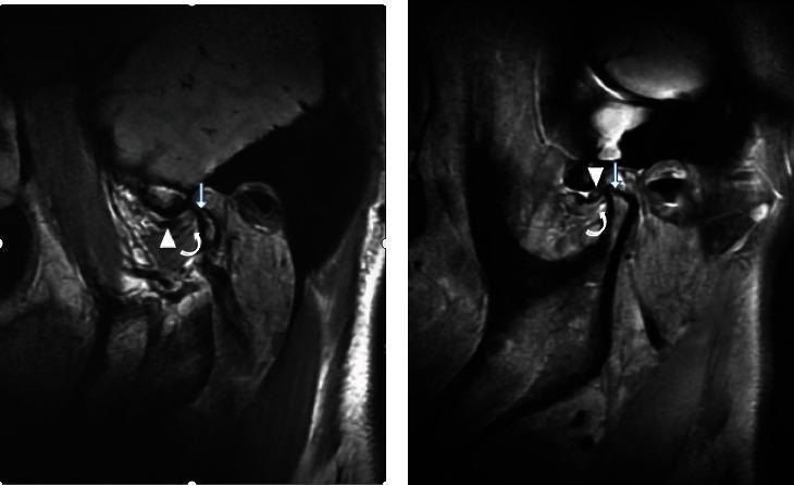
This diagnostic comparison contains two magnetic resonance imaging (MRI) views of the temporomandibular joint (TMJ) illustrating different stages of internal derangement. Image (a) demonstrates an irreducible anterior articular disc displacement. Specific pathology includes the anteriorly displaced disc (arrow), notable thickening at the attachment point of the inferior branch of the lateral pterygoid muscle (curved arrow), and secondary degenerative changes evidenced by condylar hyperosteogeny (straight arrow). Image (b) shows the contralateral side presenting with a reducible anterior articular disc displacement. While the disc is also displaced anteriorly (arrow), the lateral pterygoid muscle attachment point (curved arrow) appears normal in thickness, and there is an absence of the hyperosteogeny seen in the irreducible state. This comparison serves as an educational reference for identifying radiological signs of chronic TMJ disc displacement and associated muscle/bone remodeling, emphasizing the anatomical differences between reducible and irreducible internal derangements.
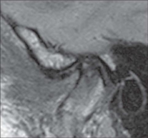
This diagnostic image is a sagittal proton density (PD)-weighted MRI of the temporomandibular joint (TMJ) in a closed-mouth position. The scan illustrates advanced internal derangement characterized by anterior disc displacement. The articular disc, which should normally reside in a biconcave configuration between the mandibular condyle and the temporal bone's articular eminence, is displaced anteriorly. Significant morphological changes indicative of degenerative disc disease are visible, including a loss of the normal biconcave shape, acquiring a more biconvex or rounded contour. The disc demonstrates thinning (tapering) of the anterior band and surface irregularities. Educational focus includes the identification of anatomical landmarks of the TMJ (mandibular condyle, articular eminence, and external auditory canal) and the assessment of disc positioning and morphology for diagnosing temporomandibular disorders (TMD). This content is relevant for intermediate to advanced medical learners in radiology, maxillofacial surgery, and dentistry.
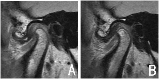
This diagnostic image displays side-by-side parasagittal MRI scans of the human temporomandibular joint (TMJ), categorized as a comparison of treatment outcomes for internal derangement. Panel A demonstrates Disc Displacement with Reduction (DDwR) before treatment; the articular disc is positioned anteriorly relative to the mandibular condyle, with a noticeable increase in the posterior joint space. Panel B shows the same joint after functional appliance therapy, illustrating successful disc recapture. In this post-treatment view, the intermediate zone of the biconcave disc is correctly interposed between the superior aspect of the condyle and the posterior slope of the articular eminence. Notably, the condylar morphology remains stable between both images, showing no evidence of degenerative changes or significant osseous remodeling. This comparison serves as an educational example of a 'good outcome' in orthodontic or maxillofacial management of TMJ disorders, highlighting the restoration of anatomical disc-condyle relationships.
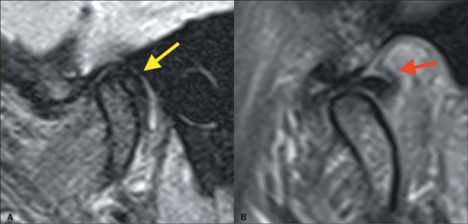
This Comparison Chart features side-by-side sagittal proton density (PD)-weighted MRI images of the temporomandibular joint (TMJ) in closed-mouth (A) and open-mouth (B) positions, illustrating internal derangement. Image A demonstrates rare posterior displacement of the biconcave articular disc (yellow arrow) relative to the mandibular condyle. Secondary signs include thickening of the posterior bilaminar zone (red arrow) and joint effusion, characterized by hyperintense signal within the joint space. Image B shows the dynamic progression where the disc is recaptured, returning to its physiological position between the condyle and the temporal bone's articular eminence as the jaw opens and the condyle translates anteriorly. The educational focus is on the diagnostic imaging features of disc displacement with reduction (recapture). This content is relevant for radiology and dentistry residents learning to interpret TMJ arthropathy, emphasizing the spatial relationship between the fibrocartilaginous disc and bony landmarks.
temporomandibular joint internal derangement disc displacement
Internal Derangement of the Temporomandibular Joint (TMJ)
Definition
- Grainger & Allison's Diagnostic Radiology, p. 1614
- Rheumatology (Firestein/Kelley), p. 762
Anatomy Relevant to ID
- Articular disc (meniscus): A biconcave fibrocartilaginous structure (bow-tie shaped on MRI) interposed between the condyle and glenoid fossa. It is avascular and aneural.
- Retrodiscal tissue (bilaminar zone): Lies behind the disc; highly vascular and well-innervated - the main source of pain when compressed.
- Lateral pterygoid muscle: Inserts into the anterior disc and condylar neck - its hyperactivity can pull the disc anteriorly.
- Joint compartments: The disc divides the joint into superior (translational) and inferior (rotational) compartments.
- Innervation: Auriculotemporal, masseteric, and deep temporal branches of V3.
- Rheumatology (Firestein/Kelley), p. 762
Classification of Internal Derangement
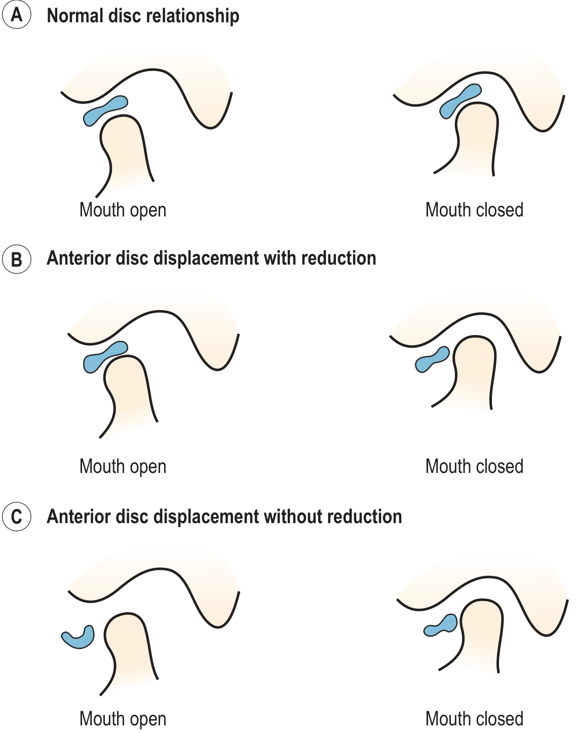
1. Disc Displacement WITH Reduction (DDwR)
- Disc is anteriorly displaced at rest (closed mouth)
- On mouth opening, the condyle slides under the disc, which snaps back to normal position = audible/palpable click
- A second click may occur on closing (reciprocal click)
- Mandible deviates toward the affected side on opening, then corrects when the disc reduces
2. Disc Displacement WITHOUT Reduction (DDwoR) - "Closed Lock"
- Disc remains anteriorly displaced throughout the range of motion
- No click (the disc blocks forward condylar translation)
- Acute: Sudden onset, severe restricted mouth opening (<25-30 mm), deviation toward affected side that does not correct
- Chronic: Disc undergoes fibrous change and perforates; progressive restriction may improve over time as the disc becomes deformed/perforated
Wilkes Classification (Staging)
| Stage | Features |
|---|---|
| I (Early) | Slight anterior disc displacement, clicking, no pain, normal radiograph |
| II (Early/Intermediate) | Occasional catching, clicking, mild pain; slight disc deformity on MRI |
| III (Intermediate) | Frequent pain, joint tenderness, restricted opening, significant disc deformity; closed lock possible |
| IV (Intermediate/Late) | Chronic pain, significant restriction, disc deformity and perforation; early bony changes |
| V (Late) | Crepitus, chronic pain, gross disc deformity/perforation, osteoarthritic bony changes |
Clinical Features
- Pain - preauricular, temporal, or facial; worsened by chewing; often referred to ear (mimics otitis)
- Joint sounds - clicking (DDwR), crepitus (late degenerative)
- Restricted mouth opening - normal inter-incisal distance is >35 mm; <25 mm in a young adult without systemic illness strongly suggests acute closed lock
- Mandibular deviation - toward affected side on opening in DDwoR
- Malocclusion - if joint collapse occurs (bilateral disease)
- Rheumatology (Firestein/Kelley), p. 763
Imaging
MRI (Investigation of Choice)
- Best modality for soft tissue/disc assessment
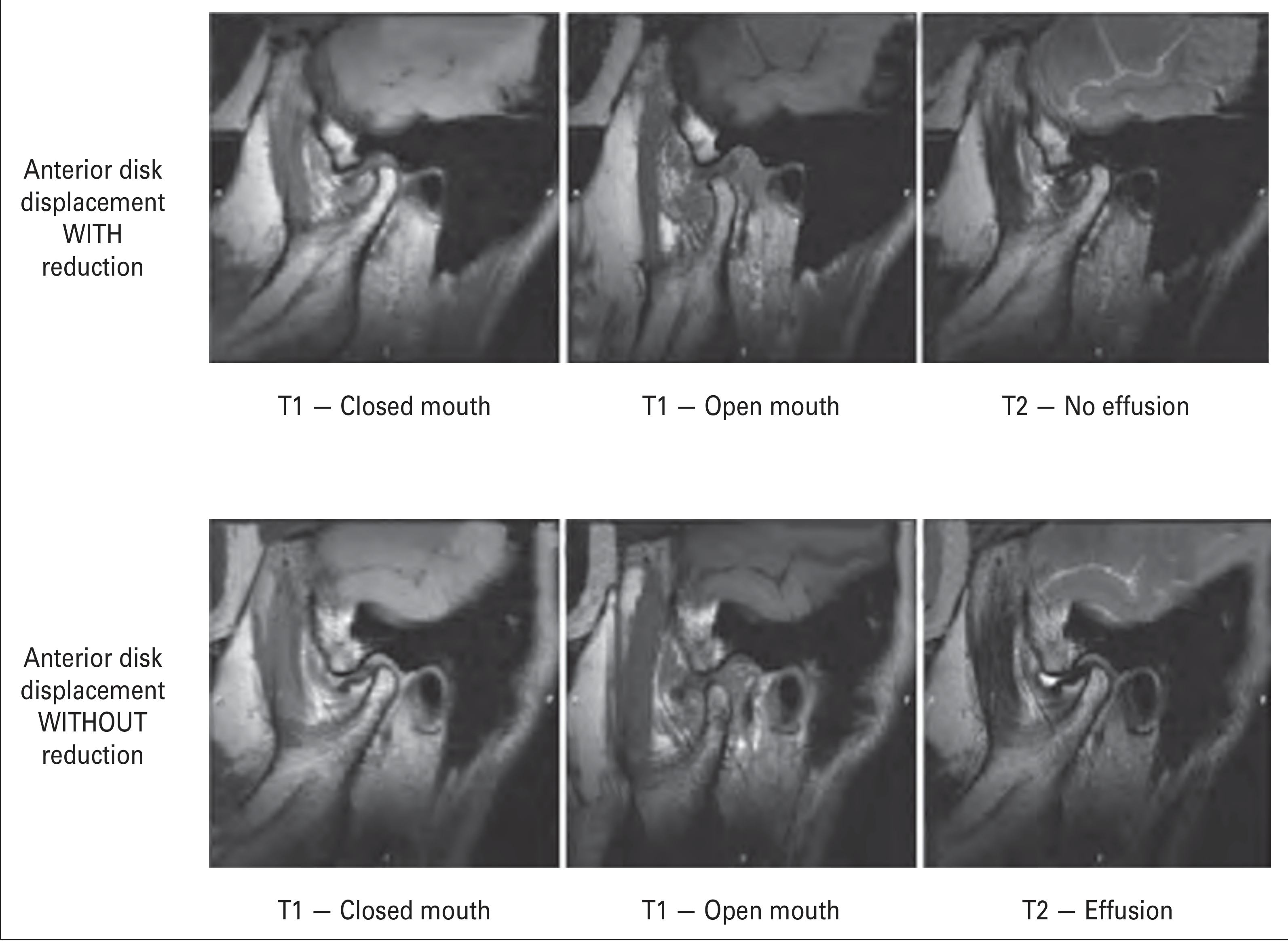
- Protocol: Proton density (PD) or T1-weighted 3 mm parasagittal slices in closed and open mouth positions; T2 for effusion/synovitis
- Normal disc: biconcave (bow-tie shaped), low signal, seated on top of the condyle
- DDwR: disc anteriorly displaced on closed-mouth images, returns to normal on open-mouth images
- DDwoR: disc remains anteriorly displaced on both closed and open mouth images; disc may become deformed/biconvex; effusion more likely
- T2 effusion is associated with more advanced stages (nonreducing discs) and represents synovitis
- Coronal views: show medial/lateral displacement and articular surface degeneration
CT
- Best for bony changes - condylar erosions, flattening, osteophytes, subchondral sclerosis in OA
- Useful for surgical planning (total joint replacement)
Plain Radiographs
-
Low predictive value for disc displacement
-
Panoramic OPG: useful screening; may show condylar flattening, erosion, or developmental asymmetry
-
Grainger & Allison's Diagnostic Radiology, pp. 1614-1615
Differential Diagnosis
-
Myofascial pain dysfunction (no joint pathology on MRI)
-
Trigeminal neuralgia / atypical facial pain
-
Odontogenic infection (molar region)
-
Sinus or ear infection (otitis media)
-
Salivary gland dysfunction (parotid)
-
Rheumatoid / psoriatic arthritis affecting TMJ
-
Ankylosis
-
Tumors (osteochondroma, condylar hyperplasia)
-
Rheumatology (Firestein/Kelley), Box 85.2, p. 763
Management
Goals
- Reduce pain and inflammation
- Improve range of motion and function
- Reduce joint loading forces
Conservative (First-Line)
| Modality | Details |
|---|---|
| Occlusal splint | Anterior repositioning splint for DDwR; stabilization splint for bruxism; a 2025 systematic review (PMID 41091263) confirms splint therapy is effective |
| Physical therapy | Jaw exercises, manual therapy, postural correction |
| Behavioral modification | Restrict wide mouth opening, diet modification (soft foods), stress reduction |
| Pharmacologic | NSAIDs (piroxicam, diclofenac, ibuprofen), muscle relaxants (cyclobenzaprine, clonazepam), tricyclics (amitriptyline), gabapentin for chronic pain |
| Topical agents | Capsaicin cream, topical diclofenac (equal efficacy to oral) |
- NSAIDs > muscle relaxants/benzodiazepines > antidepressants/antiepileptics > immune modulators (infliximab ± methotrexate for inflammatory arthropathy)
Minimally Invasive Procedures
- Arthrocentesis: Joint washout under pressure; first-line for acute restricted opening (anchored disc phenomenon); particularly effective in young adults; success 75-95%; early treatment is curative, delay can cause permanent restriction
- Arthroscopy: Diagnostic AND therapeutic; can perform lysis, lavage, disc repositioning; success 75-95%; significantly reduced need for open surgery
Surgical (5-10% of patients)
Relative indications: Pain and dysfunction directly from the TMJ refractory to all non-surgical measures
- Arthrocentesis
- Arthroscopic surgery (disc repositioning, lysis/lavage)
- Open arthroplasty with disc repositioning or discectomy
- Total joint replacement (alloplastic) - for end-stage OA; FDA-approved devices now available with long success records
- Rheumatology (Firestein/Kelley), pp. 762-763
- Scott-Brown's Otorhinolaryngology, p. 916
Epidemiology
- ~30% of the population will experience TMJ problems at some point; ~10% have active signs/symptoms at any given time
- Annual incidence of TMJD: 2-4%; >5.3 million U.S. adults seek treatment annually
- Female predominance (F:M ratios reported 2:1 to 10:1); higher risk in women
- Peak age: under 40; degenerative disease increases with age over 40
- Up to 20% progress to a persistent pain state
Key Points Summary
- Internal derangement = disc displacement, most commonly anteromedial
- Two types: DDwR (click, self-correcting on opening) vs. DDwoR/closed lock (no click, restricted opening)
- MRI is the gold standard for diagnosis - done in closed AND open mouth positions
- Mouth opening <25 mm in a young adult = presume acute closed lock; refer urgently for early arthrocentesis
- Management is stepwise: conservative → arthrocentesis → arthroscopy → open surgery
- Splint therapy remains the most studied and supported nonsurgical intervention (2025 systematic review)