
Myocardial infraction
Reading File
Now let me search the medical library for comprehensive content on myocardial infarction in parallel:
Finding Sources
Searching PubMed
"myocardial infarction"[MeSH Terms] AND "management"[MeSH Terms]
Excellent. Now let me read the most comprehensive sources in parallel - Braunwald's (gold standard cardiology), Robbins (pathology), Ganong (physiology), and Goldman-Cecil (clinical medicine):
Finding Sources
Reading File
Reading File
Reading File
Now let me read the Robbins Pathology content (pathological changes), Goldman-Cecil (clinical features & management), and Rosen's Emergency Medicine (acute management) in parallel:
Reading File
Reading File
Reading File
The Goldman-Cecil offset was off. Let me now read the Robbins Pathologic Basis of Disease (full MI pathology section), the correct Goldman-Cecil MI content, and Guyton physiology in parallel:
Reading File
Reading File
Reading File
Excellent. Now let me fetch two key diagrams and read the Katzung pharmacology section (treatment) in parallel:
Loading Image
Reading File
Reading File
Let me now get the treatment/management section and the morphological changes section from Robbins, plus an ECG/diagnosis section:
Reading File
Finding Sources
Loading Image
Now let me get the histological images and the complications section from Robbins, plus the Braunwald management section:
Loading Image
Reading File
Reading File
I now have comprehensive data from multiple authoritative textbooks. Let me compile the full, detailed response.
Myocardial Infarction (MI)
Definition
Myocardial infarction (MI), commonly called a "heart attack," is death of cardiac muscle due to prolonged ischemia. The American College of Cardiology (ACC) and European Society of Cardiology (ESC) Fourth Universal Definition specifies that MI requires:
-
Acute myocardial injury (cardiac troponin rise/fall with at least one value above the 99th percentile upper reference limit), AND
-
At least one of:
- Symptoms of myocardial ischemia
- New ECG changes (ST/T wave changes or new Q waves)
- Imaging evidence of new loss of viable myocardium or wall motion abnormality
- Angiographic or autopsy evidence of coronary thrombus
-
Rosen's Emergency Medicine, 10th Ed.
Epidemiology
-
Approximately 800,000 MIs occur per year in the United States - nearly one every 40 seconds
-
Causes almost 400,000 deaths annually
-
10% occur in people under 40 years; 45% under 65 years
-
The major underlying cause is atherosclerosis
-
Male sex increases relative risk through middle age; females are generally protected during reproductive years due to estrogen; postmenopausal women see rapid acceleration of coronary artery disease (CAD) due to decreased estrogen, increased inflammatory markers, cholesterol, and blood pressure
-
Post-menopausal hormone replacement therapy has NOT been shown to be protective and may be detrimental (pro-thrombotic effect)
-
Robbins, Cotran & Kumar Pathologic Basis of Disease
Types of MI (Universal Classification)
| Type | Description |
|---|---|
| Type 1 | Spontaneous MI from plaque rupture/erosion/fissuring + thrombus (the "true ACS event") |
| Type 2 | MI secondary to supply-demand mismatch (spasm, embolism, severe anemia, arrhythmia, hypotension) |
| Type 3 | Sudden cardiac death before biomarker sampling; thrombus found at autopsy/angiography |
| Type 4 | MI associated with PCI (>3x 99th percentile URL defines it) |
| Type 5 | MI associated with CABG (>5x 99th percentile URL + new Q waves, new LBBB, or new occlusion) |
- Rosen's Emergency Medicine
Pathogenesis
Coronary Arterial Occlusion (Typical Sequence - ~90% of Cases)
- Atheromatous plaque erosion or rupture - exposes subendothelial collagen and necrotic plaque contents
- Platelet adhesion, aggregation, and activation - release of thromboxane A2, ADP, serotonin → further aggregation + vasospasm
- Coagulation cascade activation via tissue factor → growing thrombus
- Complete occlusion of the coronary artery lumen within minutes
- When angiography is performed within 4 hours of onset, coronary thrombosis is demonstrated in nearly 90% of cases. At 12-24 hours (without intervention), only 60% still show thrombosis (spontaneous lysis occurs in some)
Non-Atherosclerotic Causes (~10% of cases)
-
Vasospasm with or without atherosclerosis (e.g., cocaine, ephedrine)
-
Embolism - from left atrial thrombus (in AF), endocarditis vegetations, prosthetic material, or paradoxical embolism via patent foramen ovale
-
Uncommon causes: small vessel vasculitis, sickle cell disease, amyloid deposition, coronary artery dissection
-
Robbins, Cotran & Kumar Pathologic Basis of Disease
Cellular & Biochemical Response
The temporal progression of ischemic injury:
| Event | Timeframe |
|---|---|
| Onset of ATP depletion | Seconds |
| Loss of contractility | <2 minutes |
| ATP reduced to 50% normal | 10 minutes |
| ATP reduced to 10% normal | 40 minutes |
| Irreversible cell injury (necrosis) | 20-40 minutes |
| Microvascular injury | >1 hour |
| Full infarct extent | 6-12 hours |
- The first consequence is cessation of aerobic metabolism within seconds, leading to accumulation of lactic acid and depletion of creatine phosphate and ATP
- Only severe ischemia (flow ≤10% of normal) lasting 20-40 minutes leads to irreversible necrosis
- Irreversible necrosis first occurs in the subendocardial zone (most vulnerable - last to receive blood, exposed to highest intramural pressures), then spreads outward as a "wavefront" toward the epicardium
- Sarcolemmal disruption allows intracellular proteins (troponins, CK-MB) to leak into the circulation - the basis for biomarker testing
Cardiac muscle requires ~1.3 mL O₂/100 g/min just to survive; if even 15-30% of normal resting coronary flow is preserved, muscle will not die. The center of a large infarct with near-zero collateral flow dies.
- Robbins, Cotran & Kumar; Guyton & Hall Textbook of Medical Physiology
Patterns of Infarction
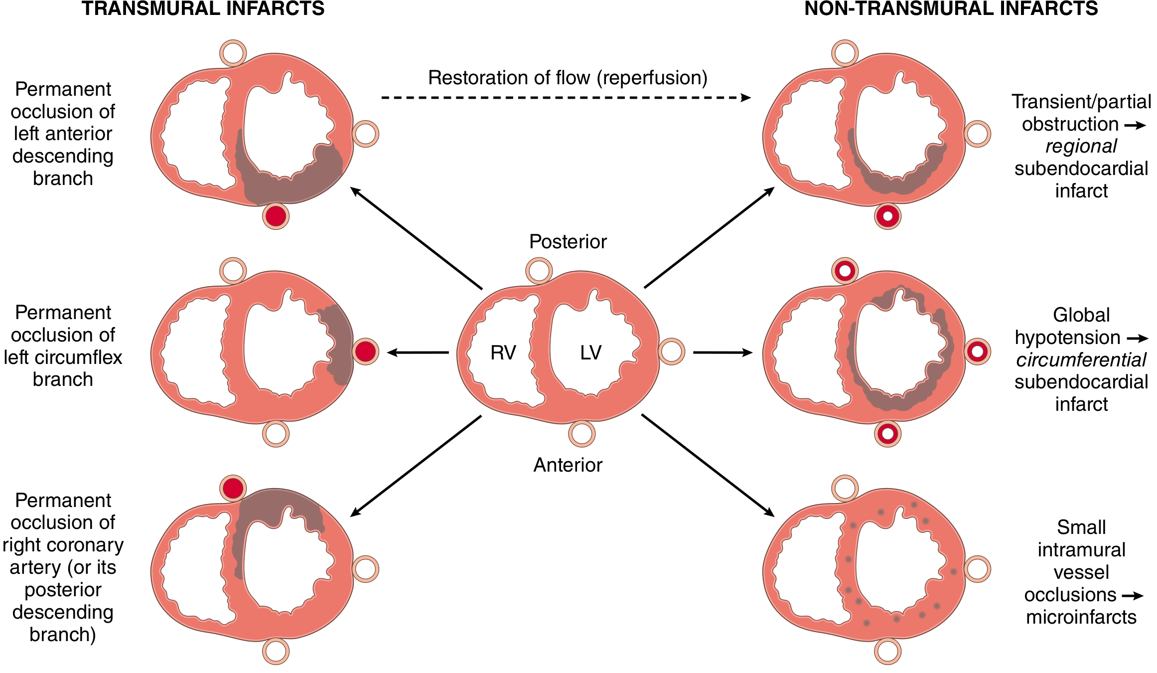
Fig. Distribution of MI patterns based on coronary artery involved (Robbins, Cotran & Kumar)
Coronary Artery - Territory Correlation
| Artery | Territory |
|---|---|
| LAD | Apex, anterior LV wall, anterior 2/3 of interventricular septum |
| RCA (in ~80% right-dominant circulation) | Entire RV free wall, posterobasal LV wall, posterior 1/3 of septum |
| LCX | Lateral wall of LV |
Infarct Patterns
- Transmural infarction - Full wall thickness necrosis; caused by complete, persistent occlusion of an epicardial vessel; associated with ST-elevation MI (STEMI)
- Subendocardial (nontransmural) infarction - Involves inner layers only; caused by transient/partial occlusion or plaque thrombus that lyses before full-thickness necrosis; also seen with global hypotension (circumferential subendocardial infarct); associated with NSTEMI
- Multifocal microinfarction - Pathology of small intramural vessels (microembolization, vasculitis, catecholamine excess)
Morphological (Gross & Microscopic) Changes
Gross Changes
| Time | Gross Appearance |
|---|---|
| 0-12 hours | Usually none visible; TTC staining reveals pallor/lack of staining |
| 12-24 hours | Pallor, possible slight yellowish discoloration |
| 1-3 days | Yellow-tan softening, pallor |
| 3-7 days | Hyperemic border; soft, yellow center with maximal pallor |
| 7-10 days | Depressed, soft; gelatinous reddish-gray at margins (granulation tissue) |
| Weeks | Grey-white fibrous scar forms at borders, progresses inward |
| >6-8 weeks | Dense white/grey fibrous scar |
Microscopic (Histological) Changes
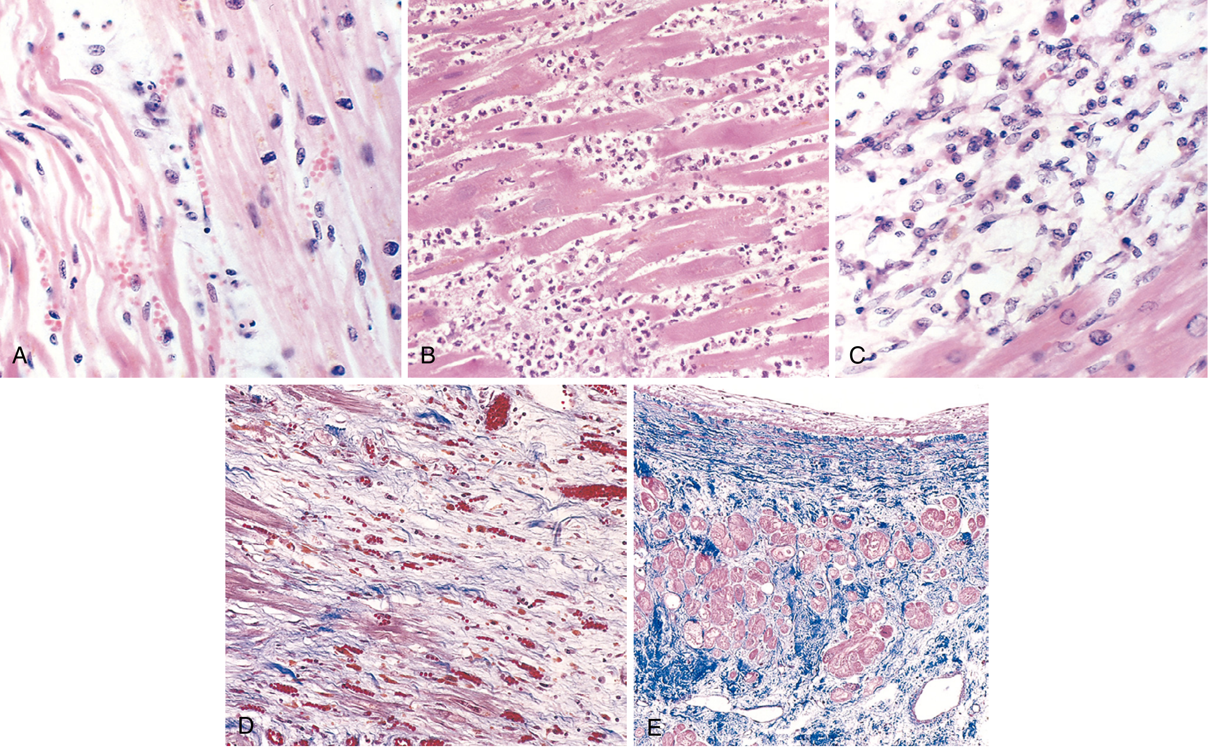
Fig. (A) Day 1: Coagulative necrosis with wavy fibers and scattered neutrophils. (B) Days 3-4: Dense neutrophilic infiltrate. (C) Days 7-10: Macrophage phagocytosis of necrotic debris. (D) Granulation tissue with loose collagen and new capillaries. (E) Healed infarct - dense collagenous scar with compensatory hypertrophy of residual muscle. (Robbins, Cotran & Kumar)
Key sequence:
- Coagulative necrosis with "wavy fibers" at infarct margins (Day 1)
- Neutrophilic infiltrate peaks at 1-3 days
- Macrophage cleanup peaks 3-7 days
- Granulation tissue forms 7-10 days (infarct heals from borders inward)
- Dense collagenous scar complete by ~6 weeks
- Note: Healed scars cannot be dated - an 8-week and a 10-year-old scar look identical
Reperfusion Injury
Restoration of flow after ischemia can cause additional injury:
- Contraction band necrosis - pathognomonic of reperfusion injury; irregular dense transverse bands of hypercontracted sarcomeres
- Gross appearance: Hemorrhagic infarct due to vascular leakage in the zone of reperfusion
- Despite this, reperfusion is still highly beneficial - the sooner achieved, the greater the myocardium salvaged
ECG Changes (Ganong's Physiology)
Three membrane abnormalities occur in acute MI:
| Defect in Infarcted Cells | Current Flow | ECG Change (leads over infarct) |
|---|---|---|
| Rapid repolarization (accelerated K⁺ channel opening) | Out of infarct | ST segment elevation |
| Decreased resting membrane potential (loss of intracellular K⁺) | Into infarct | TQ depression (recorded as ST elevation) |
| Delayed depolarization | Out of infarct | ST segment elevation |
Hallmark: ST segment elevation in leads overlying the infarct; ST depression in reciprocal leads.
- After days-weeks: ST abnormalities subside; dead muscle becomes electrically silent
- Dead area appears negative relative to normal myocardium during systole → pathologic Q waves develop (or R-wave regression in anterior leads)
- Non-Q-wave (NSTEMI) infarcts tend to be less severe but carry high risk of reinfarction
Clinical Presentation
Classic Symptoms
- Crushing, heavy, squeezing chest pain - typically substernal, radiating to left arm, jaw, neck, or back
- Pain lasting >20-30 minutes (distinguishes from stable angina)
- Diaphoresis, nausea, vomiting
- Dyspnea, anxiety, sense of impending doom
Atypical / Anginal Equivalent Presentations
-
One-third of ED patients with confirmed AMI have NO chest pain on presentation
-
Risk factors for atypical presentation: diabetes mellitus, older age (especially >85 years), female sex, nonwhite ethnicity, dementia, prior stroke/CHF
-
In patients >85 years, 60-70% present with anginal equivalents, especially dyspnea
-
Women: more likely to present with dyspnea, indigestion, weakness, unusual fatigue, cold sweats, anxiety
-
Diabetic patients: "silent MI" (medically unrecognized) occurs in ~40% vs. 25% in non-diabetics
-
Presentations that can mimic GERD, GI upset, or anxiety should prompt consideration of ACS
-
Rosen's Emergency Medicine
Biomarkers
| Marker | Rise | Peak | Return to Normal |
|---|---|---|---|
| Cardiac Troponin I/T (cTnI, cTnT) - preferred | 2-4 hours | 24-48 hours | 7-10 days |
| CK-MB | 3-8 hours | 18-24 hours | 48-72 hours |
| Myoglobin | 1-3 hours | 6-9 hours | 24 hours (least specific) |
-
Troponins begin to rise in 2-4 hours, peak at 24-48 hours, remain elevated 7-10 days
-
With reperfusion: troponin levels peak earlier and may be higher (rapid washout)
-
"Troponin leak" (low-level elevation) is seen in CHF, PE, renal failure, sepsis - serial measurements and clinical context help distinguish
-
Robbins, Cotran & Kumar
Causes of Death
- Decreased cardiac output - systolic stretch of non-contracting infarcted muscle further reduces effective pump function; in large infarcts (>40% of LV mass) → cardiogenic shock
- Pulmonary edema - LV failure → damming of blood in pulmonary vasculature
- Ventricular fibrillation - most common cause of sudden death early post-MI; due to:
- Abnormal action potentials at infarct border zone
- Re-entrant circuits from slowed conduction around infarcted area
- Ventricular dilation increasing conduction pathway length
- Cardiac rupture - occurs days 3-10 as necrotic muscle degenerates and thins; free wall rupture → cardiac tamponade → sudden death; septal rupture → VSD; papillary muscle rupture → acute mitral regurgitation
- Other arrhythmias: complete heart block (especially RCA occlusion affecting AV node), ventricular tachycardia
- Guyton & Hall Textbook of Medical Physiology
Complications
| Complication | Timing | Notes |
|---|---|---|
| Arrhythmias | Immediate - days | Most common cause of early death; VF, VT, complete heart block |
| Cardiogenic shock | Hours | LV dysfunction with low output |
| Papillary muscle dysfunction/rupture | Days 3-7 | Acute mitral regurgitation |
| Ventricular septal defect | Days 3-7 | RV ← LV shunt via septal rupture |
| Free wall rupture | Days 3-10 | Tamponade; usually fatal |
| LV thrombus | Days-weeks | Can embolize → stroke |
| Pericarditis (Dressler syndrome) | Weeks-months | Autoimmune post-MI pericarditis |
| Ventricular aneurysm | Weeks-months | Paradoxical motion on echo; persistent ST elevation |
| Heart failure (chronic) | Long-term | Remodeling, dilated cardiomyopathy |
Treatment
Initial (Acute) Management
- Oxygen - for hypoxia or respiratory distress (target SpO₂ ≥94%)
- Aspirin (antiplatelet) - immediately; inhibits TXA₂-mediated platelet aggregation
- P2Y12 inhibitor (clopidogrel, ticagrelor, or prasugrel) - dual antiplatelet therapy
- Anticoagulation - heparin (UFH or LMWH) or bivalirudin
- Nitroglycerin - for pain relief and preload reduction (avoid in RV infarct, hypotension, phosphodiesterase inhibitor use)
- Beta-blockers - reduce heart rate, oxygen demand; decrease risk of VF; start early if no contraindications (avoid in cardiogenic shock, severe bradycardia)
- Morphine - pain relief; used with caution (may mask symptoms, possible harm in NSTEMI)
Reperfusion (Primary Goal)
- STEMI: Primary percutaneous coronary intervention (PCI) is the gold standard; target door-to-balloon time <90 minutes
- If PCI not available within 120 minutes: thrombolysis (fibrinolytics: streptokinase, tPA, tenecteplase)
- Reperfusion is most beneficial when achieved quickly; benefits of early thrombolysis/angioplasty are well established
- NSTEMI/UA: Risk-stratify; early invasive strategy (angiography ± PCI) vs. conservative strategy based on TIMI/GRACE score
Long-term Management
-
ACE inhibitors (or ARBs) - reduce remodeling; especially in reduced EF
-
Beta-blockers - continued long-term; mortality benefit
-
Statins - high-intensity statin therapy regardless of baseline LDL
-
Dual antiplatelet therapy (DAPT) - for 12 months post-ACS/stent
-
Aldosterone antagonists (eplerenone, spironolactone) - in HFrEF post-MI
-
Cardiac rehabilitation - improves outcomes and quality of life
-
Robbins, Cotran & Kumar; Guyton & Hall; Rosen's Emergency Medicine
Recovery & Healing
-
After a large MI, dead fibers in the infarct center enlarge over days
-
Marginal collateral channels expand progressively, salvaging peri-infarct zone
-
Most nonfunctional muscle around the infarct eventually recovers over days to 3 weeks
-
In non-necrotic ischemic zones, pumping power typically returns to near-normal
-
The central necrotic area is replaced by fibrous scar tissue (complete by 6-8 weeks)
-
The remaining healthy muscle undergoes compensatory hypertrophy to maintain output
-
If >40% of LV mass is infarcted, adequate compensatory hypertrophy may be impossible, leading to progressive HF
-
Guyton & Hall Textbook of Medical Physiology
Sources: Robbins, Cotran & Kumar - Pathologic Basis of Disease | Guyton & Hall Textbook of Medical Physiology | Rosen's Emergency Medicine | Ganong's Review of Medical Physiology | Robbins & Kumar Basic Pathology
This is a shared conversation. Sign in to Orris to start your own chat.