
Cellulitis
"cellulitis"[MeSH Terms] AND "therapy"
cellulitis lower leg erythema clinical photograph
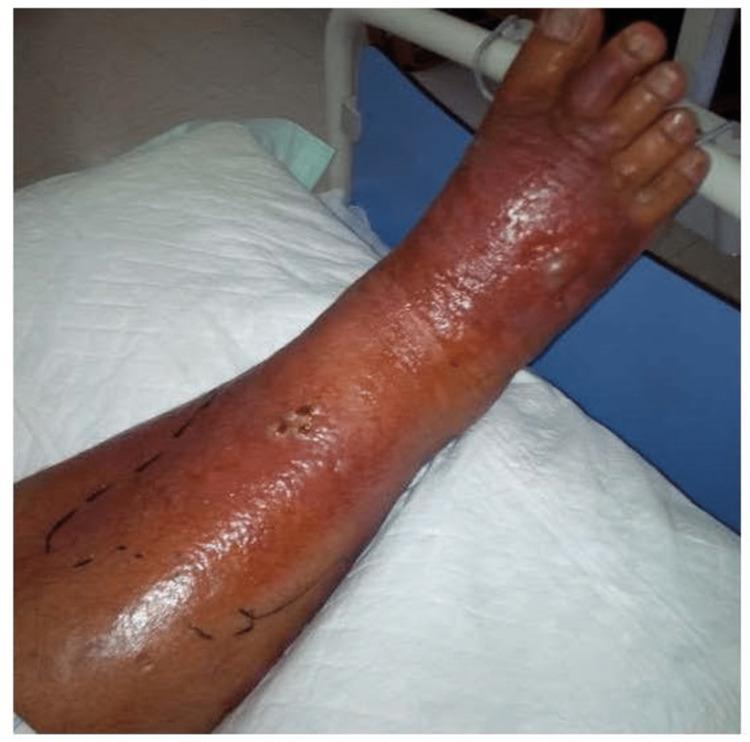
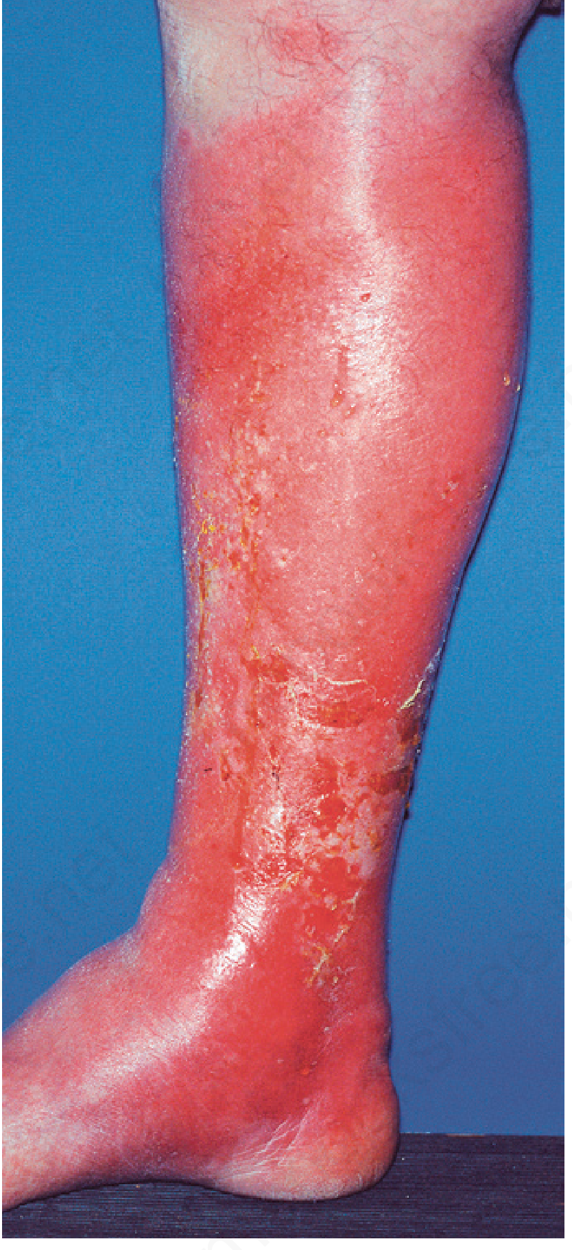
Clinical photograph of a human lower limb demonstrating severe cellulitis. The image shows extensive, confluent erythema and inflammation involving the anterior aspect of the lower leg and the dorsum of the foot. The skin appears shiny, taut, and edematous. Key pathological findings include a central area with small punctate lesions or skin openings with dark discoloration on the anterior leg, and distal cyanotic changes (dusky reddish-purple discoloration) on the toes, suggesting significant vascular congestion or severe inflammatory response. Indelible ink markings are visible on the skin, outlining the advancing borders of the erythema to monitor disease progression. This visual provides a classic representation of lower limb soft tissue infection, useful for teaching diagnostic signs of bacterial cellulitis, including edema, erythema, and the clinical practice of marking infectious boundaries in an acute care or surgical setting.
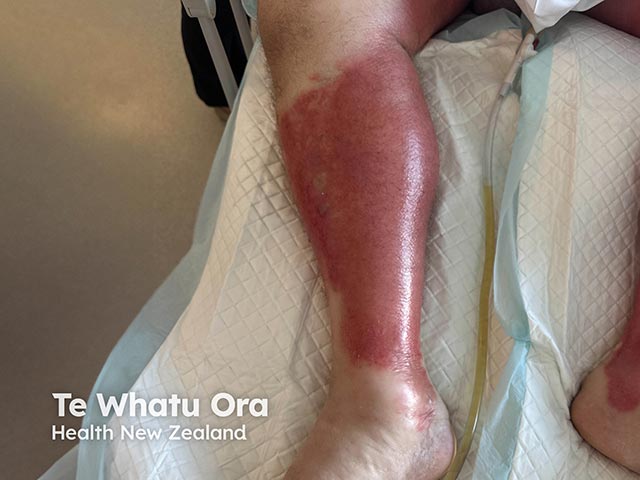
This clinical photograph depicts unilateral acute inflammatory edema of the calf. Modality and techniques: digital color photograph captured in standard clinical lighting; no contrast or staining. Anatomical context: distal thigh to ankle region of the lower limb, with emphasis on the posterior and medial calf. Visual features: bright, diffuse erythema over a swollen soft-tissue envelope, with tense, shiny skin and subtle subcutaneous edema. Palpable warmth is suggested by the clinical appearance; there is no visible necrosis, ulceration, or purpura in the field of view. Impression: the image demonstrates the hallmark appearance of acute inflammatory edema, which can accompany cellulitis, phlebitis, or other inflammatory processes in the lower leg. The photograph lacks deep radiologic detail but documents distribution, severity, and progression of edema over time. Clinical significance: unilateral erythema with edema raises suspicion for infectious etiologies such as cellulitis or inflammatory edema; differential diagnoses include deep vein thrombosis, venous insufficiency, lymphedema, or trauma-related edema. Potential clinical use cases: educational illustration of lower-leg edema, triage aid for suspected cellulitis, monitoring response to therapy, and multimodal correlation with ultrasound to exclude DVT. Recommended clinical correlation with fever, leukocytosis, and vascular imaging as indicated.
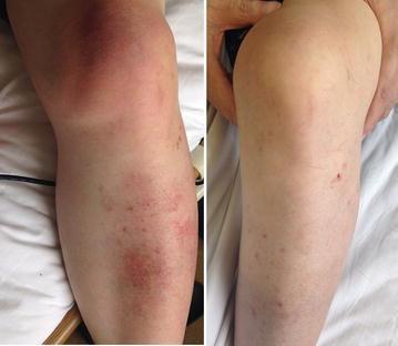
A clinical comparison photograph showing a side-by-side view of a patient's left knee and lower leg, illustrating the progression and resolution of cellulitis. The left image (pre-treatment) demonstrates multifocal, salmon-pink skin discoloration with diffuse erythema across the anterior knee and pretibial region. Significant localized soft tissue swelling (edema) is visible around the patellar and lower leg areas. The right image displays the same anatomical region after six weeks of targeted antibiotic therapy. It shows a near-complete resolution of the salmon-pink erythema and a marked reduction in localized swelling, with the skin returning to a more normal tone and contour. The visual comparison serves as an educational tool for identifying the clinical presentation of bacterial cellulitis (specifically associated with Helicobacter cinaedi bacteremia in an immunocompromised patient) and assessing its therapeutic response to antibiotics.
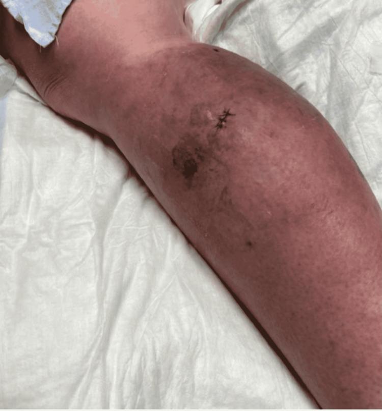
Clinical photograph of a right lower extremity (RLE) demonstrating dermatological manifestations of cellulitis and atypical mycobacterial infection. The image shows diffuse, circumferential erythema extending from the lower leg towards the knee. Notable features include patchy, violaceous to hyperpigmented areas and focal dark-colored lesions, indicative of deep-seated inflammation. A fresh, sutured surgical biopsy site is visible on the lateral aspect of the leg, surrounded by a localized area of ecchymosis. The skin appears edematous and taut. This visual material illustrates the clinical presentation of Mycobacterium chelonae infection, highlighting that while it may mimic standard bacterial cellulitis, it often presents with persistent symptoms, skip lesions, and poor response to conventional beta-lactam therapy. This image is relevant for medical education on infectious disease, dermatology, and atypical cutaneous infections.
IDSA cellulitis guideline 2025 update antibiotic duration 5 days MRSA coverage
Cellulitis
Definition
Causative organisms
- Most common: group A streptococci (S. pyogenes) and Staphylococcus aureus, alone or together. - Fitzpatrick's Dermatology, Microbiology of Cellulitis
- Erysipelas: mainly group A streptococci. The skin is bright, fiery red and sharply demarcated, often on the lower limb or face. Lymphangitic streaks may be present. - Textbook of Family Medicine 9e, p. 265
- Cellulitis other than erysipelas: the border is blurred and lymphangitic streaks are seldom present. - Textbook of Family Medicine 9e, p. 265
- Streptococcal versus staphylococcal: the two cannot reliably be told apart by appearance. Associated purulence points to S. aureus, including MRSA. - GOLDMAN-CECIL MEDICINE
| Setting | Organism |
|---|---|
| Freshwater wound | Aeromonas hydrophila |
| Saltwater or brackish water wound | Vibrio vulnificus |
| Fish, shellfish, meat or poultry handlers | Erysipelothrix rhusiopathiae |
| Diabetes or immunocompromise | Gram-negative bacilli |
| Alcoholism or immunocompromise | S. pneumoniae |
| Immunosuppressive drugs | Cryptococcus neoformans |
| Frail elderly | Non-group A β-hemolytic streptococci |
| Children | H. influenzae |
Risk factors
- Skin barrier breaks: tinea pedis, trauma, ulcers, surgical wounds, injection drug use
- Lymphedema or venous insufficiency, and obesity
- Diabetes and immunosuppression
- Prior cellulitis, which predisposes to recurrence
Clinical features
- The skin is red, warm, swollen and tender, and may look like peau d'orange. It is usually unilateral, most often on the lower leg.
- Fever, chills and malaise may be present. Lymphangitis and regional lymphadenopathy can occur.
- Bullae, abscess or purulent drainage can develop.
Diagnosis
- The diagnosis is clinical. Routine blood and swab cultures have low yield.
- Take blood cultures if there is systemic toxicity, immunocompromise, an unusual exposure or extensive disease.
- Culture any pus or abscess.
- Use ultrasound to look for abscess or to exclude DVT.
- Use MRI or CT if you suspect deep infection.
- DVT
- Stasis dermatitis
- Contact dermatitis
- Lipodermatosclerosis
- Gout
- Erythema migrans
- Drug reaction
- Necrotizing fasciitis (must not be missed)
- Pain out of proportion to the skin findings
- Systemic toxicity
- Rapid spread, bullae, skin necrosis or crepitus
- Overlying skin that may look normal - GOLDMAN-CECIL MEDICINE
Management
- Give an oral agent active against streptococci: cephalexin, amoxicillin, dicloxacillin, penicillin V, or clindamycin.
- Adding MRSA coverage to a β-lactam gave no extra benefit in pure cellulitis. TMP-SMX plus cephalexin was no better than cephalexin alone.
- Add MRSA coverage if there is penetrating trauma, known MRSA, injection drug use, SIRS or systemic toxicity, or failure to respond.
- Drain the abscess first, then cover for MRSA if indicated.
- Options are TMP-SMX or doxycycline, usually with a β-lactam, or clindamycin alone.
- Give IV therapy with MRSA coverage, such as vancomycin, linezolid, daptomycin or telavancin.
- Add broader coverage for diabetic, immunocompromised or necrotizing infection.
- Treat for 5 days if there is clear improvement by day 5. Extend only if the infection has not improved.
- Elevate the limb, treat tinea pedis and other portals of entry, and control edema (compression once the acute phase settles).
- Treat the predisposing factors.
- Consider prophylaxis with penicillin V or benzathine penicillin. It works only while it is taken.
Complications
- Abscess
- Bacteremia and sepsis
- Necrotizing fasciitis
- Osteomyelitis
- Lymphedema from recurrence
- Septic thrombophlebitis
Recent evidence check
- Anti-inflammatories as adjunct treatment (PMID 38796807)
- Diagnostic stewardship and dermatology consultation (PMID 39614879)
- Non-pharmacological measures to prevent recurrence in guidelines (PMID 40530782)
Pathology
cellulitis histopathology dermal edema neutrophilic infiltrate
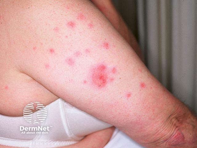
Imaging modality: Clinical photography of the cutaneous upper arm. This photograph shows multiple tumid, erythematous plaques consistent with Sweet syndrome (acute febrile neutrophilic dermatosis). Lesions are raised and well demarcated, with red to pink coloration and a taut, edematous surface. Distribution is localized to the proximal upper arm and displays variable plaque size from a few millimeters to several centimeters. There is no obvious ulceration or necrosis; the plaques appear smooth and non-vesicular. The image illustrates the characteristic appearance of inflammatory neutrophilic dermatoses in a single anatomical region, useful for pattern recognition and educational documentation. Clinically, Sweet syndrome often presents with fever and leukocytosis; biopsy (if performed) reveals a dense neutrophilic dermal infiltrate with papillary dermal edema and typically no vasculitis. The combination of acute, tender skin lesions and systemic symptoms supports the diagnosis, while differential considerations include erythema nodosum, erythema multiforme, cellulitis, urticaria, and other neutrophilic dermatoses. This image is valuable for teaching dermatology students about cutaneous neutrophilic processes, correlating clinical features with histopathology, and monitoring treatment response to corticosteroids or immunomodulatory therapy. Educationally, this image supports recognition of neutrophilic dermatoses across age groups and highlights the need for clinical correlation with fever, laboratory data, and possible associated underlying conditions such as malignancy or infection.
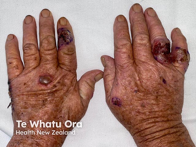
Clinical photograph demonstrating bilateral hands with oedematous, haemorrhagic, and blistering lesions on the dorsal surfaces, a recognized neutrophilic dermatosis of the hands (a somatic subset of Sweet syndrome). High-resolution color imaging captured under uniform lighting to preserve tissue hue and lesion morphology. The dorsal hand surfaces show tender, erythematous-to-violaceous plaques with pronounced edema, vesiculopustular components, and hemorrhagic crusts, extending to the fingers in several focal areas. Lesions are well-circumscribed but merge in places, with superficial epidermal disruption and surrounding erythema. The pattern is typically acral and symmetric, favoring extensor aspects and dorsal digits, and may be bilateral. Clinically, this appearance is characteristic of neutrophilic dermatosis of the dorsal hands, enabling differentiation from infectious etiologies such as cellulitis or erysipelas. Histopathology would commonly reveal a dense neutrophilic infiltrate within the dermis and dermal edema; absence or minimal leukocytoclastic vasculitis supports the neutrophilic dermatosis diagnosis. The image is valuable for dermatology education, case-based teaching, and differential diagnosis exercises, illustrating the dorsal-hand Sweet syndrome variant. Potential clinical implications include prompt recognition guiding work-up for systemic disease, hematologic malignancy, and therapeutic planning with anti-inflammatory or immunosuppressive agents. Documentation of this pattern supports multidisciplinary care and timely management.
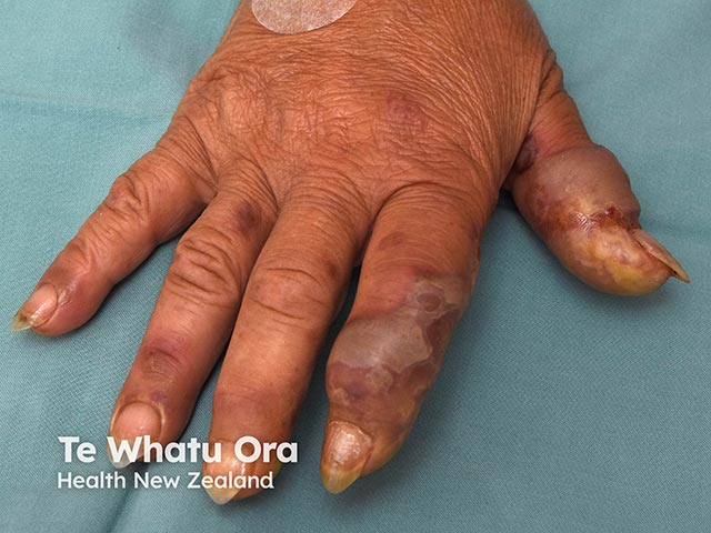
Imaging modality: Clinical photography of the integumentary system; technique: standardized close-up views of the hands with color-balanced illumination to preserve natural skin tones. Anatomical location: digits of the hand, dorsal aspect, including index through pinky, with minimal palmar involvement. Primary subject: oedematous, violaceous plaques and patches on the dorsal fingers consistent with acute febrile neutrophilic dermatosis (Sweet syndrome). Visual features: diffuse digit edema with firm, tender-appearing swelling; violaceous to pink-purple plaques along the dorsal surfaces; some overlying skin appears taut and shiny; a focal lesion on the thumb shows superficial tissue disruption/cutaneous ulceration. Notable features: acral distribution on the dorsal hand with sparing of the palmar skin in places; abundant edema without obvious purulence in this still image. Context: clinical appearance is typical for Sweet syndrome in its neutrophilic dermatosis spectrum; differential diagnoses include cellulitis, erythema multiforme, and vasculitic ulcers. Clinical correlation: often accompanied by fever and hematologic or inflammatory comorbidities; histopathology would show dense dermal neutrophilic infiltrate with edema, usually no vasculitis. Potential utility: educational reference for dermatology and infectious disease training; aids differential diagnosis and therapeutic decision-making (steroids, immunomodulators). This image supports recognition of acral edema patterns in Sweet syndrome and informs biopsy, treatment planning, and educational discussion.
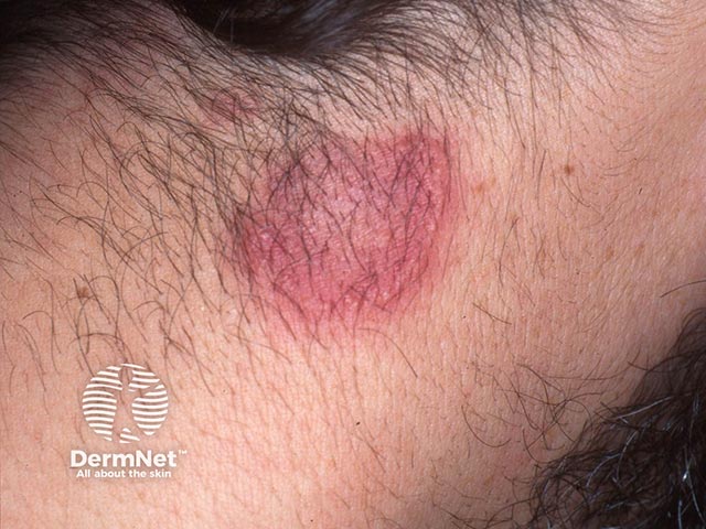
Clinical photography of a solitary annular erythematous plaque on the neck, captured in a close-up anterior view to document surface features. The lesion displays central clearing with a vivid pink-to-red halo and a notably elevated, pseudovesicular edge consistent with acute febrile neutrophilic dermatosis (Sweet syndrome). The plaque is well circumscribed, mildly edematous, and lacks a distinct scale; the edge appears vesicle-like or blistering in continuity with surrounding erythema. Correlating clinical data would anticipate fever, neutrophilia, and possible systemic symptoms; histopathology if obtained would typically reveal a dense neutrophilic dermal infiltrate with possible mild papillary dermal edema and little or no vasculitis. The image demonstrates the characteristic inflammatory border that differentiates Sweet syndrome from infectious cellulitis or erysipelas, and from other annular dermatoses such as erythema migrans or granulomatous conditions. Clinically, recognition supports prompt systemic corticosteroid therapy and evaluation for underlying triggers including hematologic malignancy or inflammatory disorders; corticosteroid response is usually rapid and dramatic. This image is relevant for dermatology, dermato-rheumatology, and medical education; useful for differential diagnosis practice, case-based discussion, and image-based teaching materials focusing on neutrophilic dermatoses and neck-lesion morphology. Understanding such presentations improves diagnostic acumen for non-infectious neck lesions and supports timely, targeted management decisions.
Pathology of cellulitis
Site of involvement
- Cellulitis centers on the deep dermis and subcutaneous fat. Erysipelas is more superficial, involving the upper dermis with prominent lymphatic involvement. - Dermatology 2-Volume Set 5e, p. 1526
- Andrews' describes cellulitis as a suppurative inflammation of the subcutaneous tissue. It often follows a wound, and tinea pedis is a common portal of entry on the leg. - Andrews' Diseases of the Skin
Pathogenesis
- Entry: bacteria cross a break in the skin barrier. Examples are tinea pedis fissures, trauma, ulcers, surgical wounds and eczema.
- Predisposing local factors: lymphedema and venous insufficiency impair lymphatic drainage and local immune surveillance. Risk factors include a preceding skin break, venous hypertension edema, lymphedema and previous cellulitis. - GOLDMAN-CECIL MEDICINE
- Spread: organisms multiply in the dermis and subcutis and spread along tissue planes and lymphatics.
- Host response: neutrophil recruitment, vasodilation and increased vascular permeability produce erythema, heat, edema and pain.
- Bacterial toxins and enzymes:
- Streptococci produce streptolysins, hyaluronidase and streptokinase, which help the infection spread through tissue.
- S. aureus produces toxins, notably Panton-Valentine leukocidin in community MRSA strains, which is associated with more skin and soft tissue infection. - GOLDMAN-CECIL MEDICINE
- Recurrence: each episode damages lymphatics. Repeated bouts cause lymphatic obstruction, chronic lymphedema and eventually fibrosis (elephantiasis nostras), which sets up a cycle of further infection. - Andrews' Diseases of the Skin
Histopathology
- Dermal edema.
- Neutrophilic infiltrate in the dermis. - GOLDMAN-CECIL MEDICINE; Dermatology 5e, Fig. 74.13
- Mixed infiltrate: a mild to moderate infiltrate of lymphocytes and neutrophils through the dermis, often extending into subcutaneous fat.
- Vascular and lymphatic changes: dilated lymphatics and small blood vessels.
- Epidermis: severe edema can lead to subepidermal bullae.
- Organisms: the causative organism may be identified with special stains, but is often not seen. - Dermatology 2-Volume Set 5e, p. 1526
- Intense, spreading erythema with induration that may pit.
- A central nodular area that may become vesicular and discharge pus and necrotic material.
- Lymphangitic streaks to regional nodes.
- Gangrene, metastatic abscesses and severe sepsis in the worst cases.
Histologic differential
| Condition | Key histologic difference |
|---|---|
| Sweet syndrome | Similar to cellulitis (dense neutrophilic dermal infiltrate with edema), so culture tissue to exclude infection |
| Familial Mediterranean fever, erysipelas-like lesions | Superficial dermal edema with perivascular and interstitial neutrophils, no vasculitis |
| Eosinophilic cellulitis (Wells syndrome) | Dermal eosinophils instead of neutrophils |
| Contact dermatitis, erythema migrans, inflammatory carcinoma, toxic erythema of chemotherapy | Different infiltrates and features, which a biopsy can distinguish |
Pathology of important variants
- Necrotizing fasciitis: infection extends along fascial planes into deeper layers and muscle, with tissue necrosis. The skin may look normal early on. - GOLDMAN-CECIL MEDICINE
- Bullous and hemorrhagic cellulitis: these occur with severe disease, for example with streptococci or Vibrio vulnificus.
- Chronic lipodermatosclerosis: inflammation followed by fibrosis of subcutaneous fat. It is a common mimic, and the skin never returns to normal color even after superimposed cellulitis is treated. - GOLDMAN-CECIL MEDICINE
Lab and culture correlates
- The white cell count is often normal or only mildly raised.
- Blood cultures are almost always negative in immunocompetent hosts. The exception is H. influenzae cellulitis, which usually has leukocytosis with a left shift and positive blood cultures. - Dermatology 2-Volume Set 5e, p. 1526
- Streptococci cause about 75% of cases and staphylococci most of the rest. - Andrews' Diseases of the Skin
Pathology
histopathology of cellulitis dermal edema neutrophils H&E
Pathology of cellulitis (expanded)
1. Entry and spread
- Microbes enter through breaks in the skin and reach the deep dermis and subcutaneous tissue. From there they spread into lymphatics, blood vessels and interstitial spaces.
- A portal of entry can be found in up to 62% of patients. Toe web infection is the most common. - Fitzpatrick's Dermatology, Etiology and Pathogenesis
- Disseminated infection with secondary skin seeding is a rare cause, mostly in immunocompromised patients.
- The infection is often paucimicrobial. Much of the clinical picture reflects the host inflammatory response to killed bacteria and bacterial exotoxins, not a heavy live organism burden. This is why cultures are so often negative. - Fitzpatrick's Dermatology
2. Why the pattern differs: cellulitis versus erysipelas
| Feature | Erysipelas | Cellulitis |
|---|---|---|
| Level | Upper dermis, superficial lymphatics | Deep dermis and subcutaneous fat |
| Border | Sharp, raised | Ill-defined |
| Color | Bright, fiery red | Pink hue |
| Lymphangitis | Common | Less common |
- Dermatology 2-Volume Set 5e; GOLDMAN-CECIL MEDICINE; Kanski's Clinical Ophthalmology
3. Microscopic findings
- Dermal edema.
- A neutrophilic infiltrate in the dermis.
- A mild to moderate mixed infiltrate of lymphocytes and neutrophils through the dermis, often reaching subcutaneous fat.
- Dilated lymphatics and small blood vessels.
- Occasional subepidermal bullae from severe edema.
- Organisms visible only with special stains, and often not seen. - Dermatology 2-Volume Set 5e, p. 1526; GOLDMAN-CECIL MEDICINE
4. Why the signs appear
| Sign | Mechanism |
|---|---|
| Erythema and warmth | Vasodilation from inflammatory mediators |
| Edema | Increased vascular permeability and lymphatic obstruction |
| Pain and tenderness | Mediator release and tissue tension |
| Fever and leukocytosis | Cytokine release (IL-1, IL-6, TNF) |
| Abscess or pus | Suppurative necrosis, mostly with S. aureus |
5. Organism and host determinants
- Group A streptococci: associated with the more severe, necrotizing variant. - GOLDMAN-CECIL MEDICINE
- Streptococcal cellulitis is usually nonpurulent. Purulent cellulitis is most often staphylococcal. - GOLDMAN-CECIL MEDICINE
- Risk factors: impaired lymphatic drainage, venous insufficiency, edema, obesity and skin barrier disruption. - GOLDMAN-CECIL MEDICINE
- Polymicrobial or atypical infection: surgical site infections, pressure or diabetic ulcers, and cellular immunodeficiency. Examples are enterococci, Pseudomonas, Gram-negative anaerobes, mycobacteria and cryptococcus. - Fitzpatrick's Dermatology
- Bite wounds: Pasteurella, Eikenella and Capnocytophaga canimorsus.
- Aquatic trauma: Aeromonas, Erysipelothrix, Mycobacterium marinum and Vibrio vulnificus.
6. Consequences of repeated infection
- Each episode damages lymphatics. Recurrent cellulitis leads to impaired drainage, permanent swelling, dermal fibrosis and epidermal thickening.
- This end result is called elephantiasis nostras, and it predisposes to further attacks. - Tintinalli's Emergency Medicine; Andrews' Diseases of the Skin
7. Histologic mimics and when to biopsy
- Biopsy is rarely needed. It can exclude contact dermatitis, erythema migrans, inflammatory carcinoma, toxic erythema of chemotherapy and Wells syndrome (dermal eosinophils). - GOLDMAN-CECIL MEDICINE
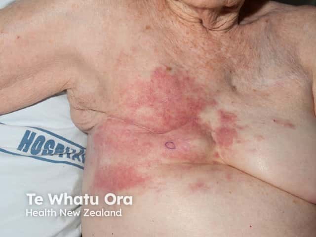
- Sweet syndrome and familial Mediterranean fever erysipelas-like lesions can also show a neutrophilic dermal infiltrate. Culture lesional tissue to exclude infection. - Fitzpatrick's Dermatology
- Carcinoma erysipeloides (dermal lymphatic invasion by breast or other adenocarcinoma) can look like erysipelas and responds poorly to antibiotics.
Management and treatment
"cellulitis"[MeSH Terms] AND "anti-bacterial agents"[MeSH Terms]
IDSA skin and soft tissue infection guideline cellulitis recommendations duration 5 days MRSA risk
Management and treatment of cellulitis
1. Principles
- Treatment is empiric, aimed at streptococci and staphylococci. No regimen has been proven better than another. Choice depends on purulence, systemic symptoms, risk factors and local MRSA rates. - Fitzpatrick's Dermatology, Management
- Outpatient oral therapy suits hemodynamically stable patients with no systemic infection. Admit patients who are seriously ill, immunocompromised, or who fail outpatient therapy. - Fitzpatrick's Dermatology
- Cultures are not recommended routinely because of poor yield. - Tintinalli's Emergency Medicine
2. Antibiotics by severity (Fitzpatrick's Table 151-5)
| Severity | First line | Alternatives |
|---|---|---|
| Mild nonpurulent (no systemic disease, oral) | Cephalexin, dicloxacillin, penicillin V | Clindamycin, macrolides |
| Moderate (2 or more SIRS criteria, or outpatient failure; IV) | Cefazolin, ceftriaxone, penicillin G | Clindamycin. Consider vancomycin if IV drug use or known MRSA colonization or infection |
| Severe (rapid progression, hypotension, end-organ damage) or immunocompromised; IV | Broad spectrum, such as vancomycin plus piperacillin-tazobactam, narrowed by culture and sensitivities | - |
3. When to add MRSA coverage
- Add it if the patient is diabetic or immunosuppressed, has peripheral vascular disease or a history of IV drug use, or needs hospitalization. - Tintinalli's Emergency Medicine
- Other triggers are suspected MRSA, high fever, hypotension, or recent or prolonged hospitalization. - Fischer's Mastery of Surgery
- Oral options are TMP-SMX, doxycycline or minocycline, or clindamycin. Linezolid is another oral option.
- Severe cases need IV MRSA-active therapy such as vancomycin.
- For nonpurulent cellulitis without these risk factors, a β-lactam alone is enough. The IDSA guideline summaries I found state that adding MRSA coverage gives no extra benefit in typical cases.
4. Purulent cellulitis or abscess
- Incision and drainage is the main treatment. A furuncle with surrounding cellulitis from MRSA should be drained and given systemic antibiotics. - GOLDMAN-CECIL MEDICINE, Fig. 408-2
- Use MRSA-active agents for purulent disease. Culture the pus.
5. Duration
- The IDSA 2014 guideline recommends 5 days. Extend only if the infection has not improved in that time (strong recommendation, high-quality evidence). - IDSA SSTI guideline
- The same guideline recommends elevating the affected area and treating predisposing factors such as edema or underlying skin disorders. Both are strong recommendations.
- Hospitalized patients often get longer courses in practice (about 2 weeks in one cited retrospective study). The IDSA text notes that failure rates did not differ with broader spectrum.
- Diabetic foot infections are a separate problem. IWGDF/IDSA advise 1 to 2 weeks for soft tissue infection.
6. Supportive and adjunctive care
- Elevation of the limb, and treatment of edema.
- Treat the portal of entry: toe web infection and tinea pedis, and barrier defects.
- Monitoring: outlining the erythema is useful, but indistinct margins, skip areas and transient progression can mislead. Serial photographs are a reliable alternative. - Fitzpatrick's Dermatology
- Anti-inflammatories:
- NSAIDs may shorten time to regression of inflammation, but there are concerns about their effect on neutrophil chemotaxis.
- Prednisolone added to antibiotics shortened healing in uncomplicated erysipelas, with a trend toward fewer relapses.
- The IDSA guidelines say to consider systemic corticosteroids in nondiabetic adults with cellulitis. - Fitzpatrick's Dermatology
- Analgesia and hydration as needed.
7. Suspected necrotizing infection
- Clues are pain out of proportion, systemic toxicity, rapid spread and bullae or necrosis.
- Give broad-spectrum IV antibiotics and call surgery urgently for debridement. Do not delay for imaging. - GOLDMAN-CECIL MEDICINE; Fitzpatrick's Dermatology
8. If the patient is not improving
- Pseudocellulitis: stasis dermatitis, lipodermatosclerosis, DVT, gout or contact dermatitis. The ALT-70 score helps separate them. - Dermatology 5e
- Undrained abscess: consider ultrasound.
- Resistant or atypical organism: MRSA, or exposure-specific organisms such as Aeromonas, Vibrio or mycobacteria.
- Deep infection: necrotizing fasciitis or osteomyelitis.
- Host factors: diabetes, immunosuppression, lymphedema.
9. Prevention of recurrence
- Recurrence occurs in more than 10% of patients, and the risk rises with each previous episode. - Fitzpatrick's Dermatology
- Treat lymphedema, toe web infections, skin barrier defects and underlying medical conditions. MRSA decolonization is controversial and unproven for preventing cellulitis. - Fitzpatrick's Dermatology
- Antibiotic prophylaxis is generally considered for 2 or more episodes a year despite risk factor control, using penicillin V or benzathine penicillin. This comes from my own knowledge, not the retrieved textbooks. It works only while it is taken.
- Compression therapy once the acute phase settles.
10. Prognosis
- Treated cellulitis has a good prognosis and negligible mortality in uncomplicated cases.
- Recovery takes several weeks. About 20% of patients are not back to normal activity at 30 days. - Fitzpatrick's Dermatology
- Untreated, the infection can reach the bloodstream and become life-threatening.
Recent evidence check (PubMed, last 3 years)
- A network meta-analysis of first- and second-line antibiotics for cellulitis and erysipelas, 2024 (PMID 39240378).
- A meta-analysis of anti-inflammatories as an adjunct, 2024 (PMID 38796807).
- A systematic review of secondary prevention, 2026 (PMID 41275489).
- A pilot RCT of high-dose cephalexin, 2023 (PMID 36592299).