
Axillary artery
Reading File
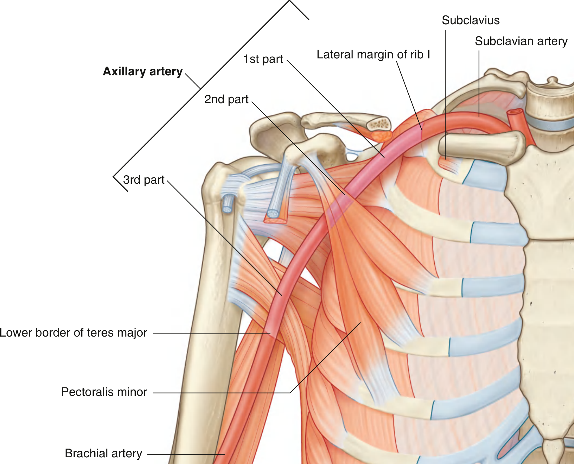
The axillary artery is the main artery of the axilla, running from the outer border of the first rib (where it continues from the subclavian artery) to the lower border of teres major (where it becomes the brachial artery). It is divided into three parts by the pectoralis minor muscle.
Searching sources...
Finding Sources
Finding Sources
Excellent sources found. Let me read the most comprehensive anatomy sections in parallel.
Reading File
Reading File
Reading File
Good data. Now let me fetch the key diagrams and read more about relations and clinical points.
Loading Image
Loading Image
Reading File
I now have comprehensive material. Let me also get the relations of each part and the brachial plexus relationship.
Reading File
Reading File
I now have all the information needed for a comprehensive answer. Let me compile it.
Axillary Artery
Overview
The axillary artery is the main arterial trunk of the upper limb, running through the axilla. It begins as the continuation of the subclavian artery at the lateral margin of the first rib and ends at the inferior border of teres major, where it continues as the brachial artery.
Parts (Divided by Pectoralis Minor)
The pectoralis minor muscle crosses anterior to the axillary artery and divides it into three parts. A useful mnemonic for the number of branches per part is 1-2-3:
| Part | Position | No. of Branches |
|---|---|---|
| 1st | Proximal to pectoralis minor | 1 |
| 2nd | Posterior to pectoralis minor | 2 |
| 3rd | Distal to pectoralis minor | 3 |
Branches
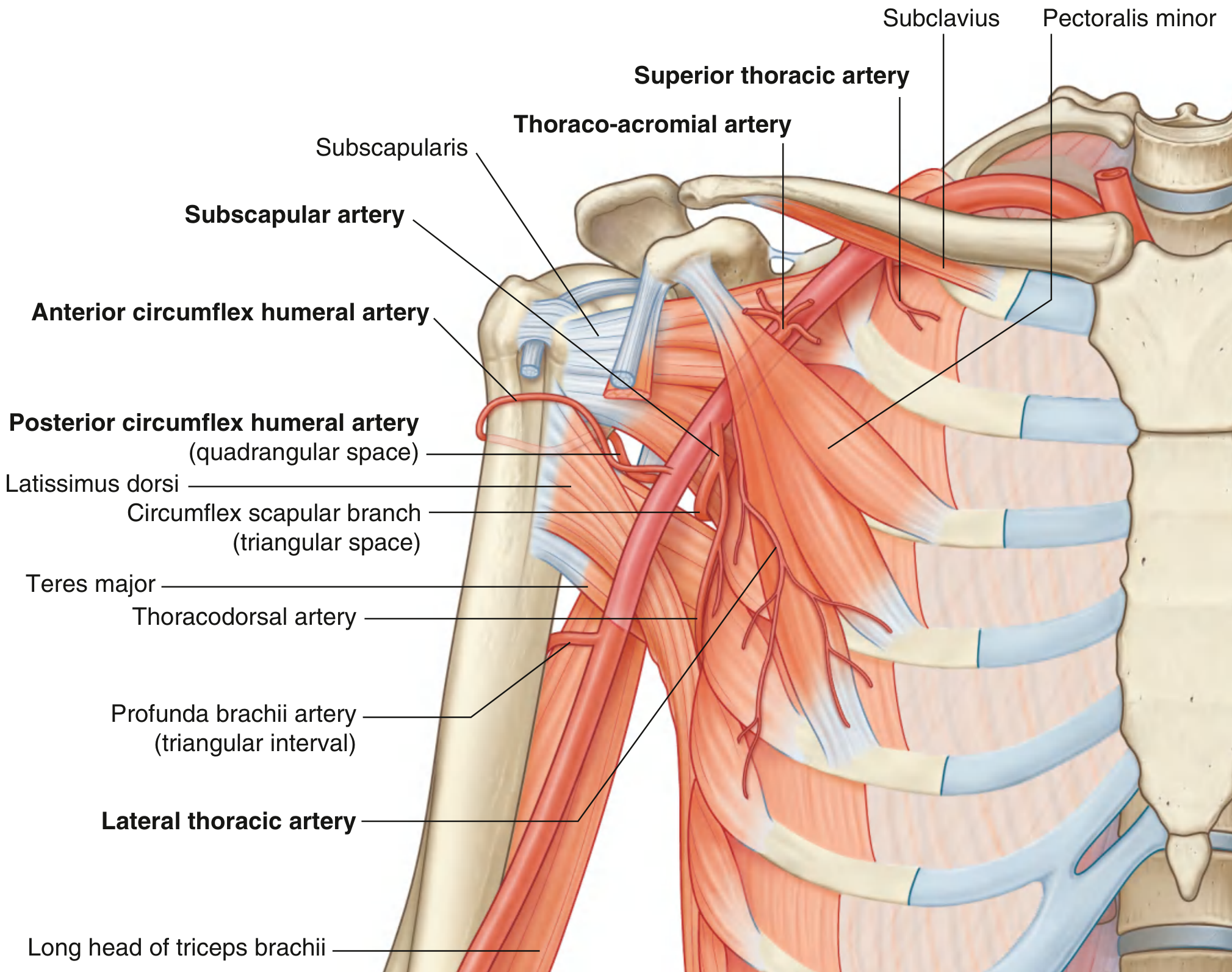
Six branches arise in total:
1st Part (1 branch)
- Superior thoracic artery - small; arises from the anterior surface; supplies the upper medial and anterior axillary walls
2nd Part (2 branches)
- Thoraco-acromial artery - short trunk arising near the superior margin of pectoralis minor; pierces clavipectoral fascia; divides into 4 sub-branches:
- Pectoral (also supplies the breast)
- Deltoid (accompanies the cephalic vein in the clavipectoral triangle)
- Clavicular
- Acromial
- Lateral thoracic artery - follows the lateral border of pectoralis minor to the thoracic wall; supplies the medial and anterior axillary walls and the breast (serratus anterior, pectoral muscles)
3rd Part (3 branches)
- Subscapular artery - the largest branch; divides into:
- Circumflex scapular artery (passes through the triangular space)
- Thoracodorsal artery (supplies latissimus dorsi)
- Anterior circumflex humeral artery - winds around the front of the surgical neck of the humerus
- Posterior circumflex humeral artery - larger; passes through the quadrangular space with the axillary nerve; winds around the surgical neck posteriorly
Relations to the Brachial Plexus
The three cords of the brachial plexus are named by their relation to the 2nd part of the axillary artery:
| Cord | Position |
|---|---|
| Lateral cord | Lateral to 2nd part |
| Medial cord | Medial to 2nd part |
| Posterior cord | Posterior to 2nd part |
Collateral Circulation
A rich peri-scapular anastomotic network connects branches of the 1st part of the subclavian artery with the 3rd part of the axillary artery. Key vessels involved include the transverse cervical, dorsal scapular, suprascapular, and subscapular arteries. This anastomosis is clinically significant - even complete occlusion of the axillary artery rarely causes complete limb ischemia.
Anatomical Variants
The branching pattern is quite variable, primarily involving the subscapular and posterior circumflex humeral arteries (~66% of cases):
- High origin of the subscapular artery from the 2nd part (29-36%)
- Common origin of the subscapular and posterior circumflex humeral arteries from the 2nd or 3rd part (12-42%)
- Thoracodorsal artery arising directly from the axillary artery (3-10%)
- Early bifurcation into superficial and deep brachial arteries
Clinical Relevance
1. Anterior shoulder dislocation
Anterior dislocation of the humeral head can compress the axillary artery causing occlusion. Rarely causes complete ischemia due to collateral flow, but surgical reconstruction may be needed. The intimately related brachial plexus may also be damaged.
2. First rib fracture
Rapid deceleration injuries can fracture rib I, compromising the distal subclavian or 1st part of the axillary artery. Collateral anastomoses around the scapula protect against complete ischemia.
3. Subclavian/axillary venous access
What clinicians call the "subclavian route" for central venous access actually enters the 1st part of the axillary vein. The puncture should be at or lateral to the midclavicular line to avoid inadvertent arterial puncture.
4. Axillary nerve block
The axillary artery is palpated with the arm abducted and externally rotated to locate the brachial plexus cords for axillary brachial plexus block. Ultrasound identifies the "U-shaped" distribution of cords surrounding the artery.
5. Axillary artery aneurysms
Rare; may result from trauma, thoracic outlet syndrome, or repetitive shoulder injury (e.g., in athletes or crutch users).
Sources: Gray's Anatomy for Students; Imaging Anatomy - Bones, Joints, Vessels and Nerves (THIEME); General Anatomy and Musculoskeletal System (THIEME Atlas); Current Surgical Therapy 14e; Gray's Anatomy for Students, p. 853-854
This is a shared conversation. Sign in to Orris to start your own chat.