

1000118916.jpg
Explain this
analyse_media
Reading File
Now let me search the medical library and for images to give a thorough explanation.
Finding Sources
Finding Sources
Searching Images
hypopigmented white macule skin arm idiopathic guttate hypomelanosis
Reading File
Reading File
Loading Image
Here is a full explanation of what is visible on the arm.
What Is That White Spot?
The image shows a forearm with a single small, flat, white-to-hypopigmented macule (roughly 1-3 mm) that has well-defined borders and sits flush with the surrounding skin - no scaling, no raised edges, no redness.
This is a very common and almost always benign finding. The most likely explanations, from most to least probable:
1. Idiopathic Guttate Hypomelanosis (IGH) - Most Likely
This is the textbook match. IGH is a common, acquired, benign disorder characterized by small (2-5 mm), hypopigmented, sharply defined macules that appear preferentially on sun-exposed areas - especially the forearms and shins, exactly where this is located.
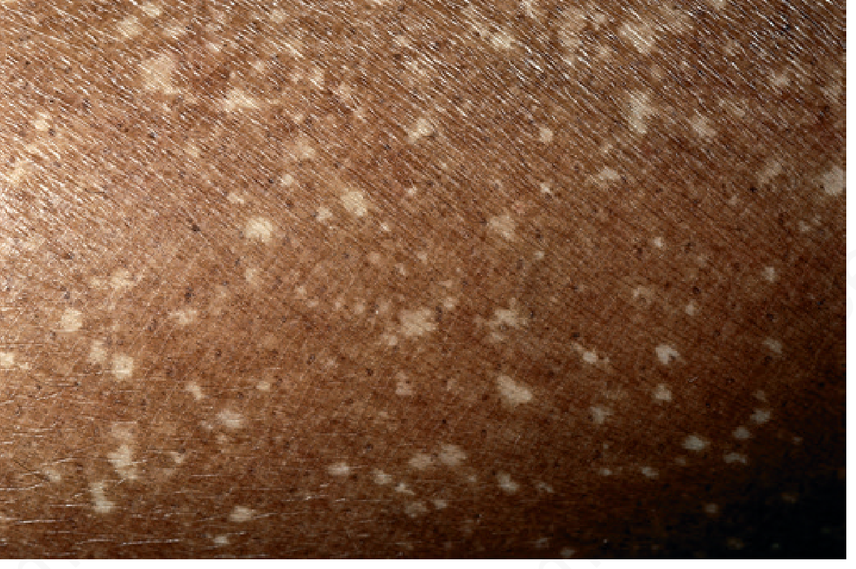
Key facts about IGH:
-
More common in women than men
-
Prevalence rises sharply with age (over 90% in Koreans above age 60)
-
Caused by reduced, hypoactive melanocytes under the skin surface
-
Not dangerous, not contagious, not a sign of systemic disease
-
Related to chronic sun exposure and photoaging
-
Histology shows epidermal atrophy and reduced melanocytes
-
Andrews' Diseases of the Skin, p. 1000
2. Post-Inflammatory Hypopigmentation
If there was a prior insect bite, scratch, minor burn, or pimple in that exact location, the healing process can temporarily or permanently deplete local melanin. This leaves a flat, pale macule that looks identical to the one in the image.
3. Tiny Scar
A healed micro-abrasion or scratch leaves a flat hypopigmented mark with sharp borders. No special significance.
4. Early / Isolated Vitiligo (Less Likely)
Vitiligo is an autoimmune loss of melanocytes that produces chalk-white, completely depigmented patches. A single small spot on the forearm is possible but less typical - vitiligo tends to be more dramatically depigmented (pure white), bilateral, and progressive. A Wood's lamp exam (UV light) can distinguish: vitiligo fluoresces bright white, while IGH/post-inflammatory changes do not.
5. Tinea Versicolor (Much Less Likely)
A superficial yeast infection (Malassezia) that causes pale patches - but these are usually slightly scaly, multiple, and appear more on the trunk than the forearms.
Bottom Line
A single, small, flat, white, asymptomatic spot on the forearm is almost certainly IGH or post-inflammatory hypopigmentation - both harmless. No treatment is required unless it is a cosmetic concern.
See a dermatologist if:
- The spot grows significantly
- New similar spots appear rapidly in clusters
- It becomes itchy, scaly, or inflamed
- You want confirmation with a Wood's lamp or dermoscopy
Disclaimer: This is educational information only and not a clinical diagnosis. A dermatologist can examine the spot in person and use specialized tools (dermatoscope, Wood's lamp) for an accurate assessment.
This is a shared conversation. Sign in to Orris to start your own chat.