
Management of epistaxis in flowchart
epistaxis management flowchart algorithm anterior posterior 2024
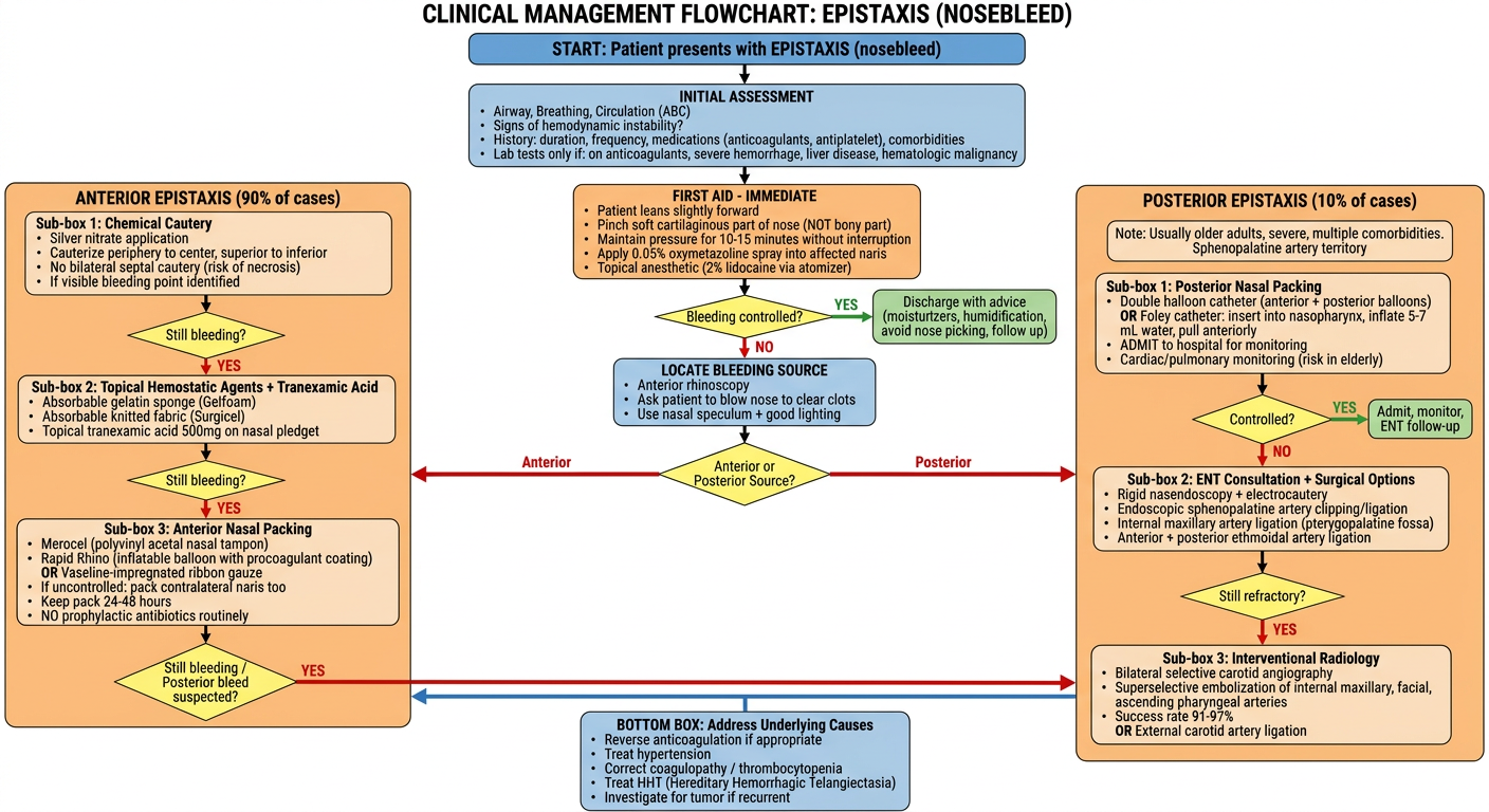
A detailed clinical management flowchart for epistaxis (nosebleed). The flowchart should be professional, clean, and organized in a top-down format with the following structure: START: Patient presents with EPISTAXIS (nosebleed) INITIAL ASSESSMENT (box): - Airway, Breathing, Circulation (ABC) - Signs of hemodynamic instability? - History: duration, frequency, medications (anticoagulants, antiplatelet), comorbidities - Lab tests only if: on anticoagulants, severe hemorrhage, liver disease, hematologic malignancy FIRST AID - IMMEDIATE (box): - Patient leans slightly forward - Pinch soft cartilaginous part of nose (NOT bony part) - Maintain pressure for 10-15 minutes without interruption - Apply 0.05% oxymetazoline spray into affected naris - Topical anesthetic (2% lidocaine via atomizer) Then DECISION DIAMOND: "Bleeding controlled?" YES arrow goes to: Discharge with advice (moisturizers, humidification, avoid nose picking, follow up) NO arrow goes down LOCATE BLEEDING SOURCE (box): - Anterior rhinoscopy - Ask patient to blow nose to clear clots - Use nasal speculum + good lighting DECISION DIAMOND: "Anterior or Posterior Source?" LEFT BRANCH - ANTERIOR EPISTAXIS (90% of cases): Sub-box 1: Chemical Cautery - Silver nitrate application - Cauterize periphery to center, superior to inferior - No bilateral septal cautery (risk of necrosis) - If visible bleeding point identified DECISION DIAMOND: "Still bleeding?" YES arrow down: Sub-box 2: Topical Hemostatic Agents + Tranexamic Acid - Absorbable gelatin sponge (Gelfoam) - Absorbable knitted fabric (Surgicel) - Topical tranexamic acid 500mg on nasal pledget DECISION DIAMOND: "Still bleeding?" YES arrow down: Sub-box 3: Anterior Nasal Packing - Merocel (polyvinyl acetal nasal tampon) - Rapid Rhino (inflatable balloon with procoagulant coating) - OR Vaseline-impregnated ribbon gauze - If uncontrolled: pack contralateral naris too - Keep pack 24-48 hours - NO prophylactic antibiotics routinely DECISION DIAMOND: "Still bleeding / Posterior bleed suspected?" YES arrow to RIGHT BRANCH RIGHT BRANCH - POSTERIOR EPISTAXIS (10% of cases): Note: Usually older adults, severe, multiple comorbidities Sphenopalatine artery territory Sub-box 1: Posterior Nasal Packing - Double balloon catheter (anterior + posterior balloons) - OR Foley catheter: insert into nasopharynx, inflate 5-7 mL water, pull anteriorly - ADMIT to hospital for monitoring - Cardiac/pulmonary monitoring (risk in elderly) DECISION DIAMOND: "Controlled?" YES: Admit, monitor, ENT follow-up NO arrow down: Sub-box 2: ENT Consultation + Surgical Options - Rigid nasendoscopy + electrocautery - Endoscopic sphenopalatine artery clipping/ligation - Internal maxillary artery ligation (pterygopalatine fossa) - Anterior + posterior ethmoidal artery ligation DECISION DIAMOND: "Still refractory?" YES arrow down: Sub-box 3: Interventional Radiology - Bilateral selective carotid angiography - Superselective embolization of internal maxillary, facial, ascending pharyngeal arteries - Success rate 91-97% - OR External carotid artery ligation BOTTOM BOX: Address Underlying Causes - Reverse anticoagulation if appropriate - Treat hypertension - Correct coagulopathy / thrombocytopenia - Treat HHT (Hereditary Hemorrhagic Telangiectasia) - Investigate for tumor if recurrent Use color coding: blue for assessment boxes, green for controlled/success outcomes, orange for treatment boxes, red for refractory/escalation boxes, yellow for decision diamonds. Include clear arrows. Make it look like a high-quality medical textbook flowchart suitable for clinical reference.
Management of Epistaxis - Summary
Key Facts
| Feature | Anterior (90%) | Posterior (10%) |
|---|---|---|
| Source | Kiesselbach's plexus / Little's area | Sphenopalatine artery / Woodruff plexus |
| Age group | Children, young adults | Elderly |
| Severity | Usually mild, self-limited | More severe, harder to control |
| Setting | Outpatient/ED | Inpatient admission required |
Step-by-Step Management
- Assess ABCs; check for hemodynamic instability
- Patient leans forward (prevents blood pooling in pharynx)
- Pinch the soft cartilaginous part of nose (not the bony bridge) for 10-15 minutes without interruption
- Apply 0.05% oxymetazoline spray to vasoconstrict
- Apply topical 2% lidocaine for anaesthesia
- Ask patient to blow nose to clear clots
- Anterior rhinoscopy with nasal speculum and good lighting
Anterior Epistaxis - Stepwise Escalation
-
Chemical cautery - Silver nitrate, applied periphery to center, superior to inferior. Never cauterize both sides of septum simultaneously (risk of necrosis).
-
Topical hemostatic agents - Gelfoam (absorbable gelatin sponge), Surgicel (absorbable knitted fabric), or topical tranexamic acid (500 mg on nasal pledget) - shown to reduce re-bleeding at 7-10 days.
-
Anterior nasal packing - Merocel tampon or Rapid Rhino inflatable balloon (preferred). Vaseline-impregnated ribbon gauze also acceptable. Pack stays 24-48 hours. Prophylactic antibiotics are NOT routinely recommended.
Posterior Epistaxis - Management
- Posterior nasal packing - Double balloon catheter (posterior balloon inflated in nasopharynx, then anterior balloon inflated). Foley catheter (5-7 mL water) is an alternative if commercial device unavailable.
- Admit to hospital - Cardiac and pulmonary monitoring especially in elderly (packing can cause hypoxia via nasopulmonary reflex).
Refractory / Intractable Epistaxis
- ENT consultation - Rigid nasendoscopy + electrocautery
- Endoscopic sphenopalatine artery ligation/clipping - Effective for significant posterior epistaxis not responding to packing
- Surgical ligation - Internal maxillary artery (via pterygopalatine fossa), anterior/posterior ethmoidal arteries, or external carotid artery above lingual artery origin
- Interventional radiology embolization - Bilateral selective carotid angiography + superselective embolization; success rate 91-97%, complication rate 0-3%
Address Underlying Causes (Always)
- Reverse anticoagulation if clinically appropriate (warfarin, rivaroxaban, aspirin/clopidogrel)
- Control hypertension (while not causally proven, persistent hypertension worsens bleeding)
- Correct coagulopathy, thrombocytopenia (haemophilia, von Willebrand's, leukaemia)
- Investigate for HHT (Osler-Weber-Rendu disease) in recurrent multifocal bleeding
- Rule out nasal tumors / juvenile angiofibroma in young males with recurrent severe epistaxis